High Risk Breast Cancer Screening Image vs Low Risk: Differences Explained

A screening image does not look inherently “high risk” or “low risk”; risk determines which imaging tests and schedule are used. Mammography remains the main screening test for most people, while breast MRI adds sensitivity for selected higher-risk groups.
Key Takeaways
- A screening image does not look inherently “high risk” or “low risk”; risk determines which imaging tests and schedule are used.
- Mammography remains the main screening test for most people, while breast MRI adds sensitivity for selected higher-risk groups.
- Dense breast tissue can reduce mammogram sensitivity, but density alone does not automatically mean a person needs MRI.
- Abnormal screening findings do not diagnose cancer; further imaging or biopsy may be needed for clarification.
- Personal and family history should be reviewed regularly because breast cancer risk and screening needs can change over time.
High-risk breast cancer screening usually uses more frequent or additional imaging, most often annual breast MRI alongside mammography, because the chance of cancer is higher and early changes may be harder to detect. Average-risk screening commonly relies on mammography at intervals recommended by the person’s healthcare team and local guidelines.
Overview: High Risk Breast Cancer Screening Image vs Low Risk
In a high risk breast cancer screening image vs low risk comparison, the important difference is usually not how the image appears but why it was ordered and how it is interpreted. People at average risk are generally screened with mammography, while people at higher risk may have mammography plus contrast-enhanced breast MRI, sometimes with ultrasound in selected situations.
Screening aims to find breast cancer before symptoms develop. High-risk pathways are designed to improve the chance of detecting small cancers early, particularly in people with inherited cancer-risk variants, a strong family history, prior chest radiation, or certain previous breast biopsy findings.
Every imaging method has strengths and limitations. Mammography is particularly helpful for seeing calcifications, which can be associated with ductal carcinoma in situ (DCIS). MRI is more sensitive for many invasive cancers but can also identify noncancerous changes, leading to extra tests or biopsies.
How Screening Pathways and Images Differ
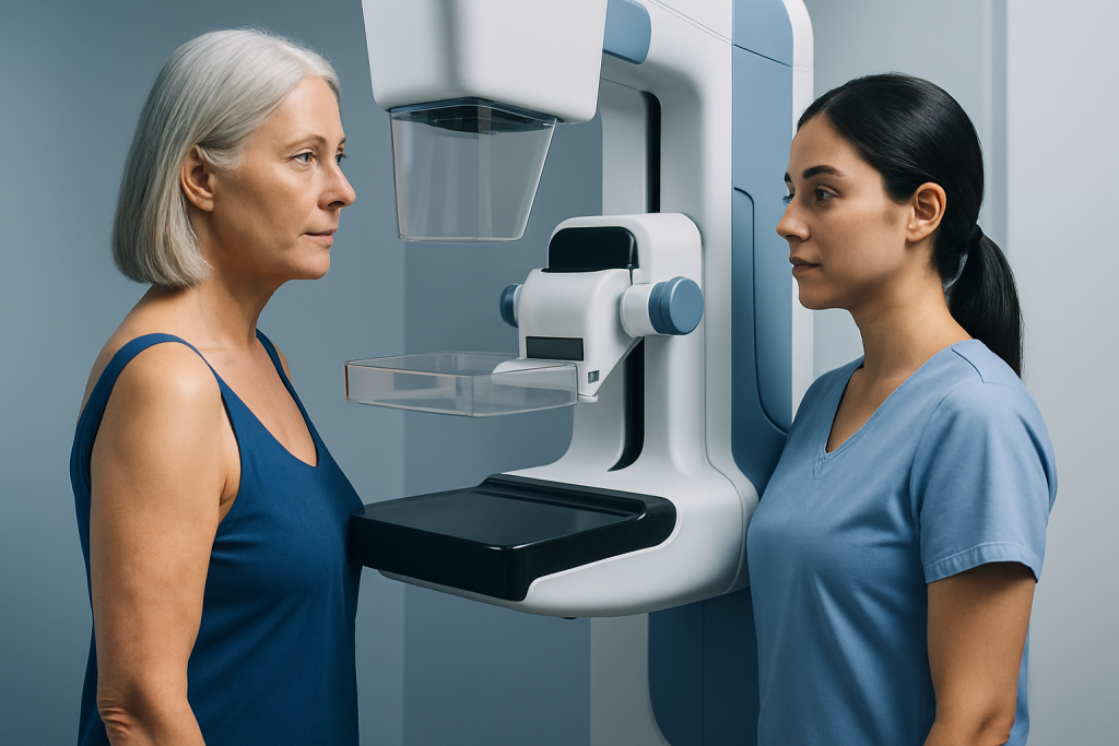
A screening mammogram uses low-dose X-rays to produce images of each breast, usually from more than one angle. Radiologists assess the breast tissue for masses, asymmetries, architectural distortion and calcifications. The images obtained may be technically similar for average- and high-risk patients, but a high-risk patient may have additional imaging and closer follow-up.
For high-risk screening, breast MRI is commonly performed with intravenous contrast. The scan evaluates blood flow and enhancement patterns in breast tissue, which may reveal cancers not visible on mammography, especially in dense breasts. MRI does not replace mammography because mammography can identify some calcifications that MRI may not show as clearly.
Screening breast ultrasound is not routinely used as the sole screening tool for average-risk patients. It may be used to examine a specific finding, to assess a palpable lump, or as an additional test when MRI is unsuitable. The most appropriate plan should be individualized after discussion with a breast specialist or radiologist.
Who Is Considered High Risk for Breast Cancer Screening?

People are often considered high risk when their estimated lifetime chance of breast cancer is approximately 20% or higher using an appropriate risk-assessment model, or when they have another major risk factor. A clinician can calculate risk using personal history, family history and, where relevant, genetic information.
Higher-risk groups may include people with a BRCA1 or BRCA2 variant or another inherited cancer predisposition; those with a first-degree relative carrying a known harmful variant who have not yet been tested; and people who received radiation therapy to the chest at a young age. A history of lobular carcinoma in situ, atypical ductal hyperplasia or atypical lobular hyperplasia may also affect screening decisions.
A strong family history can be important even if genetic testing is negative or has not been done. For example, multiple close relatives with breast, ovarian, pancreatic or prostate cancer may warrant formal risk assessment and genetic counseling. Risk is not determined by family history alone; age, reproductive history, breast density, prior biopsies and other factors can also contribute.
- Average risk does not mean no risk; routine screening is still valuable.
- Breast density is common and should be discussed in the context of overall risk.
- Screening recommendations may differ according to age, sex assigned at birth, hormone exposure and previous breast cancer treatment.
What Type of Imaging Test Is Most Effective for Detecting Breast Cancer?
No single imaging test is best for every person. For population screening, mammography is the established main test because it can detect cancers before they can be felt and has evidence for reducing breast cancer mortality in appropriate age groups. Digital breast tomosynthesis, often called 3D mammography, may improve detection in some patients and reduce recall rates compared with standard 2D mammography.
For people at high risk, contrast-enhanced breast MRI is generally the most sensitive screening examination. It can find cancers that mammography misses, particularly in dense breast tissue. However, its higher sensitivity also means that it can lead to false-positive results, additional imaging and sometimes biopsies of areas that are ultimately benign.
Ultrasound can be a useful targeted examination, but it is operator-dependent and may produce more false-positive findings when used broadly for screening. A specialist team considers the expected benefit, individual risk, breast density, medical history and ability to receive contrast when selecting imaging.
What Is the Gold Standard for Breast Imaging?
Mammography is considered the standard first-line imaging test for breast cancer screening in average-risk populations. It is widely available, efficient and especially useful for detecting suspicious calcifications. However, it is not a perfect test, and cancers can occasionally be obscured by dense breast tissue or be difficult to distinguish from normal structures.
There is no universal single “gold standard” test for all breast imaging questions. For high-risk screening, the combination of annual mammography and contrast-enhanced breast MRI is often the preferred evidence-based approach. For evaluating a new symptom, such as a lump, doctors may use diagnostic mammography, targeted ultrasound and sometimes MRI depending on age and clinical findings.
When imaging identifies a suspicious area, biopsy is the only way to confirm whether cancer is present. Image-guided biopsy takes a small tissue sample for examination by a pathologist. The imaging test guides where to sample; it does not by itself provide a final cancer diagnosis.
What's the Difference Between High Grade and Low Grade DCIS?
DCIS, or ductal carcinoma in situ, is a non-invasive breast condition in which abnormal cells are found inside milk ducts and have not invaded surrounding breast tissue. It is commonly detected on mammography because it may produce clusters of calcifications. DCIS is not the same as invasive breast cancer, but it requires careful evaluation and management.
Grade describes how abnormal the DCIS cells look under a microscope and how quickly they may be expected to grow. Low-grade DCIS cells tend to look more similar to normal cells and generally grow more slowly. High-grade DCIS cells appear more abnormal and are more likely to be associated with features such as comedo necrosis, although grade alone does not determine every person’s treatment plan.
Management may involve breast-conserving surgery, radiation therapy after surgery in some cases, endocrine therapy for hormone receptor-positive disease, or mastectomy when appropriate. Decisions depend on the size and location of DCIS, margins after surgery, grade, receptor status, personal preferences and overall health. A multidisciplinary breast team can explain the expected benefits and trade-offs of each option.
What to Expect During Breast Screening
Before mammography, the imaging center may ask about pregnancy, breastfeeding, breast symptoms, implants, previous biopsies and family history. During the examination, each breast is briefly compressed between imaging plates to keep it still and obtain clear pictures. Compression can be uncomfortable for a short time but should not cause lasting harm.
For breast MRI, patients usually lie face down on a padded table with the breasts positioned in openings designed for imaging. An intravenous line is used to administer contrast material, and the table moves into the MRI scanner. The examination is painless, but the scanner is noisy and remaining still is important. People with severe claustrophobia, certain implanted devices, significant kidney disease or a previous contrast reaction should tell the care team in advance.
There is no recovery period after a mammogram or breast MRI, and most people return to normal activities immediately. A radiologist reviews the images and the center communicates the result. If additional views, ultrasound or MRI-guided assessment are recommended, this usually means more information is needed rather than that cancer has been diagnosed.
Benefits, Limitations and When to Seek Medical Care
The main benefit of tailored screening is earlier detection, when treatment may be less extensive and outcomes may be better. MRI can add important information for people at high risk, while mammography remains essential for many screening programs. The limitations include false-positive findings, anxiety while awaiting results, exposure to a small amount of radiation from mammography, and contrast-related considerations with MRI.
People should seek medical care promptly if they notice a new breast or underarm lump, persistent focal breast pain, skin dimpling, nipple inversion that is new, nipple discharge that is bloody or spontaneous, a rash or scaling of the nipple, or a change in breast size or shape. These symptoms are often caused by noncancerous conditions, but they should not be ignored and are evaluated differently from routine screening.
A person with a strong family history, a known genetic variant, previous chest radiation or an abnormal breast biopsy should ask their doctor for a formal risk review. Acibadem International’s multidisciplinary specialists and JCI-accredited hospitals support international patients with breast assessment, imaging and individualized treatment planning, including breast cancer treatment when it is needed.
Frequently asked questions
Does a high-risk breast screening image look different from a low-risk screening image?
The mammogram itself may look similar because the same basic imaging technique is used. The difference is that high-risk screening often includes breast MRI and may be performed on a more intensive schedule. Risk information also helps the radiologist and clinical team decide how to interpret findings and plan follow-up.
At what age should high-risk breast cancer screening start?
The starting age depends on the reason a person is considered high risk. Some people with inherited cancer-risk variants or previous chest radiation begin MRI and mammography earlier than average-risk screening populations. A breast specialist or genetics professional can recommend an age-appropriate schedule.
Can dense breasts make mammograms less accurate?
Yes. Dense tissue can make it harder to see some abnormalities on mammography because both dense tissue and many cancers can appear white on the image. Density should be considered alongside a person’s complete risk profile when deciding whether additional imaging may help.
Is breast MRI better than mammography?
Breast MRI is more sensitive for detecting many cancers, particularly in high-risk patients, but it is not automatically better for everyone. It can find harmless changes that require additional testing, and it may miss some calcifications best seen on mammography. For many high-risk patients, the tests are complementary rather than interchangeable.
Does an abnormal screening result mean breast cancer?
No. Many abnormal screening findings are caused by benign cysts, normal tissue overlap, hormonal changes or other noncancerous conditions. Additional views, ultrasound, short-term follow-up or biopsy may be recommended to clarify the finding.
Can DCIS be found by breast MRI?
MRI can detect some cases of DCIS, particularly when there is enhancement associated with abnormal blood vessels. However, mammography is especially important for detecting calcifications that may be linked to DCIS. The imaging approach is selected according to the clinical question and individual risk.
References
- American Cancer Society
- American College of Radiology
- National Comprehensive Cancer Network
- World Health Organization
- U.S. Food and Drug Administration
This article is for general information only and is not a substitute for professional medical advice. Please consult a qualified doctor about your individual situation.
Breast Cancer in Turkey — costs, top hospitals & a free quote
JCI-accredited · board-certified surgeons · reply within 24h
Add us as a Preferred Source to see more of our trusted health content across Google Search, AI Overviews and Discover.

Medical Oncology Department
Medical treatment of cancer with chemotherapy, immunotherapy and targeted therapies under a multidisciplinary tumor board.
60 specialists in this unit