How Is a Stroke Diagnosed? Brain Scans, Exams, and Emergency Tests

Stroke diagnosis is an emergency because treatment depends on how quickly the stroke type is identified. A CT scan is often the first brain scan because it can quickly show bleeding.
Key Takeaways
- Stroke diagnosis is an emergency because treatment depends on how quickly the stroke type is identified.
- A CT scan is often the first brain scan because it can quickly show bleeding.
- Doctors also use neurological exams, blood tests, and heart tests to look for the cause of stroke symptoms.
- Ischemic stroke and hemorrhagic stroke need different treatments, so accurate diagnosis is essential.
- Anyone with sudden face drooping, arm weakness, speech trouble, or other stroke warning signs should seek emergency care immediately.
Stroke diagnosis must happen quickly to tell whether symptoms are caused by a blocked artery, bleeding in the brain, or another condition. Doctors use a combination of medical history, physical and neurological exams, brain imaging, heart checks, and blood tests to guide safe treatment without delay.
Overview: Why stroke diagnosis is urgent
Stroke diagnosis is the process doctors use to confirm whether a person is having a stroke, identify the type of stroke, and decide on the safest treatment. This is a medical emergency because brain cells can be injured within minutes when blood flow is interrupted or when bleeding occurs in the brain.
There are two main types of stroke: ischemic stroke, caused by a blocked blood vessel, and hemorrhagic stroke, caused by bleeding in or around the brain. A transient ischemic attack, often called a TIA or “mini-stroke,” can cause similar symptoms for a short time and is also a warning sign that needs urgent evaluation.
Because these conditions can look similar at first, doctors do not rely on symptoms alone. They use a structured approach that may include a neurological examination, urgent brain imaging, blood tests, and heart tests. This helps them rule out other causes of sudden neurological symptoms, such as seizures, low blood sugar, migraine, infection, or brain tumors.
Fast, accurate diagnosis can improve outcomes. In many cases, it allows stroke teams to decide whether the person may benefit from time-sensitive treatments such as stroke treatment or other emergency interventions.
Symptoms and signs that prompt emergency testing
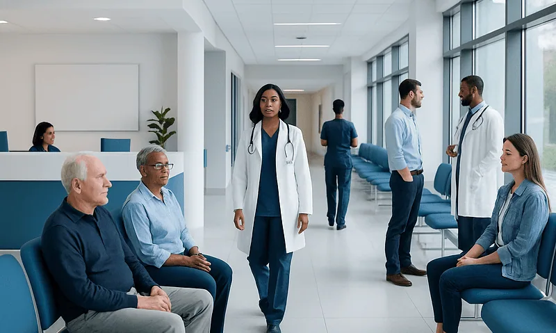
Stroke symptoms usually start suddenly. The most common warning signs include face drooping, weakness or numbness on one side of the body, trouble speaking, slurred speech, difficulty understanding language, vision changes, dizziness, loss of balance, and a severe sudden headache.
Emergency teams often use simple screening tools, such as the FAST approach: Face drooping, Arm weakness, Speech difficulty, Time to call emergency help. Some people may also have confusion, trouble walking, double vision, difficulty swallowing, or decreased alertness.
Symptoms can vary depending on which part of the brain is affected. For example, a stroke in the back of the brain may cause imbalance, vertigo, or coordination problems rather than obvious weakness. This is one reason stroke diagnosis requires a full medical assessment and not just a quick symptom check.
- Sudden numbness or weakness, especially on one side
- Sudden trouble speaking or understanding speech
- Sudden vision loss or blurred vision
- Sudden severe headache, especially with vomiting or confusion
- Sudden dizziness, difficulty walking, or loss of coordination
The first medical evaluation in the emergency department
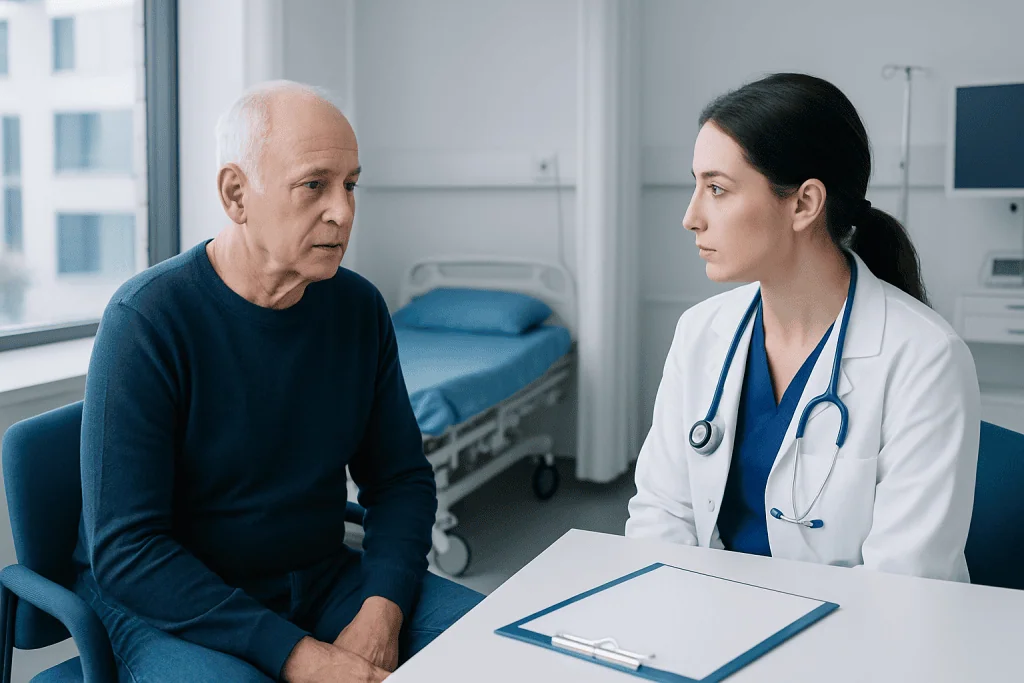
When a person arrives at the emergency department with possible stroke symptoms, the medical team begins with a rapid assessment. They check the time symptoms started or the last time the person was known to be well, because this timing affects treatment choices. If the exact time is unknown, the team gathers as much information as possible from the patient, family, or witnesses.
Doctors and nurses measure vital signs such as blood pressure, heart rate, breathing rate, temperature, and oxygen levels. They also check blood sugar right away, since very low or very high glucose can sometimes mimic stroke symptoms.
A focused neurological examination follows. This may include testing strength, sensation, coordination, speech, vision, reflexes, and level of consciousness. Many stroke teams use a standardized scoring system, such as the National Institutes of Health Stroke Scale, to describe how severe the symptoms are and to track changes over time.
The patient’s medical history also matters. Doctors ask about previous strokes or TIAs, high blood pressure, diabetes, smoking, atrial fibrillation, blood-thinning medicines, recent surgery, head injury, and any conditions that may increase bleeding risk. These details help the team interpret scan results and choose safe treatment.
Brain scans used to diagnose stroke
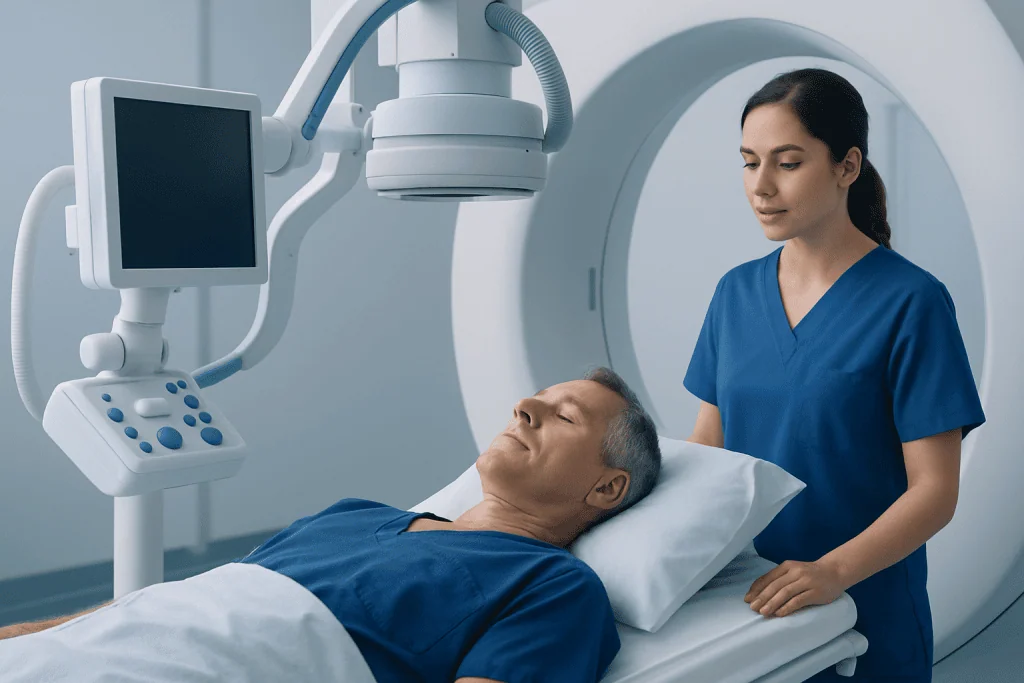
Brain imaging is one of the most important parts of stroke diagnosis. A non-contrast CT scan of the head is often the first test because it is fast and widely available. Its main early role is to show whether there is bleeding in the brain, which would point to a hemorrhagic stroke rather than an ischemic stroke.
If the CT scan does not show bleeding, doctors may look for signs of a blocked blood vessel. Additional imaging may include CT angiography, which uses contrast dye to show the brain’s blood vessels, and CT perfusion, which can help estimate how much brain tissue is at risk but may still be saved.
MRI can also be used in stroke diagnosis. It is especially helpful for detecting smaller strokes, early ischemic changes, or strokes in parts of the brain that may be harder to assess on CT. MRI is not always the first test in an emergency, but it can provide detailed information when the diagnosis is uncertain or when more precision is needed. Many patients benefit from advanced MRI and CT scan imaging during the diagnostic process.
In some cases, doctors may also order vascular imaging of the neck and brain arteries to look for narrowing, blockage, or dissection. This helps identify the cause of the stroke and guides decisions about procedures, medication, and long-term prevention.
Blood tests, heart tests, and other investigations
Stroke diagnosis is not based on brain scans alone. Blood tests help doctors look for medical problems that can mimic stroke, increase bleeding risk, or affect treatment choices. Common tests may include blood glucose, complete blood count, kidney function, electrolyte levels, and blood-clotting studies.
Doctors also evaluate the heart because some strokes happen when a blood clot forms in the heart and travels to the brain. An electrocardiogram can detect abnormal heart rhythms such as atrial fibrillation. Continuous heart rhythm monitoring may be used in the hospital if the cause is not immediately clear.
Some patients may need an echocardiogram, an ultrasound of the heart, to look for clots, valve problems, or structural conditions that may increase stroke risk. Ultrasound of the carotid arteries in the neck can help identify narrowing that limits blood flow to the brain.
Additional tests depend on the person’s age, symptoms, and medical history. If doctors suspect another condition rather than stroke, they may investigate for seizure, infection, migraine, multiple sclerosis, or a brain tumor. If symptoms were brief and resolved, the evaluation may focus on whether the event was a transient ischemic attack that still requires urgent prevention planning.
How doctors tell ischemic stroke from hemorrhagic stroke
The key goal of emergency stroke diagnosis is to distinguish ischemic stroke from hemorrhagic stroke. Although both can cause sudden neurological symptoms, their treatments are very different. A treatment that helps an ischemic stroke could be harmful if the patient actually has bleeding in the brain.
In ischemic stroke, a blood clot blocks an artery that supplies part of the brain. Brain imaging may show no bleeding at first, while vessel imaging can reveal a blockage. If the patient arrives within the right time window and meets safety criteria, doctors may consider clot-busting medication or a catheter-based procedure to remove the clot.
In hemorrhagic stroke, brain imaging shows bleeding inside the brain or around it. Treatment focuses on controlling blood pressure, managing bleeding risk, relieving pressure on the brain, and sometimes surgery or intensive neurocritical care. The diagnostic pathway may also include tests to look for the cause of bleeding, such as an aneurysm or malformed blood vessel.
Doctors also consider conditions that can mimic stroke. For example, seizures, complicated migraine, low blood sugar, infection, and medication effects can sometimes look similar in the early stages. Careful examination and imaging help reduce the chance of misdiagnosis.
What happens after the diagnosis is confirmed
Once stroke is confirmed, treatment planning begins immediately. The next steps depend on the stroke type, how severe it is, when symptoms began, and the patient’s overall health. The care team explains the findings and may start emergency treatment without delay if the person is eligible.
After the acute phase, doctors try to identify why the stroke happened so they can reduce the risk of another one. This may involve checking for high blood pressure, diabetes, high cholesterol, carotid artery disease, clotting problems, or heart rhythm disorders. Some patients need more detailed vascular imaging, longer heart monitoring, or specialist consultations.
Recovery planning is also important. Depending on the effects of the stroke, patients may need physical therapy, speech therapy, occupational therapy, swallowing assessment, or rehabilitation support. Ongoing follow-up helps adjust medicines and monitor recovery.
In centers with multidisciplinary stroke teams, evaluation and treatment are closely coordinated among emergency physicians, neurologists, radiologists, cardiologists, neurosurgeons, and rehabilitation specialists. Acibadem International’s multidisciplinary specialists and JCI-accredited hospitals diagnose and treat stroke for international patients, with access to imaging, acute care, and rehabilitation services in one system.
When to seek medical help and how to prepare
Anyone with sudden stroke-like symptoms should seek emergency medical help immediately, even if the symptoms improve quickly. A person should not wait to see if the symptoms go away, because a TIA can be a warning sign and some stroke treatments work only within limited time windows.
If possible, family members or bystanders should note when the person was last seen well, what symptoms started first, and whether the symptoms changed. Bringing a list of medicines, especially blood thinners, and a summary of medical conditions can help the emergency team work faster.
Stroke cannot always be prevented, but reducing risk factors can lower the chance of future events. Helpful steps include controlling blood pressure, managing diabetes and cholesterol, avoiding smoking, limiting alcohol, staying physically active, eating a balanced diet, and taking prescribed medicines regularly.
Regular checkups are important, especially for people with atrial fibrillation, vascular disease, or previous stroke or TIA. Prompt evaluation of warning signs and good long-term prevention can make a meaningful difference in brain health and recovery.
Frequently asked questions
What is the first test usually done for stroke diagnosis?
A non-contrast CT scan of the head is often the first test. It is fast and helps doctors quickly see whether there is bleeding in the brain. That information is essential because treatment depends on the stroke type.
Can a stroke be diagnosed without a brain scan?
Doctors may strongly suspect a stroke based on symptoms and the neurological exam, but brain imaging is usually needed to confirm the diagnosis and identify the type. Scans also help rule out other causes of sudden neurological symptoms. This makes treatment safer and more accurate.
Why are blood tests done during stroke evaluation?
Blood tests help doctors look for conditions that can mimic stroke and identify factors that affect treatment. They may show problems such as abnormal blood sugar, infection, anemia, kidney issues, or clotting abnormalities. These results support safe decision-making in the emergency setting.
Is MRI better than CT for diagnosing stroke?
MRI can be more sensitive for some early or small ischemic strokes, but CT is often used first because it is faster and very effective for detecting bleeding. The best test depends on the situation, the timing of symptoms, and what information the medical team needs urgently. In many cases, both CT and MRI have useful roles.
How do doctors know whether a stroke is caused by a clot or bleeding?
Doctors mainly use brain imaging to make this distinction. A CT scan can quickly show bleeding, while other imaging tests can reveal blocked blood vessels or areas of reduced blood flow. The exam findings and medical history also help guide interpretation.
What should someone do if stroke symptoms go away after a few minutes?
They should still seek urgent medical care right away. Brief symptoms can be caused by a transient ischemic attack, which may be an early warning sign of a future stroke. Even if the person feels normal again, prompt evaluation is important.
This article is for general information only and is not a substitute for professional medical advice. Please consult a qualified doctor about your individual situation.
Explore treatments in Turkey — costs, top hospitals & a free quote
JCI-accredited · board-certified surgeons · reply within 24h
Add us as a Preferred Source to see more of our trusted health content across Google Search, AI Overviews and Discover.
Check your numbers in seconds
BMI, calories, due date, blood pressure and 30+ more clinical calculators — free, instant, doctor-reviewed ranges.

More from the Health Library
Ischaemic Cerebrovascular Accident: Diagnosis and Emergency Treatment Options
Swallowing Problems After Stroke: Aspiration Risk, Testing, and Safe Eating
Ischemic Stroke Explained: Symptoms, Diagnosis, and First-Line Treatment
Cerebral Infarction: What It Means and How Recovery Is Managed
Medicines to Prevent Stroke: Who May Need Them and How They Work