How the Cerebral Cortex Is Organized: Lobes, Layers, and Key Functions
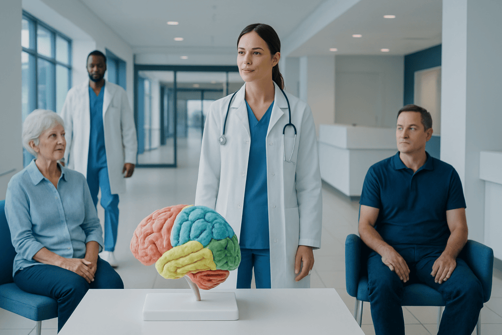
The cerebral cortex is the folded outer part of the brain involved in higher brain functions. It is divided into four main lobes: frontal, parietal, temporal, and occipital.
Key Takeaways
- The cerebral cortex is the folded outer part of the brain involved in higher brain functions.
- It is divided into four main lobes: frontal, parietal, temporal, and occipital.
- The cortex also has six microscopic layers, each with different cell types and connections.
- Many brain functions depend on networks that link multiple cortical regions rather than one isolated area.
- Symptoms from stroke, injury, seizures, or tumors often reflect which cortical area is affected.
The cerebral cortex is the brain’s outer layer, organized into specialized lobes, microscopic layers, and connected networks that support thinking, movement, language, sensation, and perception. Understanding this structure helps explain how the healthy brain works and why different symptoms can appear when specific cortical areas are affected.
Overview of the Cerebral Cortex
The cerebral cortex is the thin, wrinkled outer layer of the cerebrum, the largest part of the brain. Although it is only a few millimeters thick, it plays a central role in many of the abilities that make human behavior complex and adaptable. These include voluntary movement, sensation, language, attention, planning, memory, and conscious awareness.
Its folded surface creates ridges called gyri and grooves called sulci. This folding allows a large amount of cortical tissue to fit inside the skull. More surface area means more neurons and more connections, helping the brain process information efficiently.
Cerebral cortex organization can be understood at several levels. The broadest level is the division into lobes, each associated with certain functions. A deeper level looks at the six cellular layers of the cortex. Modern neuroscience also emphasizes networks, showing that most everyday tasks rely on communication between multiple regions rather than one single “center.”
How the Cortex Is Divided Into Lobes
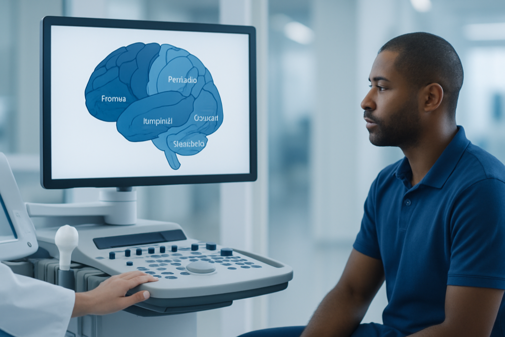
The cortex is traditionally divided into four main lobes in each hemisphere: the frontal, parietal, temporal, and occipital lobes. These divisions are based on visible landmarks on the brain’s surface. While each lobe has characteristic roles, they constantly exchange information through white matter pathways and functional networks.
The frontal lobe lies behind the forehead and is involved in planning, judgment, personality, problem-solving, and voluntary movement. It also contains important language areas in the dominant hemisphere for many people. The parietal lobe, located farther back, helps process touch, body position, spatial awareness, and aspects of attention.
The temporal lobe, found near the temples, contributes to hearing, language comprehension, emotion, and memory. The occipital lobe at the back of the head is primarily responsible for vision. In clinical practice, symptoms in one of these lobes may suggest which cortical region needs closer evaluation.
- Frontal lobe: planning, movement, behavior, executive function
- Parietal lobe: touch, spatial processing, body awareness
- Temporal lobe: hearing, memory, emotion, language understanding
- Occipital lobe: visual processing
Key Functions of Each Lobe

The frontal lobe contains the primary motor cortex, which helps initiate voluntary movements. Areas in front of it support more complex motor planning. Other frontal regions are essential for attention, self-control, decision-making, and organizing behavior over time. Damage here may affect personality, judgment, or the ability to carry out goal-directed tasks.
The parietal lobe includes the primary somatosensory cortex, which receives information about touch, pain, temperature, and body position. It also helps integrate sensory input so a person can recognize where the body is in space. Problems in this region may lead to difficulty recognizing objects by touch, neglect of one side of space, or trouble coordinating visually guided actions.
The temporal lobe contains the primary auditory cortex and areas important for understanding spoken language, especially in the dominant hemisphere. Deeper temporal structures work closely with the cortex to support memory and emotional processing. Conditions such as epilepsy can sometimes involve temporal lobe networks and affect awareness, memory, or perception during seizures.
The occipital lobe processes visual signals arriving from the eyes through the thalamus. Different occipital regions analyze features such as edges, motion, color, and form. Visual information then travels forward to temporal and parietal networks that help the brain recognize what is being seen and where it is located.
The Six Cortical Layers and Why They Matter
Under the microscope, most of the cerebral cortex is arranged into six layers. These layers are numbered from the surface inward. They differ in cell type, density, and the kinds of signals they send or receive. This layered structure helps the cortex sort information, combine it, and communicate with other parts of the nervous system.
Some layers receive incoming sensory information, especially from the thalamus. Others send signals to nearby cortical regions, to the opposite hemisphere, or to deeper structures such as the brainstem and spinal cord. For example, large pyramidal neurons in deeper layers of the motor cortex contribute to pathways that control movement.
Not every cortical area looks exactly the same. Sensory cortex, motor cortex, and association cortex have different layer patterns because they handle different kinds of information. This variation is one reason neurologists and neurosurgeons map the cortex carefully when evaluating disease or planning treatment such as awake craniotomy for selected patients.
Specialized Areas and Brain Networks
Classic brain maps describe specialized cortical regions, such as the primary motor cortex, primary visual cortex, and language areas. These maps remain useful, but they do not tell the whole story. Most human abilities depend on distributed networks that connect several cortical and subcortical areas working together in real time.
For example, speaking involves frontal language production regions, temporal language comprehension regions, motor control areas, hearing pathways, and attention networks. Memory draws on temporal lobe structures, frontal systems, and association cortex. Even a simple action like reaching for a cup requires visual, sensory, motor, and spatial systems to coordinate closely.
Modern brain imaging has helped show how these networks are organized. Techniques such as MRI and functional mapping can support diagnosis when symptoms suggest changes in cortical function. In some cases, doctors also use EEG to study electrical activity, especially when seizure-related cortical dysfunction is suspected.
How Doctors Relate Symptoms to Cortical Areas
Knowing how the cerebral cortex is organized helps doctors connect symptoms with likely brain regions. Weakness on one side of the body may suggest involvement of the motor cortex or its pathways. Numbness, altered body awareness, or problems with spatial attention can point toward the parietal lobe. Language difficulties may involve dominant frontal or temporal areas, while visual field problems often suggest occipital involvement.
Several medical conditions can affect the cortex, including stroke, head injury, infection, inflammation, tumors, and neurodegenerative disease. For example, brain tumors may cause symptoms based on location as well as size. A tumor near motor cortex may lead to weakness, while one near language cortex may affect speech or comprehension.
Cortical disorders do not always produce obvious symptoms at first. Some changes are subtle and may appear as difficulty concentrating, behavioral changes, short episodes of confusion, or sensory disturbances. Because many symptoms can have more than one cause, proper medical assessment is important whenever new neurological signs appear.
How Cortical Function Is Evaluated
Evaluation usually begins with a medical history and a neurological examination. A doctor may ask about weakness, numbness, balance, speech, memory, vision, headaches, or seizures. The examination can test strength, reflexes, sensation, eye movements, coordination, language, and higher cognitive functions, helping identify whether a cortical region may be involved.
Imaging studies are often used to look at brain structure. MRI is especially helpful for detailed views of cortical anatomy, while CT may be used in urgent settings such as suspected bleeding or acute stroke. Additional tools can include EEG, neuropsychological testing, vascular imaging, or functional imaging depending on the clinical question.
Treatment is based on the underlying cause rather than on cortical anatomy alone. Management may include medicines, rehabilitation, seizure care, surgery, or stroke treatment. Near the end of the care pathway, many patients benefit from a multidisciplinary approach; Acibadem International’s specialists in JCI-accredited hospitals evaluate and treat neurological conditions for international patients when advanced assessment is needed.
Protecting Brain Health and When to Seek Care
While cerebral cortex organization is largely determined by normal development, brain health can be supported throughout life. Good sleep, regular physical activity, balanced nutrition, blood pressure control, avoiding tobacco, and managing diabetes or high cholesterol all help protect the brain and its blood supply. Hearing and vision care, mental stimulation, and social engagement may also support cognitive health.
Prompt medical attention is important for sudden or progressive neurological symptoms. Emergency care is needed for signs such as sudden weakness, facial droop, trouble speaking, severe confusion, sudden vision loss, or a seizure. These can signal urgent conditions such as stroke or other serious brain disorders.
Non-emergency assessment is also worthwhile when symptoms are persistent, unexplained, or interfering with daily life. Ongoing memory changes, repeated headaches with neurological symptoms, episodes of altered awareness, or progressive changes in speech, behavior, or coordination should be discussed with a qualified doctor.
Frequently asked questions
What is the cerebral cortex in simple terms?
The cerebral cortex is the brain’s outer layer, often called gray matter. It helps a person think, feel sensations, move voluntarily, understand language, and interpret the world around them.
What are the four lobes of the cerebral cortex?
The four main lobes are the frontal, parietal, temporal, and occipital lobes. Each has important roles, but they work together through connected brain networks rather than acting alone.
Why does the cerebral cortex have folds?
The folds increase the surface area of the cortex. This allows more neurons and connections to fit within the skull, helping the brain handle complex functions more efficiently.
What are cortical layers?
Cortical layers are six microscopic levels of nerve cells found in most of the cerebral cortex. Different layers receive, process, and send information to other parts of the brain and nervous system.
Can one brain area control only one function?
Not usually. Some regions are specialized, but many tasks such as speech, memory, attention, and movement rely on multiple areas working together as networks.
What symptoms can happen if the cortex is affected?
Symptoms depend on the area involved and may include weakness, numbness, speech problems, vision changes, memory difficulties, seizures, or behavioral changes. New neurological symptoms should be assessed by a doctor, especially if they appear suddenly.
References
- National Institute of Neurological Disorders and Stroke
- National Institute of Mental Health
- Merck Manual Consumer Version
- MedlinePlus
- World Health Organization
This article is for general information only and is not a substitute for professional medical advice. Please consult a qualified doctor about your individual situation.
Explore treatments in Turkey — costs, top hospitals & a free quote
JCI-accredited · board-certified surgeons · reply within 24h
Add us as a Preferred Source to see more of our trusted health content across Google Search, AI Overviews and Discover.