Human Brain Anatomy: A Visual Guide to Major Regions
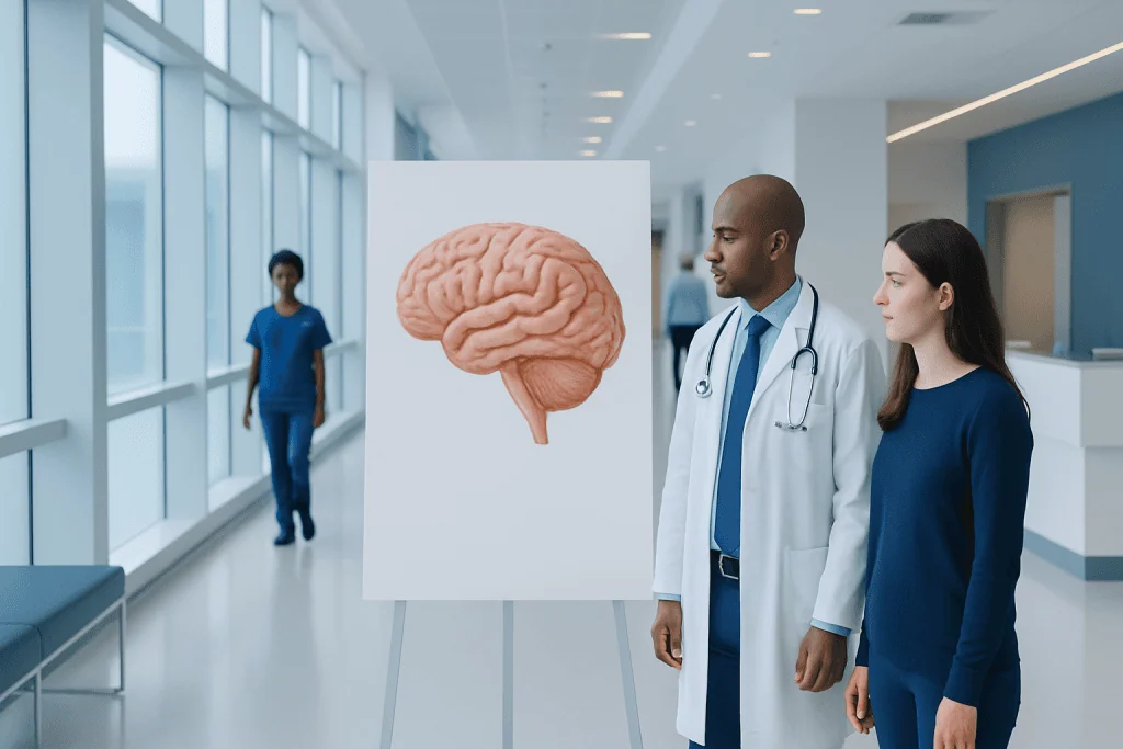
The brain is broadly divided into the cerebrum, cerebellum, and brainstem. Each brain lobe has specialized roles, including movement, sensation, language, memory, and vision.
Key Takeaways
- The brain is broadly divided into the cerebrum, cerebellum, and brainstem.
- Each brain lobe has specialized roles, including movement, sensation, language, memory, and vision.
- Deep brain structures help regulate emotion, memory, hormones, and automatic body functions.
- The right and left hemispheres communicate through nerve fibers, especially the corpus callosum.
- Imaging tests such as MRI and CT scans help doctors assess brain structure and possible disease.
Human brain anatomy can be easier to understand by looking at its major regions and what each one does. This guide explains the main parts of the brain, how they work together, and why this knowledge matters for health and diagnosis.
Overview of Human Brain Anatomy
Human brain anatomy refers to the structure of the brain and the way its different parts are organized. Although it weighs only a small portion of the body, the brain serves as the control center for thoughts, emotions, memory, movement, sensation, language, and many automatic functions such as breathing and heart rate. Understanding the major regions can make complex medical information much easier to follow.
The brain is part of the central nervous system, together with the spinal cord. It receives information from the senses, processes it, and sends signals back to the body. These messages travel through networks of nerve cells called neurons, supported by other important cells that protect and nourish brain tissue.
From a broad anatomical view, the brain has three main regions: the cerebrum, the cerebellum, and the brainstem. Inside and around these regions are additional structures with specific jobs, including the lobes of the brain, the thalamus, hypothalamus, limbic system, ventricles, and protective coverings called meninges.
A visual guide often begins with where each region sits in the skull and what it controls. This basic map helps patients better understand symptoms, test results, and conditions such as stroke, tumors, seizures, or Parkinson’s disease that may affect different brain areas in different ways.
The Cerebrum: The Largest Part of the Brain
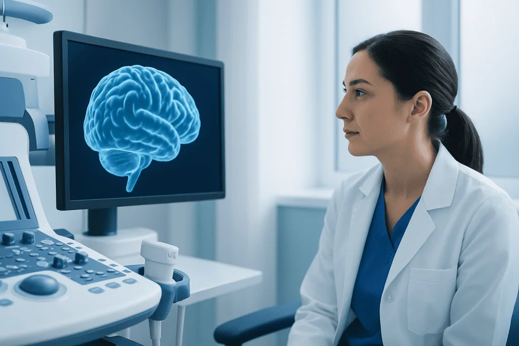
The cerebrum is the largest and most recognizable part of the brain. It fills most of the skull and is responsible for higher brain functions such as thinking, learning, memory, decision-making, voluntary movement, and interpretation of sensory information. Its outer layer is called the cerebral cortex, a folded surface that increases the area available for complex processing.
The cerebrum is divided into right and left hemispheres. In general, the left hemisphere is more involved in language for many people, while the right hemisphere often contributes more to spatial awareness and certain aspects of creativity. However, brain function is highly interconnected, and both hemispheres work together constantly.
Connecting the two hemispheres is a thick bundle of nerve fibers called the corpus callosum. This structure allows information to pass quickly from one side to the other. Smooth coordination of movement, problem-solving, and daily activities depends on this communication.
Within the cerebrum are both gray matter and white matter. Gray matter contains many neuron cell bodies and is heavily involved in processing information. White matter consists mainly of nerve fibers that connect different brain regions, helping signals travel efficiently across the brain.
The Brain Lobes and What They Do

Each cerebral hemisphere is commonly divided into four main lobes: frontal, parietal, temporal, and occipital. These lobes are not isolated compartments, but they do have well-known specialties. Recognizing them can help explain why a problem in one region may affect speech, memory, vision, or movement differently than a problem elsewhere.
The frontal lobe, located at the front of the brain, is involved in planning, judgment, attention, personality, emotional regulation, and voluntary movement. It also contains important areas related to speech production in many individuals. Damage here may affect behavior, concentration, movement, or expressive language.
The parietal lobe helps process touch, temperature, pain, pressure, and body position. It also contributes to spatial awareness and coordination. The temporal lobe, on the sides of the brain, plays a major role in hearing, language comprehension, and memory. The occipital lobe, at the back, is mainly responsible for visual processing.
- Frontal lobe: planning, decision-making, movement, behavior, speech production
- Parietal lobe: touch and sensory integration, body awareness, spatial processing
- Temporal lobe: hearing, language understanding, memory, emotion
- Occipital lobe: vision and interpretation of visual input
Deep Brain Structures: Memory, Emotion, and Regulation
Beneath the cerebral cortex lie deep brain structures that support essential functions. The thalamus acts as a relay station, directing much sensory information to the appropriate parts of the cortex. The hypothalamus helps regulate body temperature, hunger, thirst, sleep, and hormone-related processes by working closely with the pituitary gland.
The limbic system is a group of structures involved in emotion, motivation, and memory. It includes the hippocampus, which is central to forming new memories, and the amygdala, which helps process emotional responses such as fear and stress. These structures help explain why memory and emotion are closely linked.
The basal ganglia are another important set of deep structures. They help regulate movement, posture, and coordination of voluntary actions. Dysfunction in these networks is associated with movement disorders, including Parkinson disease and other neurological conditions that can affect walking, balance, or muscle control.
The brain also contains fluid-filled spaces called ventricles. These chambers hold cerebrospinal fluid, which cushions the brain, helps remove waste, and provides support. Problems with fluid flow or pressure may affect brain function and sometimes require specialized assessment.
The Cerebellum and Brainstem
The cerebellum sits at the back of the brain, below the cerebrum. Its main job is to fine-tune movement. It helps maintain balance, posture, coordination, and the smooth execution of muscle activity. Although it does not initiate movement on its own, it makes actions more accurate and controlled.
Damage to the cerebellum can lead to unsteady walking, poor coordination, tremor during movement, or difficulty with precise tasks. In some cases, speech may sound slow or less coordinated. These symptoms can result from stroke, inflammation, injury, tumors, or degenerative conditions involving the cerebellum.
The brainstem connects the brain to the spinal cord and includes the midbrain, pons, and medulla. This compact but vital region controls many automatic functions needed for life, including breathing, heart rate, blood pressure, swallowing, and wakefulness. It also contains pathways that carry signals between the brain and body.
Because the brainstem is so important, even small problems in this area can have significant effects. Doctors may use neurological examination and imaging to assess symptoms such as dizziness, double vision, weakness, swallowing difficulty, or changes in consciousness. In some situations, evaluation may involve advanced MRI scanning to study the brain in detail.
How the Brain Is Protected and Supplied
The brain is well protected by several layers. The skull forms the hard outer shield. Inside the skull are three protective membranes called the meninges: the dura mater, arachnoid mater, and pia mater. Between and around these layers, cerebrospinal fluid helps cushion the brain from everyday movement and minor impacts.
A healthy blood supply is equally important. Arteries carry oxygen-rich blood to brain tissue, and even brief interruption can damage sensitive cells. This is why stroke symptoms require urgent medical attention. The brain relies on a constant supply of oxygen and nutrients to maintain normal function.
The blood-brain barrier provides another form of protection. This specialized filtering system helps control which substances can move from the bloodstream into brain tissue. It protects the brain from many toxins and infections, while still allowing essential materials to pass through.
When doctors need to assess blood vessels or look for changes such as bleeding, blockage, or structural abnormalities, they may use imaging studies. Depending on the clinical situation, tests can include CT scanning or MRI, along with other neurological assessments.
How Doctors Study Brain Anatomy and Function
Doctors learn about brain anatomy through physical examination, neurological testing, and imaging. A neurological exam may assess strength, reflexes, sensation, coordination, speech, memory, and vision. The pattern of symptoms can offer valuable clues about which brain region may be involved.
Imaging tests play a central role in modern diagnosis. CT scans are often used in urgent situations because they can quickly show bleeding, major injury, or certain structural changes. MRI provides more detailed images of soft tissue and is especially helpful for evaluating tumors, inflammation, stroke, multiple sclerosis, and some causes of seizures or chronic headaches.
Other tests may be used when needed. Electroencephalography records electrical activity in the brain and can help assess seizure disorders. Functional studies may show which areas of the brain become active during tasks, and vascular imaging can help doctors examine blood flow or vessel abnormalities.
If a structural concern is found, treatment depends on the cause and location. Some patients may need medicine and monitoring, while others benefit from specialist care, rehabilitation, or procedures such as brain surgery when appropriate. A multidisciplinary approach helps match treatment to the specific brain region involved and the person’s symptoms.
Why Brain Anatomy Matters for Health
Knowing the major regions of the brain helps patients better understand symptoms and medical advice. For example, weakness on one side of the body may point to the motor areas of the cerebrum, while balance problems may suggest cerebellar involvement. Speech or memory changes can also be traced to specific regions, which helps guide diagnosis.
Brain anatomy is also important in conditions such as stroke, epilepsy, traumatic brain injury, dementia, infections, and tumors. The exact location of a problem often shapes both symptoms and treatment options. Even when two people share the same diagnosis, the effects may differ depending on which structures are affected.
Protecting brain health involves practical everyday steps. These include managing blood pressure, blood sugar, and cholesterol; avoiding smoking; limiting head injury risk; getting enough sleep; staying physically active; and seeking prompt medical care for sudden neurological symptoms. Learning the basics of anatomy can make it easier to recognize when something needs attention.
For people who need specialist evaluation, comprehensive neurological care may include imaging, laboratory testing, rehabilitation, and surgery when necessary. Acibadem International’s multidisciplinary specialists and JCI-accredited hospitals evaluate and treat brain-related conditions for international patients, with care tailored to the individual diagnosis and brain region involved.
Frequently asked questions
What are the three main parts of the brain?
The three main parts of the brain are the cerebrum, cerebellum, and brainstem. The cerebrum handles higher functions such as thinking and voluntary movement, the cerebellum helps with balance and coordination, and the brainstem controls many automatic life-sustaining functions.
What do the brain lobes control?
The frontal, parietal, temporal, and occipital lobes each have important roles. Together they help manage movement, sensation, language, memory, behavior, hearing, and vision, although most tasks involve several regions working at the same time.
What is the difference between gray matter and white matter?
Gray matter mainly contains nerve cell bodies and is heavily involved in processing information. White matter is made largely of nerve fibers that connect brain regions, allowing signals to travel efficiently from one area to another.
Why is the brainstem so important?
The brainstem is essential because it helps control breathing, heart rate, blood pressure, swallowing, and alertness. It also acts as a major communication pathway between the brain and the rest of the body.
How do doctors see brain anatomy?
Doctors commonly use imaging tests such as CT and MRI to look at brain structure. They also use neurological examinations and, when needed, additional tests such as EEG or vascular imaging to understand how the brain is functioning.
Can symptoms show which part of the brain is affected?
Often, yes. For example, vision changes may suggest occipital lobe involvement, balance problems may point to the cerebellum, and speech difficulty may relate to areas in the frontal or temporal lobes. A doctor’s assessment is still important because symptoms can overlap.
References
- World Health Organization
- National Institute of Neurological Disorders and Stroke
- National Institutes of Health
- MedlinePlus
- Radiological Society of North America
This article is for general information only and is not a substitute for professional medical advice. Please consult a qualified doctor about your individual situation.
Joint and spine care in Turkey — expert assessment & treatment
JCI-accredited · board-certified surgeons · reply within 24h
Add us as a Preferred Source to see more of our trusted health content across Google Search, AI Overviews and Discover.
Check your numbers in seconds
BMI, calories, due date, blood pressure and 30+ more clinical calculators — free, instant, doctor-reviewed ranges.