Hypoechoic Nodule: What Patients Need to Know

A hypoechoic nodule is an ultrasound description, not a final diagnosis. Many hypoechoic nodules are benign, but some need closer evaluation.
Key Takeaways
- A hypoechoic nodule is an ultrasound description, not a final diagnosis.
- Many hypoechoic nodules are benign, but some need closer evaluation.
- The meaning depends on where the nodule is found, such as the thyroid, breast, liver, kidney, or soft tissue.
- Ultrasound features such as margins, calcifications, blood flow, and growth help guide next steps.
- Further evaluation may include repeat imaging, blood tests, or a needle biopsy.
- Patients should seek medical review if a nodule is new, enlarging, painful, or linked with warning symptoms.
A hypoechoic nodule is a darker-than-surrounding area seen on an ultrasound scan. By itself, it does not confirm cancer or any single disease; doctors interpret it together with the nodule’s size, shape, location, and other imaging and clinical findings.
Overview: what a hypoechoic nodule means
A hypoechoic nodule is a term used in ultrasound reports. It describes an area that appears darker than the surrounding tissue because it reflects fewer ultrasound waves. This is a visual pattern, not a diagnosis on its own.
Hypoechoic nodules can be found in different parts of the body, including the thyroid, breast, liver, kidneys, lymph nodes, and soft tissues. Some are harmless and related to common benign changes such as cystic change, inflammation, or noncancerous growths. Others may need more testing to clarify what they are.
The most helpful way to understand a hypoechoic nodule is to look beyond the single word “hypoechoic.” Doctors also consider the nodule’s location, size, shape, borders, internal pattern, blood flow, and whether it has changed over time. The patient’s age, symptoms, medical history, and physical examination also matter.
For many patients, the next step is careful interpretation rather than urgent treatment. In some cases, a clinician may recommend monitoring with repeat ultrasound. In others, blood tests, additional imaging, or a biopsy may be advised to identify the cause more clearly.
Where hypoechoic nodules are commonly found
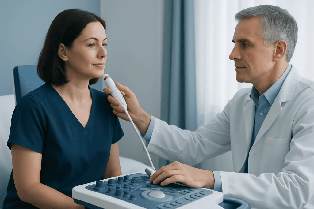
The significance of a hypoechoic nodule depends greatly on where it is seen. In the thyroid, it may represent a benign thyroid nodule, thyroiditis-related change, or less commonly a thyroid cancer. In the breast, hypoechoic nodules can correspond to fibroadenomas, cysts with complex features, areas of inflammation, or malignancy.
In abdominal organs such as the liver or kidneys, a hypoechoic nodule may reflect a benign lesion such as a focal area of altered tissue, a cyst with internal contents, or a solid mass that requires further characterization. In lymph nodes or soft tissues, hypoechoic areas may be linked to reactive change, infection, benign tumors, or other conditions.
This is why an ultrasound report is usually only one part of the evaluation. A thyroid nodule, for example, may be reviewed alongside thyroid hormone tests and, when indicated, a thyroid biopsy. A breast nodule may be interpreted with mammography, clinical examination, and sometimes breast biopsy.
If the report mentions related terms such as “solid,” “irregular margins,” “microcalcifications,” or “increased vascularity,” these details help doctors estimate how likely the finding is to be benign or whether more assessment is needed.
Symptoms and related signs

Many hypoechoic nodules do not cause symptoms and are found incidentally during imaging for another reason. When symptoms are present, they usually relate to the organ involved rather than to the ultrasound appearance itself.
For example, a thyroid nodule may cause a visible lump in the neck, pressure, trouble swallowing, hoarseness, or a feeling of fullness. A breast nodule may be felt as a lump or be associated with pain, skin changes, or nipple discharge. Liver or kidney nodules often cause no symptoms at all, though larger lesions may occasionally lead to discomfort.
Patients should also pay attention to general features that can help the doctor assess the situation. These include whether the nodule seems to be growing, whether there is pain or tenderness, and whether there are related symptoms such as fever, unexplained weight loss, persistent fatigue, or swollen lymph nodes.
- Commonly symptom-free and discovered on routine imaging
- May present as a lump, fullness, or local discomfort depending on location
- Associated symptoms can help narrow the cause
- Rapid change, persistent symptoms, or multiple warning signs deserve medical review
What causes a hypoechoic nodule and what raises concern
There is no single cause of a hypoechoic nodule. Benign causes include normal tissue variation, inflammation, scar tissue, adenomas, fibroadenomas, colloid nodules, cysts with internal debris, and other noncancerous growths. Infections and autoimmune conditions can also change tissue appearance on ultrasound.
Some nodules are more concerning because of their overall pattern, not simply because they are hypoechoic. Features that may prompt closer evaluation include irregular or blurred borders, a taller-than-wide shape in thyroid imaging, internal calcifications, marked internal blood flow, nearby abnormal lymph nodes, or documented growth on follow-up scans.
Risk factors also depend on the organ involved. For thyroid nodules, a history of neck radiation, family history of thyroid cancer, or certain inherited syndromes may matter. For breast nodules, age, personal history, family history, and hormonal factors may influence the level of concern. In the liver or kidneys, chronic disease history can shape the interpretation.
It can help to remember that concern is based on a combination of findings. A hypoechoic thyroid nodule, for instance, may still be low risk if it has smooth margins and stable size, while a different nodule with several suspicious features may need a more direct workup for thyroid cancer.
How doctors diagnose and assess a hypoechoic nodule
Evaluation usually begins with a review of the ultrasound report and the patient’s history. The clinician looks at the exact location, dimensions, shape, border definition, internal texture, and whether the area is purely solid or mixed solid and cystic. If prior imaging exists, comparing old and new studies is very helpful.
Additional tests depend on the body part involved. Thyroid nodules may be assessed with thyroid function tests and risk-stratified ultrasound criteria. Breast nodules may be correlated with mammography or MRI in selected cases. Liver, kidney, or other internal lesions may need contrast-enhanced CT or MRI to better define their nature.
When imaging cannot reliably confirm a benign cause, a needle biopsy may be recommended. Fine-needle aspiration is commonly used for the thyroid and some lymph nodes, while core needle biopsy may be preferred in breast or soft tissue lesions. If endocrine surgery becomes relevant after evaluation, treatment planning may involve specialists experienced in thyroid surgery.
The purpose of diagnosis is not only to detect serious conditions, but also to avoid unnecessary procedures when a nodule appears safely benign. A clear explanation from the care team can help patients understand why observation is appropriate in some cases and why tissue sampling is needed in others.
Treatment options and follow-up
Treatment is directed at the underlying cause, not at the word “hypoechoic” itself. If the nodule is benign and asymptomatic, no immediate treatment may be needed. Instead, the doctor may recommend observation with repeat ultrasound after a suitable interval to confirm stability.
If the nodule causes symptoms, grows, affects organ function, or has suspicious features, further treatment may be advised. This can include aspiration, medication for an associated inflammatory or hormonal condition, minimally invasive procedures, or surgery depending on the location and diagnosis.
When biopsy results show cancer or a precancerous process, care is usually planned by a multidisciplinary team. Management may vary widely between organs and between patients. For example, some thyroid cancers are treated surgically, while certain breast lesions may require surgery and other therapies based on pathology and stage; in selected settings this may be discussed within broader cancer treatment planning.
Near the end of the care pathway, patients often benefit from asking practical questions: what the report means, whether follow-up timing is important, which warning symptoms to watch for, and what type of specialist is best suited to their case. Acibadem International’s multidisciplinary specialists and JCI-accredited hospitals diagnose and treat this condition for international patients when specialist evaluation is needed.
Self-care, monitoring, and when to seek medical care
There is no home remedy that can diagnose or safely “shrink” a hypoechoic nodule without knowing its cause. The most useful self-care step is keeping follow-up appointments and bringing previous scan reports, pathology results, and a list of symptoms to the consultation. Patients should avoid assuming that internet descriptions apply to every organ or every ultrasound report.
General health measures may still support overall wellbeing. These include not smoking, following any organ-specific advice from the doctor, taking prescribed medicines as directed, and reporting new symptoms promptly. For thyroid health, it is especially important not to start iodine or hormone supplements unless a clinician recommends them.
Medical care should be sought promptly if a nodule is rapidly enlarging, newly painful, associated with hoarseness or trouble swallowing, causes breast skin or nipple changes, or is linked with fever, persistent swollen glands, or unexplained weight loss. Even when symptoms are mild, a new lump or a change in a known nodule deserves professional review.
In short, a hypoechoic nodule is a finding that needs context. Many cases turn out to be benign, but proper interpretation helps identify which nodules can simply be watched and which need tests or treatment.
Frequently asked questions
Is a hypoechoic nodule always cancer?
No. A hypoechoic nodule is an imaging description, not a diagnosis of cancer. Many are benign, and the level of concern depends on the nodule’s location, size, borders, internal features, and whether it changes over time.
What does hypoechoic mean on an ultrasound report?
Hypoechoic means the area looks darker than the surrounding tissue on ultrasound. This happens because it reflects fewer sound waves, but that pattern can be seen in both harmless and more serious conditions.
Does a hypoechoic thyroid nodule need a biopsy?
Not always. Doctors usually decide based on ultrasound features, size, symptoms, risk factors, and sometimes blood test results. Some thyroid nodules can be watched with repeat ultrasound, while others are better evaluated with needle biopsy.
Can a hypoechoic nodule go away on its own?
Some nodules remain stable for years, and some may change if they are related to inflammation or cystic processes. Others persist and simply need monitoring. Whether it can resolve depends on the cause and where it is located.
What symptoms should make someone seek medical attention sooner?
Faster medical review is wise if the nodule grows quickly, becomes painful, or causes pressure symptoms such as trouble swallowing or hoarseness. Breast skin changes, nipple discharge, fever, swollen lymph nodes, or unexplained weight loss should also be discussed promptly with a doctor.
Which doctor evaluates a hypoechoic nodule?
That depends on the organ involved. A primary care doctor may start the evaluation, but endocrinologists, radiologists, breast specialists, surgeons, gastroenterologists, urologists, or oncologists may be involved based on the location and test results.
This article is for general information only and is not a substitute for professional medical advice. Please consult a qualified doctor about your individual situation.
Oncology care in Turkey — second opinion and treatment plan
JCI-accredited · board-certified surgeons · reply within 24h
Add us as a Preferred Source to see more of our trusted health content across Google Search, AI Overviews and Discover.

More from the Health Library

Apricot Nut Benefits: An Evidence-Based Guide for Patients

Popliteal Vein: A Complete Medical Overview

How to get rid of male breast? A Doctor-Reviewed Answer

Mitochondria: A Complete Medical Overview

Garlic Pills: A Complete Medical Overview