Hypopigmentation: An Evidence-Based Guide for Patients

Hypopigmentation means areas of skin look lighter because they contain less melanin. Common causes include healed inflammation, skin injury, fungal infection, vitiligo, and certain medications or procedures.
Key Takeaways
- Hypopigmentation means areas of skin look lighter because they contain less melanin.
- Common causes include healed inflammation, skin injury, fungal infection, vitiligo, and certain medications or procedures.
- Diagnosis is based on the skin’s appearance, medical history, and sometimes tests such as Wood’s lamp examination, skin scraping, or biopsy.
- Treatment depends on the cause and may involve observation, topical medicines, light-based therapy, or managing an underlying condition.
- New, spreading, itchy, painful, or unexplained pale patches should be assessed by a qualified doctor or dermatologist.
Hypopigmentation is a term for skin that becomes lighter than a person’s usual skin tone because it has less melanin, the pigment that gives skin its color. It is a sign rather than a single disease, and causes range from minor skin irritation to chronic conditions that may need medical assessment.
Overview: what hypopigmentation means
Hypopigmentation is an area of skin that appears lighter than the surrounding skin because it contains less melanin. Melanin is the natural pigment made by specialized cells called melanocytes, and it helps determine the color of the skin, hair, and eyes. When melanin production decreases or pigment cells are damaged, the affected area may look pale, off-white, or lighter than a person’s usual complexion.
Importantly, hypopigmentation is not a diagnosis on its own. It is a visible skin change that can happen for many reasons, including inflammation, eczema, burns, infections, autoimmune conditions, and healing after cosmetic or medical procedures. In some people it is temporary and gradually improves; in others it can persist or progress depending on the underlying cause.
The appearance can vary widely. Some people notice one small patch after a rash heals, while others develop multiple spots or larger areas with clearer borders. Hypopigmentation can affect any part of the body and any skin tone, although it may be more noticeable in people with medium to dark skin.
Because the causes are different, the most helpful approach is not to focus only on the color change itself, but to understand what triggered it. A clinical evaluation can help distinguish simple post-inflammatory lightening from conditions such as vitiligo or other disorders that may need targeted treatment.
How hypopigmentation can look and feel
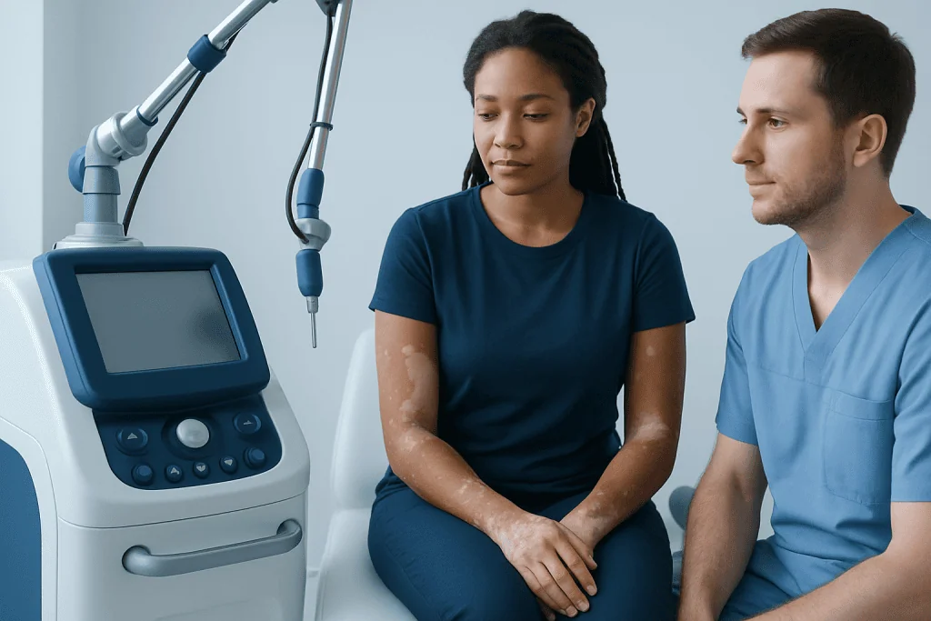
Hypopigmented areas may be lighter than the surrounding skin but not completely white. The edges may be sharply defined or more gradual. The skin may feel completely normal, or it may also be dry, scaly, sensitive, or slightly rough depending on the cause. Sometimes the hair in the area also becomes lighter.
Some people have no symptoms other than the change in color. Others notice itching, mild burning, flaking, or a previous rash in the same area. For example, after eczema, psoriasis, or minor skin injury heals, the skin may temporarily lose some pigment. In fungal infections, pale areas may be accompanied by fine scale.
Patterns can offer clues. Small scattered spots may follow a prior inflammatory rash; larger symmetrical patches may suggest an autoimmune process; and pale areas after a scratch, peel, laser treatment, or burn may point to skin injury. Children can also develop lighter facial patches from mild eczema or dryness.
Color change can affect confidence and emotional well-being, especially when it involves visible areas such as the face, hands, or neck. Even when the condition is medically harmless, it is reasonable to seek assessment and support if appearance-related distress is significant.
Common causes and risk factors

One of the most common causes of hypopigmentation is post-inflammatory change. This happens when the skin heals after eczema, acne, psoriasis, allergic reactions, friction, cuts, or burns. Inflammation can temporarily disrupt melanin production, leaving a lighter patch behind. In many cases the pigment slowly returns over time, although recovery can take months.
Infections can also lead to hypopigmentation. A common example is tinea versicolor, a superficial fungal condition that causes lighter or darker scaly patches, often on the chest, back, shoulders, or upper arms. Other causes include certain bacterial or viral skin conditions, though these are less common. When there is scale or a rash before color loss, infection is more likely to be considered.
Autoimmune and chronic skin conditions are another important group. Vitiligo occurs when pigment-producing cells are lost, leading to more clearly defined pale or white areas. Other inflammatory diseases, including eczema and some forms of dermatitis, may leave post-inflammatory hypopigmentation after the active rash settles. Genetic conditions, nutritional issues, and rare congenital disorders can also affect pigmentation.
Risk factors depend on the cause but may include a personal or family history of autoimmune disease, repeated skin irritation, prior inflammatory skin conditions, sun exposure that makes contrast more visible, and procedures that irritate the skin. Some people develop lighter patches after chemical peels, lasers, or other interventions, especially if the skin becomes inflamed during healing.
How doctors diagnose the cause
Diagnosis begins with a careful history and skin examination. A doctor will usually ask when the lighter area first appeared, whether there was a rash or injury before it, whether it is spreading, and whether there are symptoms such as itching, scale, pain, or sun sensitivity. Information about medicines, recent procedures, family history, and other health conditions can also be helpful.
The appearance and distribution of the patches often provide valuable clues. A dermatologist may use a Wood’s lamp, which emits ultraviolet light, to examine pigment changes more closely. In some conditions this can make affected areas more visible or highlight signs of infection. If a fungal cause is suspected, a skin scraping may be examined under a microscope.
Not every patient needs tests, but some do. If the diagnosis is unclear, doctors may recommend a skin biopsy to look at pigment cells and inflammation under the microscope. Blood tests may be considered when an autoimmune condition is suspected or when there are related symptoms, such as thyroid problems.
Accurate diagnosis matters because treatment differs by cause. A patch that needs only time and skin care should be managed differently from one caused by active inflammation or an autoimmune disorder. In centers with specialist dermatology services, evaluation may include dermatology assessment and, where appropriate, a structured medical check-up to look for associated conditions.
Treatment options and what improvement to expect
Treatment for hypopigmentation depends on the underlying condition, the size and location of the affected area, and how much it bothers the patient. In some cases, no active treatment is needed because pigment gradually returns on its own. This is often true for post-inflammatory hypopigmentation after eczema, minor injury, or temporary irritation.
When there is active inflammation, doctors may prescribe topical treatment to calm the skin and support recovery. If infection is the cause, antifungal or other targeted therapy may be used. For certain immune-related conditions, dermatologists may recommend medicated creams or ointments, and in selected cases light-based treatment may be discussed. Care should be individualized, especially for facial skin, children, and people with sensitive skin.
Cosmetic camouflage, tinted sunscreens, and careful sun protection can help reduce contrast between lighter and surrounding skin while natural repigmentation occurs. If the surrounding skin tans, hypopigmented areas may appear more obvious, so daily sun protection is often part of management. Patients should avoid trying harsh home remedies, scrubs, or unprescribed steroid creams, as these can worsen irritation and delay healing.
Some patients with persistent or extensive pigment loss may be referred for specialized care, including skin laser treatment or other advanced approaches when appropriate. Because results vary by cause and skin type, it is important to discuss realistic expectations. Improvement may be gradual, and complete repigmentation is not always possible.
Self-care, prevention, and living with skin color changes
Good skin care can help protect the skin barrier and reduce the chance of further irritation. Gentle cleansing, regular moisturizing, and avoiding friction are useful for many patients, especially when hypopigmentation follows eczema or dermatitis. Fragrance-free products are often preferred for sensitive skin.
Sun protection is one of the most practical self-care steps. Broad-spectrum sunscreen, shade, and protective clothing help prevent surrounding skin from darkening, which can make lighter areas stand out more. Sunburn should be avoided because it can trigger further inflammation and may worsen pigment changes.
Prevention also includes early treatment of skin conditions that cause inflammation. Managing eczema flares promptly, treating fungal infections appropriately, and following aftercare instructions after cosmetic procedures may reduce the risk of post-inflammatory pigment change. Picking, scratching, or using irritating over-the-counter products can prolong recovery.
Living with visible skin color differences can be emotionally difficult, even when the skin is otherwise healthy. Support from a doctor, dermatologist, or counselor may help if the condition affects self-image or social confidence. Near the end of the care pathway, some patients may also benefit from a multidisciplinary opinion; Acibadem International’s specialists in JCI-accredited hospitals evaluate and treat skin conditions for international patients when further assessment is needed.
When to seek medical care
Medical advice is recommended if hypopigmentation appears suddenly, spreads, or has no clear explanation. It is also sensible to seek assessment when lighter patches are associated with itching, scaling, pain, redness, hair color change, or a history of repeated recurrence. These features may point to an active skin disorder rather than a simple healing change.
Children should be evaluated if pale patches persist, increase in number, or are associated with rash or discomfort. Adults should also arrange a review if the color change affects the face, genitals, hands, or large areas of the body, or if there are signs of autoimmune disease such as other skin changes, fatigue, or thyroid-related symptoms.
People who recently had a peel, laser, burn, or other skin procedure should contact their treating clinician if they notice pronounced or worsening pigment loss. Early advice may help prevent further irritation and guide appropriate aftercare. If a diagnosis such as eczema or vitiligo is suspected, prompt evaluation can help clarify the cause and treatment options.
Urgent care is rarely needed for hypopigmentation itself, but any rapidly changing rash, severe pain, blistering, or signs of infection should be assessed without delay. When in doubt, a qualified doctor or dermatologist is the safest source of guidance.
Frequently asked questions
Is hypopigmentation the same as vitiligo?
No. Hypopigmentation is a general term for skin that becomes lighter than usual, while vitiligo is one specific cause of pigment loss. Many other conditions, including healed inflammation, fungal infection, or skin injury, can also cause lighter patches.
Can hypopigmentation go away on its own?
Sometimes it can. When hypopigmentation follows minor inflammation or injury, the skin may slowly regain pigment over weeks to months. Recovery time varies, and some cases last longer or remain permanent depending on the cause.
Does hypopigmentation mean a serious illness?
Not usually. In many people it is linked to a mild skin problem or a healing response after irritation. Still, persistent, spreading, or unexplained areas should be evaluated so that autoimmune or infectious causes are not missed.
How is hypopigmentation diagnosed?
Doctors diagnose it mainly through a skin examination and medical history. If needed, they may use a Wood’s lamp, perform a skin scraping for fungus, request blood tests, or take a small biopsy to confirm the cause.
What treatments are available for hypopigmentation?
Treatment depends on the reason the skin is lighter. Options may include observation, moisturizers, anti-inflammatory creams, antifungal treatment, sun protection, camouflage products, or specialist therapies for conditions such as vitiligo.
Can sun exposure make hypopigmentation look worse?
Yes. Sun exposure can darken the surrounding skin, making lighter patches more noticeable by contrast. Using sunscreen and protective clothing can help reduce this effect and protect sensitive skin.
This article is for general information only and is not a substitute for professional medical advice. Please consult a qualified doctor about your individual situation.
Explore treatments in Turkey — costs, top hospitals & a free quote
JCI-accredited · board-certified surgeons · reply within 24h
Add us as a Preferred Source to see more of our trusted health content across Google Search, AI Overviews and Discover.

More from the Health Library

Individual — Explained by Medical Evidence, Not Myths

Trichomoniasis: A Complete Medical Overview
Feet Flaky Skin: An Evidence-Based Guide for Patients

Extra Virgin Olive Oil Benefits — Explained by Medical Evidence, Not Myths

Thymus Gland: An Evidence-Based Guide for Patients