Idiopathic Intracranial Hypertension Treatment: How It Works, Results and What to Expect

Vision assessment is central to idiopathic intracranial hypertension treatment because sight-threatening changes may occur even when headache symptoms vary. Weight management can reduce intracranial pressure for many people with IIH when excess weight is a contributing factor.
Key Takeaways
- Vision assessment is central to idiopathic intracranial hypertension treatment because sight-threatening changes may occur even when headache symptoms vary.
- Weight management can reduce intracranial pressure for many people with IIH when excess weight is a contributing factor.
- Medicines may reduce cerebrospinal fluid production and help manage symptoms, but regular monitoring is needed.
- Surgery or venous sinus stenting may be considered when vision is worsening or symptoms remain severe despite appropriate medical care.
- New or rapidly worsening visual symptoms, severe headache, confusion or neurological changes require urgent medical assessment.
Idiopathic intracranial hypertension treatment aims to lower pressure around the brain, relieve symptoms and, most importantly, protect vision. Care is individualized and may combine weight management, medication, close eye monitoring and procedures when vision is at immediate risk.
Overview: How Idiopathic Intracranial Hypertension Treatment Works
Idiopathic intracranial hypertension (IIH) is a condition in which pressure inside the skull is raised without a brain tumor, hydrocephalus or another clear structural cause. It is also sometimes called pseudotumor cerebri. The raised pressure can affect the optic nerves, causing swelling known as papilledema and placing vision at risk.
Idiopathic intracranial hypertension treatment works by reducing pressure, addressing contributing factors and monitoring the optic nerves closely. The treatment plan is guided by the severity of visual changes, headache burden, general health, medicines being taken and imaging and spinal fluid test results. Most people are cared for by a team that may include neurology, neuro-ophthalmology, ophthalmology, radiology, nutrition specialists and, when needed, neurosurgery.
Not every person needs a procedure. For stable IIH without rapidly progressive visual loss, clinicians commonly begin with lifestyle support, medication and scheduled eye examinations. If vision is deteriorating, treatment may need to be escalated quickly to protect the optic nerves.
Symptoms, Causes and Risk Factors
Headache is common in IIH and may be daily or resemble migraine. Other possible symptoms include brief episodes of dimmed or blurred vision, double vision, blind spots, pulsating noise in the ears, neck or back pain, nausea and light sensitivity. Symptoms alone do not reliably show how much the optic nerves are affected, so formal visual testing is important.
The exact cause of IIH is not fully understood. It is more common in women of childbearing age and is associated with recent weight gain or obesity, although it can occur in people of any sex, age or body size. Certain medicines and medical conditions can also contribute to raised intracranial pressure and must be considered during assessment.
Doctors review factors such as vitamin A-related products, tetracycline-class antibiotics, steroid withdrawal, hormonal history, sleep apnea and endocrine or blood-related conditions when appropriate. Identifying a secondary cause is essential because the treatment may differ from treatment for truly idiopathic intracranial hypertension.
What triggers an IIH flare-up?
An IIH flare-up can mean worsening headache, pulsatile tinnitus or visual symptoms, but these symptoms do not always indicate a definite rise in pressure. Weight gain, missed treatment, sleep disruption, migraine, medication changes and untreated sleep apnea may contribute for some people. A new medicine or worsening symptoms should be discussed with the treating clinician rather than managed by stopping prescribed medication independently.
Assessment and Candidacy for Different Treatments

Diagnosis usually requires a careful eye examination, visual field testing and imaging of the brain, often including imaging of the veins in the brain. A lumbar puncture may then measure cerebrospinal fluid opening pressure and check that the fluid does not show another cause of illness. These tests also help rule out conditions such as a blood clot in the brain’s venous system.
People are candidates for conservative treatment when vision is stable and there is no urgent threat to sight. This approach may include a supported weight-management plan, headache care and medicine that lowers cerebrospinal fluid production. People with moderate or severe papilledema, declining visual fields or vision that worsens quickly may need urgent procedural treatment.
Selection for surgery or stenting is individualized. Specialists consider the speed of visual loss, optic nerve findings, response to medication, anatomy on imaging, overall health and the likely balance of benefit and risk. Headache alone is not usually enough reason for an invasive pressure-lowering procedure unless there are additional clinical reasons.
Treatment Options: Medicines, Weight Management and Procedures
When appropriate, gradual, sustainable weight loss is a key part of idiopathic intracranial hypertension treatment. Even a modest reduction can improve pressure and papilledema for some people, while maintaining weight loss may lower the chance of recurrence. A clinician or dietitian can help create a plan that supports nutrition, physical ability, other health conditions and long-term goals.
Medicines that reduce cerebrospinal fluid production may be prescribed to help lower pressure and protect vision. They can cause side effects, so clinicians may check blood tests and review symptoms during treatment. Headaches may also need a separate plan, especially if they have migraine-like features; frequent use of pain relievers can sometimes worsen headache patterns.
If visual function is worsening despite medical management, clinicians may recommend an operation to relieve pressure around the optic nerve, known as optic nerve sheath fenestration, or a cerebrospinal fluid shunt to divert fluid elsewhere in the body. In selected patients with a significant narrowing of a brain venous sinus and a meaningful pressure gradient, venous sinus stenting may be discussed. These approaches are intended mainly to preserve vision and are not guaranteed to eliminate headaches.
For individuals with severe obesity when non-surgical weight-management measures have not achieved sufficient results, metabolic or bariatric surgery may be considered as part of broader care. This decision requires an individual assessment of risks, benefits and readiness for long-term follow-up.
What Happens During a Procedure and Recovery
The exact steps depend on the procedure. Optic nerve sheath fenestration is performed by an experienced eye or orbital surgeon to create a small opening in the tissue around the optic nerve, allowing pressure to be relieved. A cerebrospinal fluid shunt is placed by a neurosurgeon; a thin tube diverts fluid from the brain or lower spine to another area of the body, commonly the abdomen.
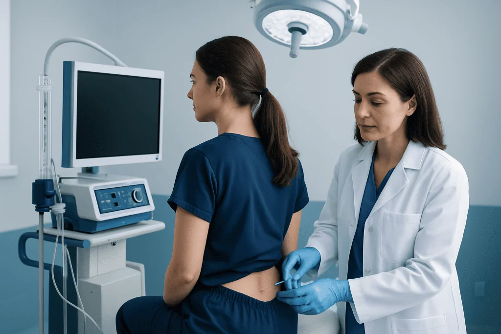
Venous sinus stenting is a catheter-based procedure performed in carefully selected cases. A specialist passes a thin tube through a blood vessel, usually from the groin or wrist, to place a small mesh tube in the narrowed venous sinus. Testing before and during the procedure helps determine whether the narrowing is clinically significant. People who receive a stent typically need medicines that reduce clotting risk for a period determined by their specialist.
Recovery varies. After a catheter-based procedure, a person may stay in hospital overnight or longer depending on symptoms and monitoring needs. Shunt or eye procedures can require a longer recovery, particularly if vision was seriously affected before surgery. Follow-up includes eye examinations, visual field testing and review of headaches, wounds, medicines and any new symptoms.
Benefits can include stabilization or improvement of vision, reduced papilledema and lower pressure-related symptoms. Risks differ by procedure and may include bleeding, infection, anesthesia complications, blood clots, shunt blockage or over-drainage, recurrent symptoms, double vision or the need for further procedures. The treating team explains the specific risks for the individual situation.
How Long Does It Take for Intracranial Hypertension to Go Away?
There is no single timeline for IIH to resolve. Some people improve over weeks to months after treatment begins, particularly when pressure falls and contributing factors are addressed. Others need treatment and monitoring for longer, and symptoms such as headache may persist even after papilledema improves.
Visual recovery depends partly on how severely and how long the optic nerves were affected. Early treatment of active papilledema gives the best opportunity to preserve sight. Follow-up should continue until clinicians confirm that optic nerve swelling and visual function are stable, and some people need longer-term review because IIH can recur.
What are the four stages of intracranial hypertension?
There is no universally accepted four-stage system for idiopathic intracranial hypertension. Clinicians instead assess severity using the appearance of papilledema, visual acuity, visual field testing, symptoms and the speed of change. Papilledema itself may be graded using the Frisén scale, which has more than four grades and is only one part of the overall assessment.
It is helpful to focus on whether IIH is stable, improving, active with mild visual effects, or rapidly threatening vision. This practical assessment guides how urgently treatment is needed and whether medical care alone is appropriate.
Vision Outlook and Ongoing Self-Care
The major concern in IIH is permanent visual loss caused by prolonged or severe optic nerve swelling. Regular eye testing can detect changes before a person notices a major loss of sight. Attending scheduled appointments, reporting new visual symptoms promptly and following the agreed treatment plan are important parts of protecting vision.
How many people with IIH go blind?
Most people with IIH do not become completely blind, especially when the condition is recognized and monitored promptly. However, a minority can develop permanent visual impairment, and the risk is higher when papilledema is severe or visual fields deteriorate quickly. Because published estimates vary according to the population studied and the definition of visual loss used, an individual’s outlook is best discussed with their eye and neurology team.
Self-care may include working toward a sustainable weight-management plan if recommended, getting adequate sleep, treating sleep apnea when present and avoiding unprescribed supplements or medicines that may affect intracranial pressure. People should not stop steroids, hormonal medication or other prescribed treatment without medical advice. Keeping a symptom record can help distinguish headache patterns from changes that need urgent eye assessment.
When to Seek Medical Care
Urgent medical assessment is needed for new or rapidly worsening blurred vision, loss of side vision, persistent double vision, a sudden marked change in vision, severe unusual headache, weakness, confusion, fainting, fever with severe headache or symptoms after a head injury. These symptoms can have causes other than IIH and should not be assumed to be a flare-up.
People already diagnosed with IIH should contact their care team promptly if visual obscurations become more frequent, headaches change substantially, pulsatile tinnitus worsens, medication side effects become difficult to manage or a planned follow-up cannot be attended. Regular ophthalmic monitoring is especially important during active disease, pregnancy planning or medication changes.
Acibadem International’s multidisciplinary specialists and JCI-accredited hospitals can assess and treat IIH for international patients, with coordinated neurology, neuro-ophthalmology, imaging and neurosurgical care when needed.
Frequently asked questions
Can idiopathic intracranial hypertension be cured?
IIH can improve substantially and may go into remission, particularly when contributing factors are addressed and intracranial pressure is controlled. However, recurrence can occur, so follow-up is important even after symptoms improve. The aim of care is to preserve vision, reduce pressure and manage ongoing symptoms safely.
Is a lumbar puncture a treatment for IIH?
A lumbar puncture is mainly used to help diagnose IIH by measuring opening pressure and testing cerebrospinal fluid. Removing fluid may provide temporary symptom relief for some people, but repeated lumbar punctures are generally not a long-term treatment strategy. A specialist can explain whether it has a role in an individual situation.
Will weight loss help idiopathic intracranial hypertension?
Weight loss can reduce intracranial pressure and improve papilledema in many people when excess weight is a relevant factor. The most suitable approach is gradual, nutritionally adequate and supported by a healthcare professional. Weight management is only one part of care and does not replace eye monitoring or prescribed treatment.
Can IIH headaches continue after pressure improves?
Yes. Some people continue to have headaches even after papilledema and intracranial pressure improve, often because headache disorders such as migraine may coexist. Headache treatment may therefore need a separate, individualized plan.
When is surgery needed for idiopathic intracranial hypertension?
Surgery is usually considered when vision is worsening quickly, papilledema remains severe or medical treatment has not adequately controlled a sight-threatening situation. The choice between optic nerve sheath fenestration, shunting and other options depends on symptoms, examination findings and local specialist expertise. Procedures have risks and require ongoing follow-up.
Can pregnancy affect IIH treatment?
IIH can occur during pregnancy, and treatment should be coordinated by obstetric, neurology and eye specialists. Some medicines may not be suitable during pregnancy or breastfeeding, while vision-threatening disease may still require urgent treatment. Anyone planning pregnancy or who becomes pregnant should contact their care team promptly.
References
- National Institute of Neurological Disorders and Stroke
- American Academy of Ophthalmology
- European Headache Federation
- Mayo Clinic
This article is for general information only and is not a substitute for professional medical advice. Please consult a qualified doctor about your individual situation.
Hypertension in Turkey — costs, top hospitals & a free quote
JCI-accredited · board-certified surgeons · reply within 24h
Add us as a Preferred Source to see more of our trusted health content across Google Search, AI Overviews and Discover.