Is an Echocardiogram an Ultrasound: Preparation, Procedure and Results

An echocardiogram is a heart ultrasound and does not use radiation. A standard transthoracic echocardiogram is painless, non-invasive and usually needs little preparation.
Key Takeaways
- An echocardiogram is a heart ultrasound and does not use radiation.
- A standard transthoracic echocardiogram is painless, non-invasive and usually needs little preparation.
- Different types of echocardiograms may be used depending on the clinical question, including Doppler, stress and transesophageal studies.
- Results are interpreted alongside symptoms, examination findings and other tests; an unexpected finding does not automatically mean urgent treatment is needed.
- Patients should seek urgent medical care for symptoms such as severe chest pain, fainting or sudden shortness of breath.
Yes, an echocardiogram is an ultrasound of the heart. It uses harmless high-frequency sound waves to create real-time images that help clinicians evaluate heart chambers, valves, pumping function and blood flow.
Overview: Is an Echocardiogram an Ultrasound?
Yes. An echocardiogram is an ultrasound examination of the heart. A trained sonographer or cardiologist places a small handheld device, called a transducer, on the chest. The device sends sound waves into the body and receives returning echoes, which a computer converts into moving images of the heart.
Unlike an X-ray or CT scan, echocardiography does not use ionising radiation. It can show the size and shape of the heart, how well its chambers contract, how heart valves open and close, and the direction and speed of blood flow. It is one of the most commonly used tests for investigating heart symptoms and monitoring known heart conditions.
The most common form is a transthoracic echocardiogram, often called a TTE or standard echo. Other forms, such as Doppler echocardiography, stress echocardiography and transesophageal echocardiography, provide additional information in selected situations.
How Echocardiography Works and What It Can Show
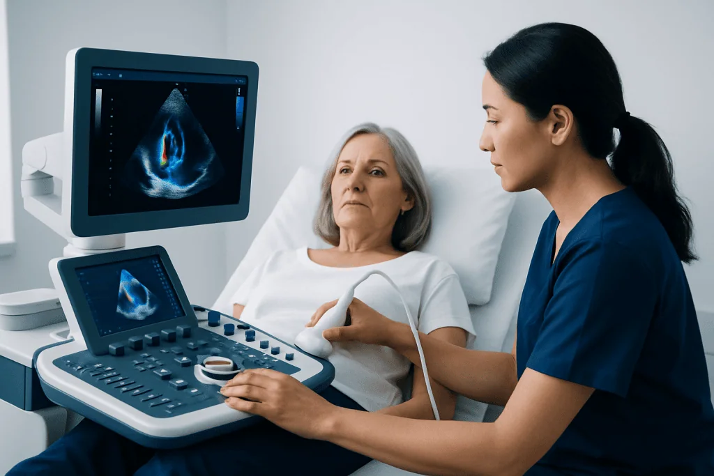
Ultrasound waves travel through body tissues differently depending on the structure they meet. The echocardiography machine processes these returning echoes to produce images of the heart in motion. Gel applied to the skin helps remove air between the transducer and the chest, allowing sound waves to travel more effectively.
Clinicians use the images to assess the heart muscle, the upper and lower chambers, the outer lining around the heart and the major blood vessels near the heart. Doppler ultrasound adds information about blood flow. It may help identify leakage through a valve, narrowing of a valve, abnormal pressure patterns or an opening between heart chambers.
An echocardiogram may support assessment of symptoms such as breathlessness, chest discomfort, palpitations, swelling in the legs, dizziness or a heart murmur. It may also be used to monitor conditions including heart valve disease, heart muscle disorders or changes after a heart attack. The test is informative, but it is only one part of a full clinical assessment.
Who May Need an Echocardiogram?

A doctor may recommend an echocardiogram when a person has symptoms that could be related to the heart or when an examination suggests an abnormal heart sound. It may also be used after an electrocardiogram, blood test or chest imaging result raises a question about heart structure or function.
People with known high blood pressure, congenital heart disease, valve conditions, heart failure or a family history of certain heart muscle conditions may need echocardiography at intervals recommended by their clinician. It can also be performed before some procedures or treatments when an up-to-date assessment of heart function is important.
The test is generally suitable for adults and children, including pregnant people, because standard ultrasound does not involve radiation. The choice of test type depends on the person’s symptoms, medical history, physical examination and the detail that the treating team needs to see.
- A transthoracic echo is usually the first examination.
- A stress echo may be used to assess heart function during exertion or with medication that mimics exercise.
- A transesophageal echo may be advised when clearer views of certain structures are required.
Do I Need to Do Anything Special to Prepare for an Echocardiogram?
For a standard transthoracic echocardiogram, most people do not need special preparation. They can usually eat, drink and take regular medicines as normal unless their doctor gives different instructions. Wearing a two-piece outfit may make it easier to undress from the waist up for the examination.
It is helpful to bring a medication list and details of prior heart tests, if available. Patients should tell the team about symptoms, allergies, pregnancy and any medical conditions that may affect the examination. They should not stop prescribed medicines without guidance from the clinician who requested the test.
Preparation differs for some specialised echocardiograms. A transesophageal echo commonly requires fasting beforehand because a probe is passed into the oesophagus, and sedation may be used. A stress echo may involve instructions about caffeine, food, exercise and selected medicines. The hospital or clinic will provide specific instructions before these tests.
Step by Step: What Happens During an Echocardiogram?
For a standard echo, the patient changes into a gown or removes clothing from the upper body and lies on an examination couch, often turned slightly onto the left side. Small sticky electrodes may be placed on the chest to record the heart rhythm during the scan. The sonographer applies warm or cool gel to several areas of the chest.
The transducer is moved gently over the chest and sometimes just below the ribcage or near the base of the neck. Pressure may be felt when images are obtained between the ribs, but the examination should not be painful. The patient may be asked to change position, breathe in or out, or briefly hold their breath to improve image quality.
Images and measurements are recorded for a cardiologist or appropriately trained clinician to interpret. The scan itself is usually quiet, although blood-flow Doppler may create whooshing sounds. A standard transthoracic echocardiogram is an outpatient test, and the patient can generally leave immediately afterwards.
How Long Does an Echogram Usually Take?
The term “echogram” is often used informally to mean an echocardiogram. A standard transthoracic echocardiogram commonly takes about 30 to 60 minutes, although the time can vary. Additional images may be needed if the heart is difficult to visualise, if detailed measurements are required or if previous results need comparison.
A stress echocardiogram usually takes longer because it includes preparation, exercise on a treadmill or bicycle, or medication administration when exercise is not suitable. A transesophageal echocardiogram also requires more time for fasting checks, sedation, monitoring and recovery.
Patients may wish to allow extra time for registration, changing clothes and discussion of next steps. The imaging professional performing the test may not always provide final results immediately, as the recorded images often need formal review by a cardiologist.
Benefits, Risks and Recovery After a Heart Ultrasound
The main benefit of echocardiography is that it provides detailed, real-time information without surgery or radiation exposure. It can help clarify whether symptoms may relate to pumping strength, valve function, fluid around the heart or blood-flow changes. It may also help a care team decide whether monitoring, further testing or treatment is appropriate.
There are no known significant risks from a routine transthoracic echocardiogram. Some people notice mild temporary discomfort from pressure on the chest or skin irritation from electrode pads. Recovery is immediate, and normal activities can usually resume straight away.
Specialised tests have different considerations. Exercise or medication used during stress echocardiography can temporarily cause symptoms such as tiredness, palpitations or breathlessness, so patients are monitored closely. Transesophageal echocardiography has small risks related to sedation and passage of the probe, including throat discomfort; patients generally need someone to accompany them home after sedation and should follow the aftercare instructions provided.
What Happens If They Find Something on an Echocardiogram?
An abnormal or unexpected result does not by itself establish a diagnosis or mean that immediate treatment is required. Echocardiogram findings must be considered together with symptoms, medical history, physical examination, blood pressure, heart rhythm and, when needed, other tests.
The report may describe findings such as changes in heart chamber size, reduced or increased pumping function, a valve that is narrowed or leaking, a thickened heart muscle, fluid around the heart or an area that needs closer assessment. Some findings are mild and are followed over time, while others may lead to blood tests, rhythm monitoring, CT or MRI imaging, or referral to a heart specialist.
Treatment depends on the underlying cause and its severity. Options can range from lifestyle measures and medicines to monitoring or procedures. If a structural heart problem needs further evaluation, a cardiology team may discuss appropriate options, including heart valve surgery when it is clinically indicated. Acibadem International’s multidisciplinary specialists at JCI-accredited hospitals evaluate and treat cardiovascular conditions for international patients.
When to Seek Medical Care
People should arrange a medical assessment for new or persistent breathlessness, reduced exercise tolerance, repeated palpitations, ankle swelling, unexplained fatigue, dizziness or a newly detected heart murmur. These symptoms have many possible causes, and an echocardiogram may or may not be needed after a clinician’s assessment.
Urgent medical care is important for severe or persistent chest pain, sudden severe shortness of breath, fainting, new weakness on one side of the body, confusion, or a rapid irregular heartbeat accompanied by feeling unwell. Emergency services should be contacted according to local guidance rather than waiting for an outpatient echocardiogram.
For people with an established heart condition, changes in symptoms should be discussed promptly with the treating clinician. Following planned appointments and taking medicines as prescribed can help the care team identify changes early and tailor follow-up appropriately.
Frequently asked questions
Does an echocardiogram count as an ultrasound?
Yes. An echocardiogram is an ultrasound scan focused on the heart. It uses sound waves, not radiation, to create moving images of heart structures and blood flow.
Is an echocardiogram painful?
A standard transthoracic echocardiogram is usually painless. Some pressure from the transducer may be felt against the chest, particularly between the ribs, but it should be brief and tolerable.
Can a person eat before an echocardiogram?
Most people can eat and drink normally before a standard transthoracic echocardiogram. However, fasting may be required for a transesophageal echocardiogram, and stress echocardiography may have separate instructions, so patients should follow the instructions from their care team.
When will echocardiogram results be available?
Timing varies by clinic and by the complexity of the study. A cardiologist may need to review the images and measurements before the final report is available, and the requesting clinician will explain what the findings mean for the individual.
Can an echocardiogram detect blocked arteries?
A resting echocardiogram does not directly show all coronary artery blockages. It may show changes in heart muscle movement that suggest reduced blood supply, while stress testing, CT coronary angiography or invasive angiography may be considered when coronary artery disease is suspected.
Can an echocardiogram show heart failure?
An echocardiogram can provide important information about pumping function, chamber size, valve function and pressure patterns that may support evaluation of heart failure. A diagnosis is based on the whole clinical picture, including symptoms, examination and other test results.
References
- American Heart Association
- American Society of Echocardiography
- National Heart, Lung, and Blood Institute
- Mayo Clinic
This article is for general information only and is not a substitute for professional medical advice. Please consult a qualified doctor about your individual situation.
Ultrasound in Turkey — costs, top hospitals & a free quote
JCI-accredited · board-certified surgeons · reply within 24h
Add us as a Preferred Source to see more of our trusted health content across Google Search, AI Overviews and Discover.
Check your numbers in seconds
BMI, calories, due date, blood pressure and 30+ more clinical calculators — free, instant, doctor-reviewed ranges.

Check-up & Preventive Medicine
Comprehensive health screenings designed for early detection and prevention.
1 specialists in this unit