Knee Images: What Patients Need to Know

Knee images may include X-ray, MRI, CT, ultrasound, and sometimes nuclear medicine scans. Different tests answer different questions: X-rays show bones well, while MRI is better for soft tissues such as ligaments and menisci.
Key Takeaways
- Knee images may include X-ray, MRI, CT, ultrasound, and sometimes nuclear medicine scans.
- Different tests answer different questions: X-rays show bones well, while MRI is better for soft tissues such as ligaments and menisci.
- Imaging is usually chosen based on symptoms, examination findings, and how the injury happened.
- Not every painful knee needs immediate imaging; a doctor may recommend rest, examination, or follow-up first.
- Urgent assessment is important after major trauma, inability to bear weight, fever, severe swelling, or a locked knee.
Knee images are tests that show the bones, cartilage, ligaments, tendons, and other structures inside and around the knee. They help doctors identify the cause of pain, swelling, stiffness, instability, or injury and choose the most appropriate treatment.
Overview: what knee images show
Knee images are medical tests used to look inside the knee joint and surrounding tissues. They can show bones, joint space, cartilage, ligaments, tendons, muscles, fluid, and signs of wear or injury. In practice, the term may refer to an X-ray, MRI, CT scan, ultrasound, or another imaging study recommended for a specific reason.
Doctors request knee images to help explain symptoms such as pain, swelling, stiffness, clicking, locking, giving way, or reduced movement. Imaging can also be useful after a fall, sports injury, twisting injury, or overuse. The goal is not simply to “take a picture,” but to answer a clinical question clearly and safely.
One important point for patients is that no single test is best for every situation. A plain X-ray may be the first choice when a fracture or arthritis is suspected, while an MRI is often preferred when the concern is a meniscus tear, ligament injury, or cartilage damage. The best test depends on the symptoms, examination findings, age, activity level, and whether the problem is sudden or long-standing.
Common types of knee images
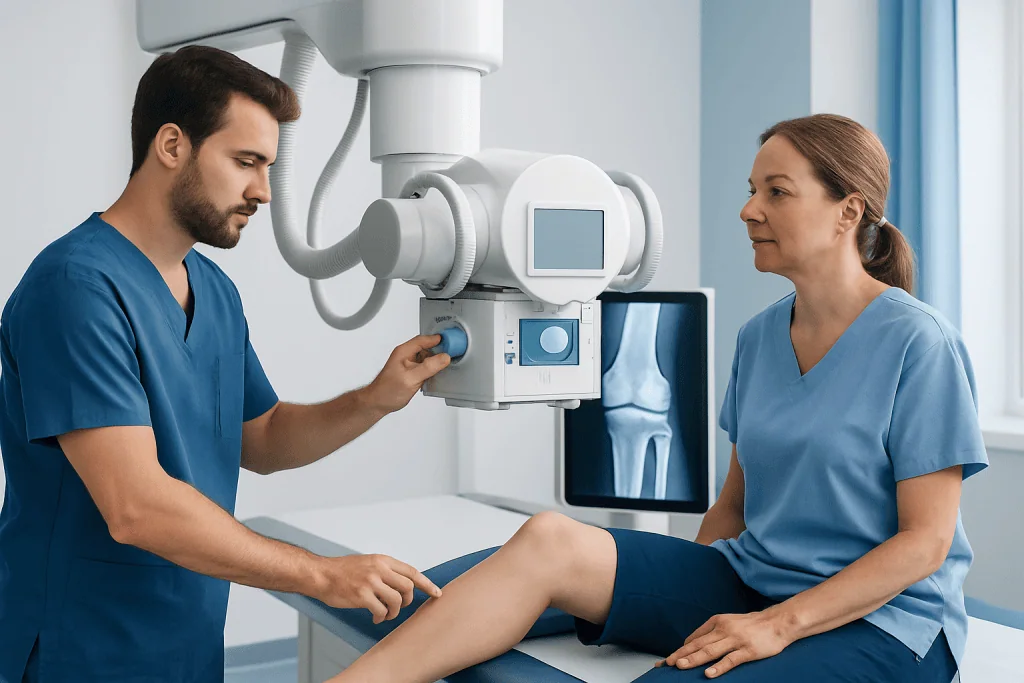
X-rays are often the starting point for knee imaging. They are quick, widely available, and very good at showing bones. A knee X-ray can help identify fractures, dislocations, joint narrowing from osteoarthritis, bone alignment problems, and some signs of inflammation or calcium deposits. However, X-rays do not show soft tissues such as ligaments and menisci well.
MRI uses magnetic fields rather than radiation. It gives detailed images of soft tissues and is especially helpful for tears of the meniscus, injuries to the anterior cruciate ligament or other ligaments, cartilage damage, tendon problems, bone bruising, and certain hidden fractures. MRI is commonly used when the physical examination suggests an internal knee injury or when symptoms continue despite initial care.
CT scans provide detailed cross-sectional images and can be useful for complex fractures or when doctors need a closer look at bony anatomy. Ultrasound can assess some soft-tissue problems around the knee, such as tendon irritation, fluid collections, bursitis, or cysts, and it can guide injections or fluid drainage. In selected cases, additional imaging may be used, but the main aim is always to match the test to the most likely cause of symptoms.
- X-ray: best for bones, fractures, alignment, arthritis
- MRI: best for ligaments, menisci, cartilage, tendons, bone marrow changes
- CT: useful for complex bone injuries and detailed bony structure
- Ultrasound: useful for fluid, tendons, bursae, and guided procedures
When doctors recommend knee imaging

Knee images are recommended when the findings from symptoms and physical examination suggest that a scan will change management. For example, imaging may be needed after a traumatic injury, if there is significant swelling, if the knee cannot fully straighten, or if the joint feels unstable. It may also be used when symptoms do not improve with time, activity changes, or conservative treatment.
In some situations, imaging helps confirm common conditions such as osteoarthritis, where X-rays may show joint-space narrowing, bone spurs, or other degenerative changes. In active individuals, imaging may be used to investigate tears involving the meniscus or ligaments, especially if there is clicking, locking, or repeated giving way. MRI is often especially informative in these cases.
Imaging can also support treatment planning. For example, a doctor may use the results to decide whether rehabilitation, bracing, injection therapy, or surgery is most appropriate. When symptoms point to advanced joint damage, imaging may help patients understand whether procedures such as knee replacement are being considered and what the likely goals of treatment would be.
What knee images can help diagnose
Knee imaging can help diagnose a wide range of problems. These include fractures, dislocations, cartilage wear, ligament tears, tendon injuries, meniscus tears, bursitis, cysts, joint effusions, and some bone or soft-tissue tumors. It can also show signs of inflammatory arthritis, infection, and complications after previous surgery.
In younger patients or athletes, MRI is often used when doctors suspect internal derangement of the knee, such as an ACL injury or a meniscus tear. In older adults, X-rays are frequently used to evaluate arthritis or chronic pain related to age-related joint changes. Sometimes imaging reveals more than one issue at the same time, such as arthritis together with a meniscal problem.
It is helpful for patients to know that imaging findings do not always match symptoms perfectly. Some people have visible changes on a scan but little pain, while others have significant symptoms with only subtle imaging findings. For that reason, doctors interpret knee images together with the history, examination, activity level, and overall health rather than relying on the scan alone.
What to expect during the test
Most knee imaging tests are straightforward. During an X-ray, the patient is asked to position the leg in one or more views while images are taken in a few minutes. CT scans are also relatively quick and involve lying still while the machine creates detailed cross-sectional images. MRI usually takes longer, often around 20 to 45 minutes depending on the study, because multiple detailed sequences are recorded.
Ultrasound is performed by moving a small handheld probe over the skin with gel. It is painless for most patients and allows the clinician to assess structures in real time. MRI does not use ionizing radiation, but patients need to remove metal items and tell staff about pacemakers, implants, or severe claustrophobia. If contrast material is considered, the team will explain why it is needed and review any relevant medical history.
Preparation is often minimal. Comfortable clothing is helpful, and patients should bring prior reports or scans if available. After the test, a radiologist reviews the images and sends a report to the referring doctor, who explains what the findings mean and whether further treatment, follow-up, or another imaging study is needed.
How imaging guides treatment and recovery
Knee images are valuable because they can direct treatment decisions more precisely. If imaging shows a simple strain or mild overuse problem, the plan may focus on rest, activity modification, ice, physiotherapy, and gradual strengthening. If it shows arthritis, treatment may include exercise therapy, weight management where appropriate, pain-relief strategies, and measures to support joint function.
When imaging identifies a structural injury such as a major ligament tear, displaced fracture, or severe cartilage damage, the next step may be referral to an orthopedic specialist. Some patients benefit from targeted procedures, including knee arthroscopy for selected internal knee problems. Others may need a structured rehabilitation program rather than surgery, even if the scan shows a tear.
Imaging also helps monitor progress after treatment. Follow-up images may be used after injury or surgery if healing is uncertain or symptoms persist. Near the end of the care pathway, some patients seek treatment at specialized centers; Acibadem International’s multidisciplinary specialists in JCI-accredited hospitals diagnose and treat knee conditions for international patients, including evaluation with MRI scan when clinically appropriate.
Limits of knee images and questions to ask
Although imaging is very useful, it has limits. A normal X-ray does not always rule out soft-tissue injury, and even MRI may show changes that are not the true cause of pain. Some findings are age-related and common in people without symptoms. This is why a scan is most helpful when it is ordered for a clear clinical reason.
Patients may want to ask what the doctor is looking for, which test is most suitable, whether radiation is involved, and how the result will affect treatment. It can also be helpful to ask whether immediate imaging is necessary or whether watchful waiting, home care, or physical therapy is reasonable first. These questions support shared decision-making and can prevent unnecessary tests.
If previous knee images have been done elsewhere, bringing them to the appointment can be very helpful. Comparing current and prior studies may show whether the problem is new, stable, or progressing. That comparison can improve diagnostic accuracy and may reduce the need for repeat imaging.
When to seek medical care
Medical care is advisable if knee pain is severe, follows an injury, or does not improve with basic measures such as rest and avoiding aggravating activity. A person should also arrange prompt assessment if the knee repeatedly gives way, becomes stuck or locked, swells significantly, or if walking and bearing weight are difficult.
Urgent evaluation is especially important after a major fall or accident, if there is obvious deformity, inability to move the knee, numbness, fever, redness, or intense warmth around the joint. These features may suggest a fracture, major ligament injury, infection, or another condition that needs timely treatment. Children, older adults, and people with underlying bone or joint disease may also need earlier review.
For milder symptoms, a primary care doctor, sports medicine physician, or orthopedic specialist can decide whether knee images are needed and which type is most appropriate. Early assessment can be helpful when symptoms interfere with daily life, work, sleep, or exercise, even if the problem seems minor at first.
Frequently asked questions
What are knee images used for?
Knee images are used to look for the cause of symptoms such as pain, swelling, stiffness, instability, or limited movement. They help doctors identify problems involving bones, cartilage, ligaments, tendons, menisci, or fluid in and around the joint.
Which is better for the knee: X-ray or MRI?
Neither is better in every situation. X-rays are usually best for fractures, bone alignment, and arthritis, while MRI is better for soft-tissue structures such as ligaments, menisci, cartilage, and tendons. The right test depends on the symptoms and what the doctor suspects.
Do all patients with knee pain need imaging?
No. Many cases of knee pain can be assessed first with a medical history and physical examination, and some improve with rest, exercise modification, or rehabilitation. Imaging is usually most helpful when symptoms are severe, persistent, follow an injury, or suggest a specific structural problem.
Are knee imaging tests safe?
Most knee imaging tests are safe when used appropriately. X-rays and CT scans involve radiation, but the amount is generally kept as low as possible, while MRI and ultrasound do not use ionizing radiation. Patients should always tell the care team about pregnancy, implants, or other relevant medical conditions.
How long does a knee MRI take?
A knee MRI often takes about 20 to 45 minutes, depending on the exact images needed. The patient usually lies still while the scanner takes detailed pictures. The test is painless, though some people may find the space or noise uncomfortable.
Can knee images show arthritis?
Yes. X-rays commonly show features of arthritis such as narrowing of the joint space, bone spurs, and changes in bone shape. MRI can also show cartilage loss, inflammation, and related soft-tissue changes when more detail is needed.
References
- American Academy of Orthopaedic Surgeons
- Radiological Society of North America
- American College of Radiology
- National Institute of Arthritis and Musculoskeletal and Skin Diseases
- National Health Service
This article is for general information only and is not a substitute for professional medical advice. Please consult a qualified doctor about your individual situation.
Joint and spine care in Turkey — expert assessment & treatment
JCI-accredited · board-certified surgeons · reply within 24h
Add us as a Preferred Source to see more of our trusted health content across Google Search, AI Overviews and Discover.

More from the Health Library

Best Exercises for Balance: An Evidence-Based Guide for Patients

Postural Vertigo Benign: A Complete Medical Overview

75 Medium Challenge: An Evidence-Based Guide for Patients

Fructose Metabolic Effects Study: An Evidence-Based Guide for Patients

Gemfibrozil: What Patients Need to Know

Methylcellulose: An Evidence-Based Guide for Patients
Orthopedic Surgery & Traumatology Specialists at Acibadem

Dr. Abdullah Yener İnce
Orthopedic Surgery & Traumatology
Dr. Adnan Abbasoğlu
Orthopedic Surgery & Traumatology
Prof. Dr. Ahmet Alanay
Orthopedic Surgery & Traumatology
Ahmet Turan
Physical Medicine & Rehabilitation