Lattice Retinal Degeneration Treatment: How It Works, Results and What to Expect

Lattice degeneration is a thinning pattern in the peripheral retina that may increase the risk of retinal tears in some people. Most people with lattice degeneration do not need preventive laser treatment and benefit from regular dilated eye examinations.
Key Takeaways
- Lattice degeneration is a thinning pattern in the peripheral retina that may increase the risk of retinal tears in some people.
- Most people with lattice degeneration do not need preventive laser treatment and benefit from regular dilated eye examinations.
- Laser photocoagulation may be considered when there is a retinal tear, concerning symptoms, or particular individual risk factors.
- New flashes, a sudden increase in floaters, or a curtain-like shadow in vision require urgent eye assessment.
- Laser treatment is usually an outpatient procedure, but follow-up remains important because tears can develop elsewhere in the retina.
Lattice retinal degeneration treatment is often careful monitoring rather than immediate treatment, because many people never develop a retinal tear or detachment. When risk is higher, an ophthalmologist may recommend preventive laser treatment to create small protective scars around the weak retinal area.
Overview: What Is Lattice Retinal Degeneration Treatment?
Lattice retinal degeneration treatment depends on a person’s individual risk of retinal tearing or detachment. In many cases, no procedure is needed: an ophthalmologist monitors the retina with dilated eye examinations and explains the warning symptoms that need urgent assessment. Preventive treatment is not routinely necessary for every area of lattice degeneration.
Lattice degeneration is a relatively common change in the far peripheral retina, the light-sensitive tissue lining the back of the eye. The affected areas are thinner than surrounding retina and may appear elongated or lattice-like during an examination. Although lattice degeneration can be associated with retinal holes or tears, most people do not develop a retinal detachment.
When treatment is appropriate, the usual approach is retinal laser photocoagulation. The laser creates small, controlled scars around a tear, hole, or selected high-risk lattice area. These scars help secure the retina to the tissue beneath it and can reduce the chance that fluid passes under the retina and causes a detachment.
How Laser Treatment Works and Who May Benefit
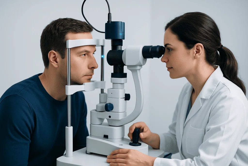
Laser photocoagulation uses focused light energy to create tiny burns around a retinal break or an area judged to be at increased risk. During healing, these spots form an adhesion between the retina and the underlying eye wall. The aim is prevention: laser does not restore thinned retinal tissue to normal or improve vision that has already been affected by another eye condition.
An ophthalmologist, often a retina specialist, considers preventive laser on a case-by-case basis. It may be recommended for a retinal tear, a symptomatic retinal break, or certain lesions with features that raise concern. Factors such as recent flashes and floaters, a history of retinal detachment in the other eye, severe short-sightedness, previous eye surgery, eye trauma, and the appearance of the retina may guide the decision.
Not every hole associated with lattice degeneration requires treatment. Treating low-risk, symptom-free areas may expose a person to procedure-related risks without clear benefit. A detailed dilated retinal examination, and sometimes retinal imaging, helps the specialist recommend observation or treatment based on the whole clinical picture.
People with conditions that can also affect the retina may need coordinated follow-up. For example, monitoring and treatment planning can be individualized alongside care for diabetic retinopathy when relevant.
What Happens During Lattice Degeneration Laser Treatment?

Retinal laser treatment is generally performed as an outpatient procedure. Before treatment, dilating drops widen the pupil so the specialist can see the peripheral retina clearly. Numbing eye drops are typically used, and a contact lens may be placed gently on the eye to focus the laser and keep the eyelids open.
The patient sits at a laser machine, similar to equipment used for some eye examinations. The ophthalmologist applies laser spots around the retinal tear, hole, or selected lattice area. Bright flashes of light may be visible, and some people notice pressure or mild brief discomfort. The procedure length varies according to the area being treated, but treatment itself is often completed within one visit.
Vision can remain blurred and light-sensitive for several hours because of the dilating drops. Patients should arrange for someone else to drive them home unless their eye team advises otherwise. The treating clinician will provide personalized instructions about normal activities, eye drops if needed, and the timing of follow-up examinations.
For people advised to have this procedure, retinal laser photocoagulation is designed to reinforce the retina around a vulnerable area. It is different from laser treatment for clouding after cataract surgery or laser procedures used to correct refractive errors.
Benefits, Risks and Results to Expect
The main potential benefit of laser treatment is lowering the risk that a known retinal tear or selected high-risk lesion progresses to retinal detachment. The laser scars usually develop over the following days to weeks. A follow-up examination confirms whether the treated area has formed an adequate barrier and whether any new retinal changes are present.
Laser treatment is generally well tolerated, but no procedure can remove all future risk. A retinal tear or detachment can still occur, including in a different part of the retina. This is why people with lattice degeneration should continue to attend recommended eye examinations and seek urgent review for new warning symptoms.
Possible side effects include temporary blurred vision, light sensitivity, eye irritation, mild ache, or a short-term increase in floaters. Less common complications can include inflammation, bleeding, damage to nearby retinal tissue, visual field effects, or the need for additional laser treatment. The specialist discusses the expected benefits and the individual risks before proceeding.
If retinal detachment develops, it requires prompt specialist treatment rather than observation. Depending on the type and extent of detachment, management may involve procedures such as vitrectomy or other retinal repair techniques.
Can Lattice Degeneration Get Better?
Lattice degeneration itself generally does not reverse or disappear. It is a structural thinning pattern of the peripheral retina, and laser treatment does not make that retinal tissue normal again. Instead, treatment can create a protective seal around a tear, hole, or selected vulnerable area.
The outlook is still often reassuring. Many people live with lattice degeneration without vision loss, retinal tears, or a need for treatment. Regular monitoring and knowing the symptoms of a possible retinal tear or detachment are the most useful ways to protect eye health.
Vision changes should not automatically be attributed to lattice degeneration. Cataracts, refractive errors, migraine-related visual symptoms, inflammation, and other retinal conditions can cause different visual complaints. A dilated examination is important to identify the cause accurately.
How Long Does It Take to Recover From Lattice Degeneration Laser Treatment?
Most people can return to routine, non-strenuous activities within a day or two after retinal laser treatment, although vision may be blurry or sensitive to light for the rest of the treatment day. The pupil-dilating drops can temporarily affect focus, especially for reading and close work.
The laser spots typically take about one to two weeks to develop a firm scar response, though healing varies. The ophthalmologist may recommend avoiding heavy lifting, vigorous exercise, or activities with a risk of eye impact for a short period, particularly if a retinal tear has been treated. Individual instructions should take priority.
A follow-up appointment is essential, even when the eye feels comfortable. The specialist checks the laser response and looks for further tears or traction elsewhere in the retina. New flashes, a sudden shower of floaters, or a shadow in vision should be assessed urgently rather than waiting for the planned visit.
How Fast Does Lattice Degeneration Progressive and Does It Get Worse With Age?
Lattice degeneration often remains stable for many years and does not usually progress quickly. It may be found incidentally during a routine dilated examination, with no symptoms and no need for treatment. Its presence does not mean that retinal detachment is inevitable.
Changes in the vitreous, the gel inside the eye, are more common with age. As the vitreous naturally separates from the retina, called posterior vitreous detachment, it can sometimes pull on vulnerable areas and cause a retinal tear. This process may produce flashes and new floaters, which is why these symptoms require prompt examination.
Lattice degeneration can appear more extensive over time in some eyes, but age alone does not mean it will become dangerous. Risk assessment depends on examination findings, symptoms, refractive error, eye history, and whether a retinal tear or detachment has occurred in either eye. People with high myopia may also benefit from discussion of their broader retinal risks during routine eye care.
When to Seek Medical Care
Anyone with known lattice degeneration should seek urgent same-day eye assessment for new flashes of light, a sudden increase in floaters, a cluster of dark dots, reduced side vision, or a gray or dark curtain moving across the visual field. These symptoms can occur with a retinal tear or detachment and should be checked promptly, even if they are painless.
A routine eye appointment is appropriate for people without urgent symptoms who have been told they have lattice degeneration and need follow-up advice. Dilated retinal examinations are particularly important after a significant eye injury, before or after certain eye procedures when advised by an ophthalmologist, or when there is a personal or family history of retinal detachment.
Acibadem International’s multidisciplinary specialists and JCI-accredited hospitals provide assessment and treatment planning for retinal conditions for international patients. A retina specialist can explain whether monitoring or laser treatment is suitable and ensure that follow-up is tailored to the individual eye findings.
Frequently asked questions
Is laser treatment necessary for all lattice degeneration?
No. Most people with lattice degeneration do not need laser treatment. An ophthalmologist may recommend monitoring when there is no retinal tear, no concerning symptoms, and no other features that substantially increase risk.
Can lattice degeneration cause blindness?
Lattice degeneration itself does not usually cause blindness. In a small proportion of people, it may be associated with a retinal tear or detachment, which can threaten vision if not treated promptly. Regular monitoring and urgent assessment of warning symptoms are important.
Are flashes and floaters always caused by a retinal tear?
No. Flashes and floaters are common and can occur with normal age-related changes in the vitreous gel. However, a sudden onset or increase can also signal a retinal tear, so a prompt dilated eye examination is needed to distinguish the cause.
Can I exercise after lattice degeneration laser treatment?
Many people can resume light daily activities quickly, but the eye specialist may advise temporarily avoiding vigorous exercise, heavy lifting, or contact sports. Advice varies depending on the reason for treatment and the condition of the retina. Patients should follow their own ophthalmologist’s instructions.
Will laser treatment improve my eyesight?
Retinal laser treatment is intended to reduce the risk of retinal detachment around a tear or high-risk area. It does not usually improve visual sharpness because it does not correct refractive error or restore already damaged retinal tissue. Any change in vision after treatment should be discussed with the treating eye team.
How often should lattice degeneration be checked?
The appropriate interval depends on symptoms, retinal findings, short-sightedness, previous tears or detachment, and other eye health factors. Some people need periodic routine dilated examinations, while others require closer follow-up after a new symptom or laser treatment. An ophthalmologist can set an individualized schedule.
References
- American Academy of Ophthalmology
- National Eye Institute
- Royal College of Ophthalmologists
- Mayo Clinic
This article is for general information only and is not a substitute for professional medical advice. Please consult a qualified doctor about your individual situation.
Explore treatments in Turkey — costs, top hospitals & a free quote
JCI-accredited · board-certified surgeons · reply within 24h
Add us as a Preferred Source to see more of our trusted health content across Google Search, AI Overviews and Discover.

More from the Health Library



Related Specialists

Prof. Dr. Kerim Sarıyılmaz
Orthopedic Surgery & Traumatology
Prof. Dr. Cahide Soydaş Çınar
Cardiology
Assoc. Prof. Dr. Özlem Özgür Gürsoy
Gynecology & Obstetrics
Ece Nur Sonuk
Physical Medicine & Rehabilitation