Left Lung — Explained by Medical Evidence, Not Myths
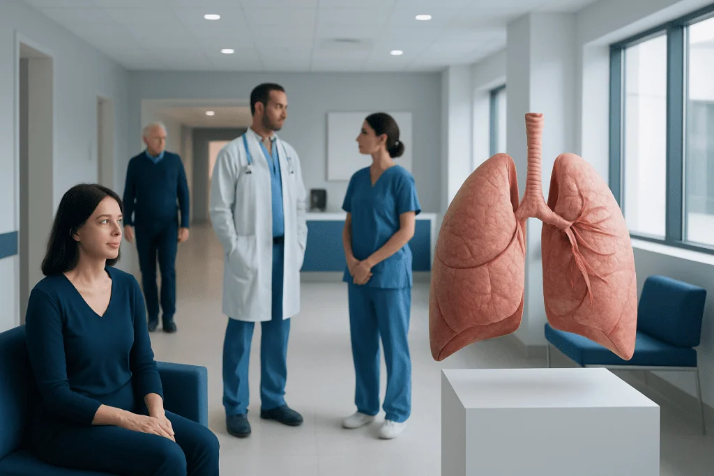
The left lung has two lobes, while the right lung has three. Its smaller size is a normal anatomical adaptation that accommodates the heart.
Key Takeaways
- The left lung has two lobes, while the right lung has three.
- Its smaller size is a normal anatomical adaptation that accommodates the heart.
- The lungs work together, but each lung has its own airways, blood vessels and lining tissues.
- Chest symptoms felt on the left side can come from the lung, heart, muscles, ribs, nerves or digestive system.
- Persistent breathing changes, coughing blood, severe chest pain or sudden shortness of breath require prompt medical assessment.
The left lung is a vital breathing organ located on the left side of the chest. It is slightly smaller than the right lung because the heart occupies space nearby, but it still performs essential oxygen and carbon dioxide exchange.
Left Lung Overview: What It Is and Why It Is Smaller
The left lung is one of the body’s two lungs, located inside the chest on the left side of the rib cage. Its central role is gas exchange: oxygen from inhaled air passes into the bloodstream, while carbon dioxide, a waste gas made by the body, moves from blood into the lungs to be exhaled. The left and right lungs work continuously with the diaphragm, chest muscles, airways and circulatory system to support normal breathing.
The left lung is normally smaller and narrower than the right lung. This is not a weakness or a sign of disease. It is an expected feature of human anatomy because the heart sits slightly to the left of the middle of the chest, taking up space in an area between the lungs called the mediastinum.
A common myth is that one lung can simply “take over” all breathing without consequence. People can often function after removal of part or, in selected circumstances, an entire lung, but this depends on their remaining lung capacity, heart health, fitness and the reason for surgery. The two lungs are designed to contribute together to breathing and overall respiratory reserve.
Anatomy of the Left Lung
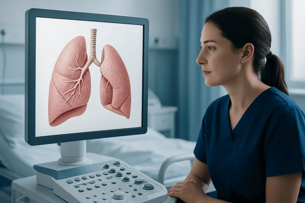
The left lung has two main sections called lobes: the superior lobe and the inferior lobe. By comparison, the right lung has superior, middle and inferior lobes. The two lobes of the left lung are divided by an oblique fissure, a natural groove in the lung tissue. Each lobe contains progressively smaller branches of airways and blood vessels.
The left lung has a characteristic indentation called the cardiac notch, where it curves around the heart. A small tongue-like extension of the upper lobe, called the lingula, lies near this notch. Although the lingula is sometimes compared anatomically with the middle lobe of the right lung, it is part of the left upper lobe.
Each lung is covered by a thin, double-layered membrane called the pleura. A tiny amount of fluid between these layers helps the lungs glide smoothly against the chest wall during breathing. The main breathing muscle, the diaphragm, sits beneath the lungs. On the left side, the diaphragm is also close to the stomach and spleen, which is why symptoms from nearby structures may occasionally be felt in the lower left chest.
The left main bronchus carries air from the windpipe, or trachea, into the left lung. It branches into lobar and smaller segmental bronchi, eventually reaching microscopic air sacs called alveoli. Around the alveoli is a dense network of capillaries, where oxygen and carbon dioxide are exchanged.
How the Left Lung Works With the Heart and Circulation
When a person inhales, the diaphragm moves downward and the rib cage expands. This creates lower pressure within the chest and draws air through the nose or mouth, down the trachea and into both lungs. Air travels through branching airways until it reaches the alveoli, which provide a very large surface area for gas exchange.
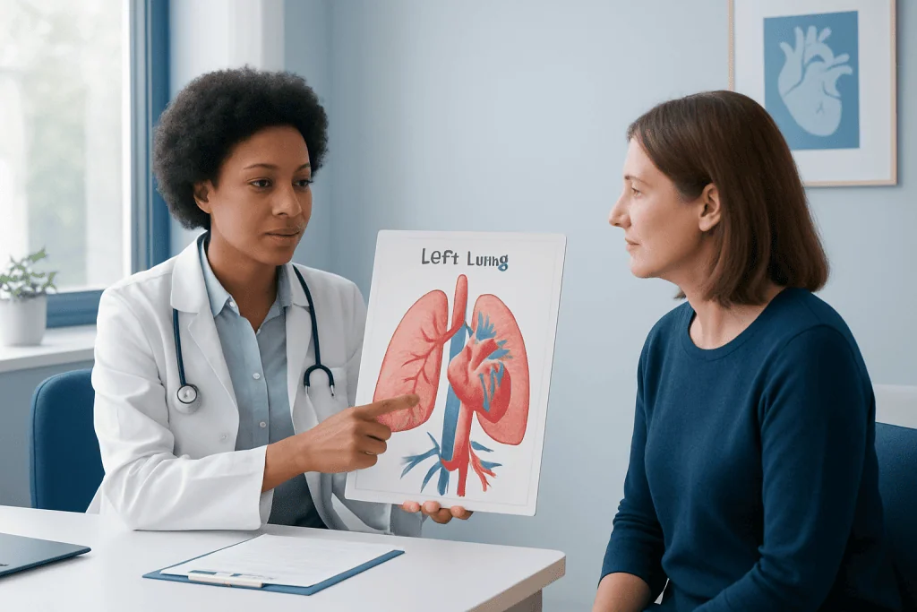
Blood that is low in oxygen travels from the heart to the lungs through the pulmonary arteries. In the alveoli, oxygen moves into the blood and carbon dioxide moves out. Oxygen-rich blood then returns to the heart through the pulmonary veins, including veins from the left lung, so the heart can pump it to the rest of the body.
Because the heart and left lung are close together, symptoms in the left chest should not automatically be attributed to the lung. For example, chest discomfort may arise from the heart, pleura, ribs, chest muscles, esophagus, stomach or nerves. The location of a symptom alone cannot identify its cause, which is why a clinician may consider breathing symptoms, heart symptoms, medical history and examination findings together.
Common Left Lung Symptoms and What They May Mean
Symptoms that seem to involve the left lung may include cough, wheezing, shortness of breath, chest tightness, pain with deep breathing, mucus production or reduced exercise tolerance. These symptoms are not specific to one side of the lung. They may result from temporary infections, airway inflammation, allergies, asthma, chronic lung conditions, blood clots, pleural conditions or other health concerns.
Pain on the left side of the chest is often assumed to be “left lung pain,” but lung tissue itself has few pain-sensitive nerves. Pain may instead come from the pleura, chest wall, muscles or ribs. Pain that becomes sharper with coughing or deep inhalation can occur when the pleura or chest wall is irritated, though medical assessment is still important when pain is new, significant or persistent.
Not every cough or brief episode of breathlessness signals a serious problem. Viral respiratory infections, physical exertion and irritants such as smoke can cause short-term symptoms. However, a cough lasting more than several weeks, recurrent wheeze, unexplained fatigue, repeated chest infections, coughing blood or unintentional weight loss should be discussed with a healthcare professional.
Conditions can affect either lung or both lungs. Examples include pneumonia, asthma, chronic obstructive pulmonary disease, pleural effusion and lung cancer. A diagnosis should be based on clinical evaluation rather than on the side where a person notices symptoms. For more information on respiratory infections, patients may also read about pneumonia.
How Doctors Assess Left Lung Concerns
Assessment usually begins with a medical history and physical examination. A clinician may ask when symptoms began, whether they are linked with exercise or infection, whether there is fever or mucus, and whether the person smokes or has been exposed to dust, chemicals or secondhand smoke. They may listen to the chest for wheezing, crackles or reduced breath sounds and check oxygen levels when appropriate.
A chest X-ray is often used to look for changes such as infection, fluid around the lung, collapse of part of the lung or an obvious mass. Depending on the symptoms and initial findings, a doctor may recommend computed tomography (CT), which can provide more detailed images of the lung tissue, airways and nearby structures. Imaging results always need interpretation in the context of the person’s symptoms and medical history.
Other tests may include spirometry and other pulmonary function tests, which measure airflow and lung volume; blood tests; mucus testing; electrocardiography for possible heart-related symptoms; or bronchoscopy in selected cases. If a pulmonary embolism, or blood clot in a lung artery, is a concern, clinicians may use specific blood tests and imaging based on the person’s likelihood of having the condition.
Finding an abnormality in the left lung does not always mean cancer or permanent damage. Infections, inflammation, old scars and benign nodules can also appear on imaging. Follow-up recommendations depend on the appearance, size and location of a finding as well as individual risk factors, and should be discussed with the treating clinician.
Protecting Lung Health and Supporting Recovery
The most effective step for protecting both lungs is avoiding tobacco smoke. This includes cigarettes, waterpipes and exposure to secondhand smoke. Stopping smoking can improve respiratory health at any age, and a healthcare professional can help identify practical behavioral support and medication options when appropriate.
Regular physical activity, within a person’s abilities and medical guidance, helps the heart and lungs work efficiently. Walking, cycling, swimming and structured pulmonary rehabilitation may improve stamina for people with chronic respiratory conditions. Gradual activity is generally preferable after an illness, especially if breathlessness is ongoing; people should seek advice before resuming demanding exercise.
Vaccination can lower the risk of certain respiratory infections or serious complications, depending on age, health conditions and local recommendations. Hand hygiene, adequate sleep, balanced nutrition and good control of conditions such as asthma can also support respiratory wellbeing. People with asthma should use their prescribed inhalers correctly and attend follow-up reviews.
Indoor air quality matters as well. Reducing exposure to smoke, poorly ventilated cooking fumes, mold and occupational dust may help limit airway irritation. A well-fitted protective mask may be advised in some workplaces. Any new or worsening breathing symptoms should be assessed rather than managed solely with over-the-counter products.
When to Seek Medical Care
Urgent medical care is needed for sudden or severe shortness of breath, chest pressure or pain, bluish lips or face, fainting, confusion, coughing up more than a small streak of blood, or symptoms that develop after a significant chest injury. Emergency evaluation is also important when chest symptoms occur with sweating, nausea, pain spreading to the arm, jaw or back, or a sense of impending collapse, because heart-related causes must be considered.
A non-emergency medical appointment is appropriate for a cough that does not improve, wheezing, repeated respiratory infections, breathlessness that limits usual activities, unexplained chest discomfort or a persistent change in mucus. People who smoke or formerly smoked may benefit from discussing individualized lung health and screening recommendations with a clinician, particularly if they meet local eligibility criteria.
For patients needing coordinated evaluation, Acibadem International’s multidisciplinary specialists and JCI-accredited hospitals diagnose and treat respiratory conditions for international patients. Care plans should be individualized after assessment of symptoms, imaging, lung function and overall health.
Frequently asked questions
Why is the left lung smaller than the right lung?
The left lung is smaller because the heart sits slightly toward the left side of the chest. The left lung has two lobes and a cardiac notch that creates space for the heart. This is a normal feature of anatomy.
How many lobes does the left lung have?
The left lung has two lobes: an upper, or superior, lobe and a lower, or inferior, lobe. The right lung has three lobes. The left lung also has a small upper-lobe extension called the lingula.
Can a person feel pain in the left lung?
The lung tissue itself has limited pain sensation, so pain described as left lung pain often comes from the pleura, chest wall, ribs, muscles or nearby organs. It may also be related to the heart or digestive system. New, severe or persistent left-sided chest pain should be medically assessed.
Is the left lung connected directly to the heart?
The left lung and heart are separate organs, but they work closely through the pulmonary circulation. Blood travels from the heart to the lungs for oxygen uptake and returns to the heart to be pumped around the body. They are also located close together within the chest.
Can pneumonia affect only the left lung?
Yes, pneumonia can affect part of one lung, an entire lobe, or both lungs. Symptoms may include cough, fever, shortness of breath and chest pain, but severity varies. A clinician may use examination and chest imaging to confirm the diagnosis and guide treatment.
What does a spot on the left lung mean?
A spot seen on a chest X-ray or CT scan can have many explanations, including infection, inflammation, scarring or a benign nodule. Less commonly, it can indicate cancer or another important condition. The next steps depend on the imaging appearance and a person’s risk factors, so follow-up with a clinician is essential.
References
- American Lung Association
- National Heart, Lung, and Blood Institute
- Centers for Disease Control and Prevention
- National Health Service
- Mayo Clinic
This article is for general information only and is not a substitute for professional medical advice. Please consult a qualified doctor about your individual situation.
Persistent digestive symptoms? Get evaluated in Turkey
JCI-accredited · board-certified surgeons · reply within 24h
Add us as a Preferred Source to see more of our trusted health content across Google Search, AI Overviews and Discover.