Leukoderma: A Complete Medical Guide — Function, Normal Levels, and Health Implications

Leukoderma means skin depigmentation or lighter patches caused by reduced melanin. It may be linked to conditions such as vitiligo, skin injury, inflammation, or chemical exposure.
Key Takeaways
- Leukoderma means skin depigmentation or lighter patches caused by reduced melanin.
- It may be linked to conditions such as vitiligo, skin injury, inflammation, or chemical exposure.
- Diagnosis usually depends on a skin examination and, when needed, tests to identify the underlying cause.
- Treatment depends on the cause and may include medicines, light-based therapy, or procedures in selected cases.
- Sun protection and early medical assessment can help protect skin health and guide effective care.
Leukoderma is a term for areas of skin that lose their normal color because melanin is reduced or absent. It is not a single disease, but a descriptive finding that can happen for several reasons, so evaluation focuses on identifying the cause and choosing the most suitable treatment or skin care plan.
Overview: what leukoderma means
Leukoderma is a general medical term used to describe white or lighter-than-normal patches on the skin. These patches appear when the skin loses melanin, the pigment that gives skin its usual color. In many people, the term is used broadly, while the exact diagnosis depends on why the pigment loss happened.
This is an important distinction: leukoderma is a description, not always a final diagnosis. Some people have depigmentation because of vitiligo, while others develop it after skin inflammation, burns, eczema, psoriasis, fungal infection, or repeated contact with certain chemicals. The appearance may look similar, but the underlying cause can differ.
Leukoderma can affect any age group and any skin tone. It may occur on the face, hands, arms, feet, or around body openings, but it can appear anywhere on the body. In darker skin tones, the contrast between normal and affected skin may be more noticeable, which can make the condition feel especially distressing even when it is not physically harmful.
Because pigment loss has several possible explanations, medical assessment is useful. A careful diagnosis helps distinguish harmless post-inflammatory changes from autoimmune depigmentation or other skin conditions, and it guides treatment choices, expectations, and follow-up.
How leukoderma looks and feels
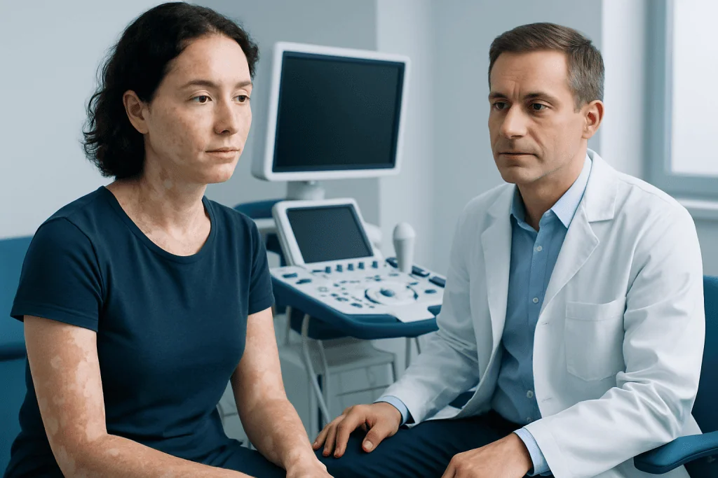
The main sign of leukoderma is one or more sharply defined or irregular pale patches on the skin. The color may range from lighter than the surrounding skin to completely white. In some cases, the hairs growing from the affected area may also turn white if pigment cells in the hair follicles are involved.
Leukoderma itself often does not cause pain, but some people notice associated dryness, mild itch, or sensitivity if the depigmentation follows an inflammatory skin problem. The skin texture is usually normal in vitiligo-related leukoderma, while scaling, redness, or thickening may point toward another diagnosis.
Patterns vary. Some patches remain stable for years, while others slowly enlarge or new areas appear over time. Common sites include:
- Face, especially around the eyes and mouth
- Hands, fingers, elbows, and knees
- Feet and ankles
- Armpits and groin
- Areas of previous rash, injury, or friction
Changes in appearance can affect confidence and emotional well-being, especially when visible areas are involved. This is a valid part of the condition and worth discussing during care, because treatment planning may include both medical management and cosmetic support.
Causes and risk factors

Leukoderma happens when melanocytes, the cells that make pigment, are damaged, reduced, or stop functioning normally. One of the best-known causes is vitiligo, an autoimmune condition in which the immune system targets pigment cells. However, not all leukoderma is autoimmune.
Another common cause is post-inflammatory hypopigmentation or depigmentation. This can develop after eczema, psoriasis, acne, burns, cuts, laser procedures, or other skin injury. Once the skin heals, pigment may return slowly over months, though sometimes the change lasts longer. In some cases, white patches can also follow irritant or allergic contact dermatitis.
Chemical leukoderma may occur after repeated exposure to certain substances that affect melanocytes, especially in the workplace or through regular contact with rubber, adhesives, dyes, detergents, or cosmetic products. Infections can also mimic leukoderma; for example, some superficial fungal conditions create lighter patches and need different treatment.
Risk factors depend on the cause, but may include:
- Personal or family history of autoimmune disease
- Previous inflammatory skin disease
- Repeated skin trauma or friction
- Occupational or household exposure to depigmenting chemicals
- A history of sunburn or skin injury
Because the term covers several possibilities, a doctor usually considers the person’s full history, including when the patches began, whether they spread, and whether there was a rash, injury, or new product before the color change appeared.
How doctors diagnose leukoderma
Diagnosis begins with a clinical skin examination. A dermatologist looks at the shape, location, borders, and distribution of the patches and asks about symptoms, timing, family history, medications, chemical exposure, and prior skin problems. This information often provides strong clues about the cause.
A Wood’s lamp examination may be used in the clinic. This special light can make areas of pigment loss more visible and may help distinguish true depigmentation from conditions that only look lighter on the surface. If there is scaling or signs of infection, a doctor may also test for fungal causes.
Sometimes additional tests are helpful. Blood tests may be considered if autoimmune disease is suspected, especially when there are symptoms suggesting thyroid disease or other immune-related conditions. In selected cases, a small skin biopsy may be needed to confirm the diagnosis or rule out uncommon disorders.
The goal of diagnosis is not only to name the condition but also to understand whether the pigment loss is stable, progressive, inflammatory, infectious, or autoimmune. That information helps predict whether repigmentation is likely and which therapies are most appropriate.
Treatment options for leukoderma
Treatment depends on the underlying cause, the size and location of the patches, how long they have been present, and how much they affect the person’s daily life. Some cases, especially after inflammation, improve gradually with time and gentle skin care. Others benefit from active treatment to reduce progression or encourage repigmentation.
For inflammatory or autoimmune causes, doctors may recommend topical medicines such as corticosteroids or calcineurin inhibitors. When leukoderma is related to vitiligo, light-based treatment may be considered to stimulate repigmentation in suitable patients, including phototherapy under specialist supervision. Cosmetic camouflage can also be helpful for visible areas and may improve confidence while medical treatment is ongoing.
Stable, localized depigmentation that does not respond to medical therapy may sometimes be treated with procedural approaches. In carefully selected cases, specialists may discuss skin grafting or other surgical repigmentation techniques. These options are generally reserved for people whose condition is stable and who have realistic expectations about results.
If the cause is an infection, irritation, or chemical exposure, treatment focuses on removing the trigger and treating the underlying problem rather than simply targeting color change. When pigmentation loss follows another skin condition, management of that original disease is important. In some people with visible white patches, supportive dermatologic care may overlap with services used in dermatology clinics for long-term skin monitoring and treatment planning.
Prevention and self-care
Not all leukoderma can be prevented, especially when it is linked to autoimmune disease. Still, practical self-care may reduce triggers, protect affected skin, and support recovery where repigmentation is possible. Daily sun protection is especially important because depigmented skin burns more easily and tanning of nearby normal skin can make patches stand out more.
Gentle skin care is recommended. Friction, scratching, harsh exfoliation, and unnecessary irritation may worsen some forms of pigment change, particularly in people whose skin reacts strongly to trauma. Using mild cleansers and fragrance-free moisturizers can help protect the skin barrier.
Helpful self-care steps include:
- Use broad-spectrum sunscreen on exposed skin
- Avoid known chemical or cosmetic triggers if one has been identified
- Manage eczema, psoriasis, or other inflammatory skin diseases promptly
- Do not self-treat persistent white patches with strong over-the-counter products
- Discuss camouflage cosmetics with a dermatologist if appearance is a major concern
It is also helpful to take photographs over time, as this can make subtle change easier to assess during follow-up visits. If patch size, number, or distribution changes, updated medical review may be needed.
When to seek medical care
Medical care is recommended for new white patches on the skin, especially if they spread, involve the face or hands, or appear after a rash, injury, or chemical exposure. Assessment is also important if the diagnosis is uncertain, because several skin conditions can look similar but need different treatment.
A person should seek prompt advice if depigmentation is accompanied by redness, pain, scale, hair loss, mouth or genital involvement, or symptoms that suggest an autoimmune problem, such as fatigue or unexplained changes in weight. Children and teenagers with persistent pigment loss should also be evaluated rather than simply observed.
Early evaluation can improve the chances of identifying reversible causes and starting treatment at an appropriate stage. Near the end of the care pathway, patients seeking specialist assessment may wish to know that Acibadem International’s multidisciplinary specialists in JCI-accredited hospitals diagnose and treat skin conditions for international patients, including depigmenting disorders and related concerns such as psoriasis when relevant.
If a patch changes quickly, becomes inflamed, or raises concern about a different skin disorder, a dermatologist can advise on next steps and whether additional testing is necessary. A calm, step-by-step evaluation usually provides the clearest path forward.
Frequently asked questions
Is leukoderma the same as vitiligo?
Not exactly. Leukoderma is a broad term that describes white patches caused by pigment loss, while vitiligo is one specific cause of that pigment loss. A doctor distinguishes between them by looking at the pattern, history, and sometimes additional tests.
Can leukoderma go away on its own?
Sometimes it can, depending on the cause. Pigment loss after inflammation or minor skin injury may gradually improve over time, while autoimmune depigmentation may persist or progress without treatment. Recovery is often slow, so follow-up is important.
Is leukoderma dangerous?
Leukoderma itself is usually not dangerous, but it should not be ignored because the cause matters. Some forms are mainly cosmetic, while others are linked to autoimmune disease, skin inflammation, or chemical exposure. A proper diagnosis helps guide safe care.
Is leukoderma contagious?
No, leukoderma itself is not contagious. It is a change in skin pigmentation, not an infection that spreads from person to person. However, a skin infection can sometimes cause light patches that resemble leukoderma, which is one reason medical evaluation is useful.
What doctor treats leukoderma?
A dermatologist is usually the most appropriate specialist. Dermatologists can assess the pattern of pigment loss, identify possible triggers, and recommend treatment or monitoring. In some cases, other specialists may be involved if an autoimmune condition is suspected.
Does sun exposure help or worsen leukoderma?
Sun exposure usually makes leukoderma more noticeable because the surrounding normal skin tans while depigmented skin does not. Affected areas are also more vulnerable to sunburn. For this reason, regular sun protection is an important part of self-care.
References
- American Academy of Dermatology
- National Institute of Arthritis and Musculoskeletal and Skin Diseases
- British Association of Dermatologists
- Mayo Clinic
- DermNet
This article is for general information only and is not a substitute for professional medical advice. Please consult a qualified doctor about your individual situation.
Explore treatments in Turkey — costs, top hospitals & a free quote
JCI-accredited · board-certified surgeons · reply within 24h
Add us as a Preferred Source to see more of our trusted health content across Google Search, AI Overviews and Discover.