Leukoplakia: What Patients Need to Know

Leukoplakia usually appears as a persistent white patch inside the mouth that does not rub off. Common triggers include tobacco, alcohol, chronic friction from teeth or dentures, and irritation in the mouth.
Key Takeaways
- Leukoplakia usually appears as a persistent white patch inside the mouth that does not rub off.
- Common triggers include tobacco, alcohol, chronic friction from teeth or dentures, and irritation in the mouth.
- Some cases are harmless, but others may show dysplasia, meaning abnormal cells that can raise cancer risk.
- Diagnosis may include an oral exam and biopsy to rule out oral cancer or other conditions.
- Treatment focuses on removing the cause, monitoring the area, and treating abnormal tissue when needed.
- Any mouth patch lasting more than two weeks should be assessed by a dentist or doctor.
Leukoplakia is a term for white or gray patches in the mouth that cannot be wiped away. It is often linked to long-term irritation, but because some patches can contain precancerous or cancerous cells, professional evaluation is important.
What leukoplakia means
Leukoplakia is a descriptive medical term for a white or gray patch that develops on the lining of the mouth and cannot be rubbed or scraped away. It is not a single disease by itself. Instead, it is a clinical finding that can have several possible causes, ranging from repeated irritation to abnormal cell changes that need closer attention.
These patches may appear on the cheeks, gums, tongue, floor of the mouth, or roof of the mouth. Some are flat and thin, while others look thicker, rougher, or slightly raised. Leukoplakia may be painless, which is one reason people sometimes delay evaluation.
Most cases are not cancer. However, some patches contain dysplasia, meaning cells that are growing abnormally, and a smaller number may already represent early oral cancer. That is why a white patch that persists should not be ignored. A careful exam helps determine whether it is simple irritation, oral cancer, or another oral condition needing treatment.
How it can look and feel

Leukoplakia often causes no symptoms beyond the visible patch. A person may notice a white area while brushing their teeth, seeing a dentist, or looking in a mirror. The patch usually does not wipe away with gauze or a toothbrush, which helps distinguish it from some infections or surface debris.
Its appearance can vary. Some patches are smooth and evenly white. Others are thickened, cracked, wrinkled, or mixed red-and-white. Red areas within a white patch can be more concerning because they may be linked with a higher chance of abnormal cells. This type is sometimes called speckled leukoplakia.
Possible features include:
- White, gray, or mixed red-and-white patches inside the mouth
- A rough, thickened, or hardened surface
- Patches on the tongue, cheeks, gums, or floor of the mouth
- Mild tenderness, burning, or sensitivity in some people
- No pain at all in many cases
A related but distinct condition is oral hairy leukoplakia, which usually appears as corrugated white patches on the sides of the tongue and is associated with Epstein-Barr virus and immune suppression. Although the names are similar, it is evaluated somewhat differently from standard oral leukoplakia.
Why leukoplakia develops
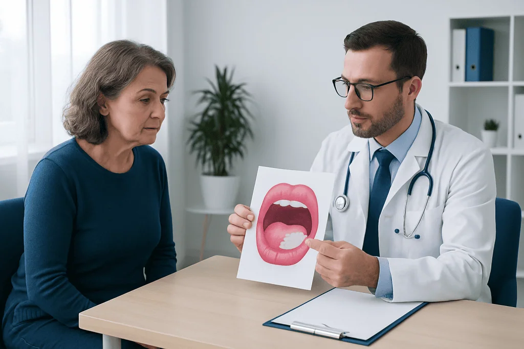
The exact cause is not always clear, but leukoplakia is commonly linked to chronic irritation of the oral tissues. Tobacco use is one of the strongest associations. This includes cigarettes, cigars, pipes, and smokeless tobacco. Alcohol may also contribute, especially when combined with tobacco.
Mechanical friction can play a role as well. A rough tooth edge, broken filling, poorly fitting denture, or habitual cheek biting may repeatedly irritate the same area. Some people develop patches in places where there is long-standing contact or pressure.
Factors that can increase risk include:
- Tobacco use in any form
- Regular alcohol use
- Chronic friction from teeth, dental restorations, or dentures
- Poor oral hygiene
- Older age
- A history of mouth cancer or precancerous oral lesions
- Immune suppression in certain cases, especially for oral hairy leukoplakia
Leukoplakia itself is not usually an infection and is not generally contagious. In some patients, no clear trigger is found. Even then, persistent patches should still be assessed because appearance alone cannot reliably show whether abnormal cells are present.
How doctors diagnose it
Diagnosis starts with a detailed oral examination. A dentist, oral medicine specialist, ENT doctor, or other qualified clinician looks at the size, texture, location, and pattern of the patch. They may also ask about tobacco use, alcohol intake, dentures, recent dental problems, medications, and any history of oral lesions.
The clinician may first try to identify and remove obvious causes of irritation. For example, smoothing a sharp tooth or adjusting a denture may be recommended. If the patch does not improve after the irritant is addressed, or if it has a suspicious appearance from the start, further testing is often advised.
A biopsy is the most important test when leukoplakia appears persistent or concerning. During a biopsy, a small tissue sample or the whole lesion is removed and examined under a microscope. This helps determine whether the patch is benign, inflamed, dysplastic, or cancerous. In some cases, the patient may be referred for biopsy testing and specialist assessment to guide next steps.
Depending on the location and findings, doctors may also recommend a more comprehensive evaluation of the mouth and throat. If there is concern about a deeper lesion or spread, further workup may include ENT evaluation or imaging, but many patients only need a careful clinical exam and biopsy.
Treatment options and follow-up
Treatment depends on what the biopsy shows and whether a clear cause is present. If the patch is related to irritation and no dysplasia is found, the first step is removing the trigger. This may include stopping tobacco, reducing alcohol, correcting dental irritation, and improving oral hygiene. Some patches shrink or disappear after the source of irritation is removed.
If the tissue shows dysplasia or if the lesion has suspicious features, removal may be advised. This can be done using conventional excision, laser treatment, or other local procedures depending on the size and location. The goal is to remove abnormal tissue and lower the chance of progression.
Even after treatment, leukoplakia can recur, especially if risk factors continue. Regular follow-up matters because new lesions can develop and previously treated areas can change over time. For patients with more extensive disease or confirmed malignancy, care may involve oral surgery, pathology review, and oncology care as part of a multidisciplinary plan.
Patients often benefit from ongoing monitoring rather than a one-time visit. Follow-up schedules vary, but doctors usually recommend periodic oral exams, especially for people with prior dysplasia, recurrent lesions, or continued exposure to tobacco or alcohol.
Self-care and prevention
Prevention focuses on lowering irritation and protecting the health of the mouth. Not every case can be prevented, but daily habits make a meaningful difference. The most important step is avoiding tobacco products. Quitting smoking or smokeless tobacco reduces ongoing exposure to substances that can damage oral tissues.
Good oral care also matters. Regular brushing, flossing, routine dental visits, and prompt repair of broken teeth or ill-fitting dentures can reduce chronic friction. Limiting alcohol and maintaining a balanced diet may support overall oral health as well.
Helpful self-care steps include:
- Stop smoking and avoid smokeless tobacco
- Limit alcohol use
- Have rough teeth, broken fillings, or loose dentures corrected
- Brush and floss regularly and attend dental checkups
- Look inside the mouth occasionally for any new or changing patches
- Seek review for any lesion that lasts longer than two weeks
People should avoid trying to diagnose or treat persistent mouth patches on their own. Over-the-counter mouth products may soothe irritation in some cases, but they do not replace a professional assessment when a lesion remains visible.
When to seek medical care
A healthcare professional should evaluate any white patch in the mouth that does not go away within about two weeks. This is especially important if the patch is thick, rough, enlarging, or mixed with red areas. Early review can clarify whether the lesion is due to irritation, infection, or a more serious problem.
Prompt assessment is also important if leukoplakia is accompanied by pain, bleeding, numbness, difficulty chewing, trouble swallowing, or a persistent sore throat. These symptoms do not always mean cancer, but they deserve attention. People with a history of tobacco use, heavy alcohol use, or previous oral lesions should be particularly careful about follow-up.
Patients often start with a dentist, primary care doctor, or ENT specialist. If more advanced assessment or treatment is needed, multidisciplinary teams can coordinate diagnosis and care. Acibadem International’s specialists in JCI-accredited hospitals diagnose and treat oral conditions for international patients, including lesions that may require pathology review or specialist follow-up.
Frequently asked questions
Is leukoplakia always cancer?
No. Many leukoplakia patches are caused by irritation and are not cancer. However, some contain precancerous changes or early cancer, which is why persistent patches should be professionally evaluated.
What does leukoplakia look like?
It usually appears as a white or gray patch inside the mouth that cannot be rubbed off. It may be flat, thickened, rough, or mixed with red areas depending on the type and cause.
Can leukoplakia go away on its own?
Sometimes it improves after the source of irritation is removed, such as stopping tobacco or fixing a rough tooth or denture. If it persists, a doctor or dentist may recommend a biopsy and ongoing monitoring.
Who is most at risk for leukoplakia?
People who smoke, use smokeless tobacco, or drink alcohol regularly have a higher risk. Chronic mouth irritation, older age, and a history of oral precancer or cancer can also increase risk.
How is leukoplakia treated?
Treatment depends on the cause and biopsy results. It may include removing sources of irritation, stopping tobacco, monitoring the patch over time, or removing abnormal tissue if dysplasia is found.
Should a painless white patch in the mouth still be checked?
Yes. Leukoplakia is often painless, so the absence of pain does not rule out an important problem. Any patch that remains for more than two weeks should be assessed by a qualified clinician.
References
- World Health Organization
- National Institute of Dental and Craniofacial Research
- American Dental Association
- Mayo Clinic
- National Cancer Institute
This article is for general information only and is not a substitute for professional medical advice. Please consult a qualified doctor about your individual situation.
Explore treatments in Turkey — costs, top hospitals & a free quote
JCI-accredited · board-certified surgeons · reply within 24h
Add us as a Preferred Source to see more of our trusted health content across Google Search, AI Overviews and Discover.