Lithotripsy procedure: Symptoms, Causes, and Treatment — Complete Patient Guide
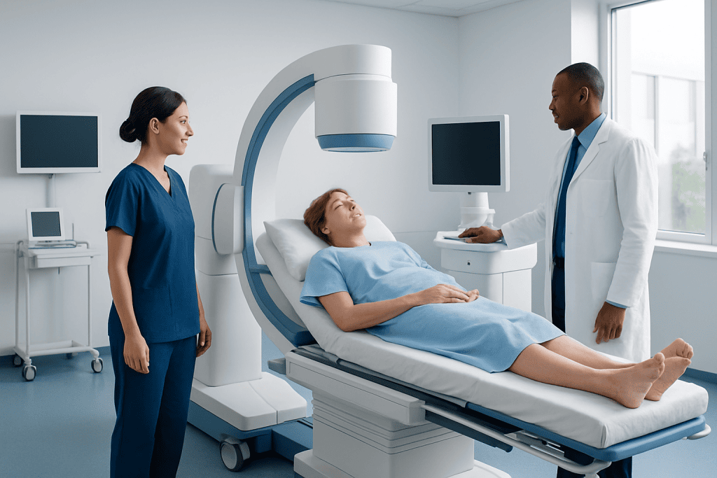
Lithotripsy is not a disease but a treatment for urinary stones, most often kidney or ureter stones. Common types include shock wave lithotripsy and endoscopic laser lithotripsy.
Key Takeaways
- Lithotripsy is not a disease but a treatment for urinary stones, most often kidney or ureter stones.
- Common types include shock wave lithotripsy and endoscopic laser lithotripsy.
- Not every stone needs lithotripsy; some small stones pass on their own with monitoring and supportive care.
- Doctors choose the method based on stone size, location, hardness, and whether there is infection or blockage.
- After treatment, mild blood in the urine, soreness, and passing stone fragments can be expected for a short time.
- Urgent medical care is needed for fever, severe pain, reduced urine output, or signs of urinary obstruction.
A lithotripsy procedure is a medical treatment used to break stones in the kidney, ureter, or bladder into smaller pieces that can pass naturally or be removed more easily. The right method depends on the stone’s size, location, composition, symptoms, and the patient’s overall health.
Overview: what a lithotripsy procedure is
A lithotripsy procedure is a treatment used to break stones in the urinary tract into smaller fragments. It is most commonly used for kidney stones and ureter stones, but some techniques may also be used for bladder stones. Once the stone is broken into smaller pieces, those fragments may pass in the urine or be removed during the procedure.
This treatment is often recommended when a stone is too large to pass comfortably, is causing repeated pain, blocks urine flow, leads to bleeding, or raises the risk of infection or kidney damage. Lithotripsy does not prevent new stones from forming, but it can relieve the immediate problem and protect the urinary system from complications.
Several different approaches may be called lithotripsy. The best-known is shock wave lithotripsy, which uses sound waves from outside the body to break the stone. Another common method is laser lithotripsy, performed with a thin scope passed through the urinary tract. In some cases, other procedures are more suitable, especially for larger or more complex stones.
Why lithotripsy may be needed
Many urinary stones do not cause symptoms right away. A small stone may pass on its own with fluids, pain control, and time. When a stone becomes lodged or starts moving through the ureter, however, it can cause sudden and intense symptoms. In these situations, the goal of treatment is to relieve pain, restore urine flow, and reduce the risk of infection or kidney injury.
Symptoms linked to a stone that may lead to evaluation for a lithotripsy procedure can include:
- Sharp pain in the back, side, lower abdomen, or groin
- Pain that comes in waves and changes in intensity
- Blood in the urine
- Nausea or vomiting
- Frequent urination or urgency
- Burning during urination
- Cloudy or foul-smelling urine
Not every person with these symptoms will need lithotripsy. Doctors look at the stone’s size and position, whether symptoms are manageable, and whether the urinary tract is blocked. If a stone is causing severe pain, repeated emergency visits, or persistent obstruction, active treatment becomes more likely.
People sometimes learn they have stones during imaging for another reason. Even then, treatment may be advised if the stone is large, likely to move, or already affecting kidney drainage. A urology team may also assess related problems such as kidney stones or recurrent stone disease.
Causes and risk factors behind urinary stones
Lithotripsy treats the result of stone formation, not the underlying cause. Urinary stones form when minerals and salts in the urine become concentrated and crystallize. Over time, these crystals can grow into stones. The exact cause may differ from person to person, and some people are more prone to stone formation than others.
Common risk factors include not drinking enough fluids, a diet high in salt, certain metabolic conditions, obesity, a family history of stones, and repeated urinary tract infections. Some medications and digestive conditions can also affect how the body absorbs calcium, oxalate, or uric acid, increasing stone risk.
Stone type matters because it can influence both prevention and treatment. Calcium oxalate stones are common, but uric acid, struvite, and cystine stones also occur. Harder stones or stones in certain positions may respond less well to shock waves and may be better treated with an endoscopic approach.
For patients with repeated stone episodes, doctors may recommend a broader evaluation after the immediate problem is treated. This can include blood tests, urine testing, and analysis of any passed stone fragments to understand why the stones formed and how to reduce the chance of recurrence.
How doctors decide which lithotripsy procedure is best
Choosing the best treatment is more individualized than many patients expect. Doctors consider the stone’s size, location, density, and likely composition. They also look at symptoms, signs of infection, the anatomy of the urinary tract, pregnancy status, blood-thinning medications, and any other health conditions that may affect anesthesia or recovery.
Shock wave lithotripsy is usually considered for certain kidney stones or upper ureter stones that are not excessively large or very hard. It is noninvasive because no scope or incision is used, but it may be less effective for some stones and may require repeat treatment. Endoscopic approaches, including ureteroscopy, are often preferred when the stone is lower in the ureter, harder, or needs direct visualization and removal.
For larger stones, especially those filling part of the kidney, other options such as percutaneous nephrolithotomy may be more appropriate than standard lithotripsy alone. If stones are in the bladder, management may differ and may involve bladder stones treatment tailored to the cause.
This decision-making process helps explain why two people with “kidney stones” may receive different recommendations. The aim is to choose the safest and most effective method for complete stone clearance while reducing the need for repeat procedures.
Diagnosis and preparation before treatment
Before a lithotripsy procedure, the doctor confirms where the stone is, how big it is, and whether it is causing obstruction. Imaging tests are central to this process. Depending on the situation, evaluation may include ultrasound, X-ray, or CT scanning. Urine tests help check for blood, crystals, and infection, while blood tests can assess kidney function and overall health.
If there is a suspected urinary infection, treatment may need to be adjusted or delayed until the infection is controlled. An obstructed urinary tract with infection can be a medical urgency, and the immediate priority may be drainage rather than stone fragmentation. This is one reason pre-procedure assessment is so important.
Preparation depends on the type of lithotripsy planned. Patients may be asked not to eat or drink for a period before the procedure, especially if sedation or general anesthesia will be used. The care team also reviews medicines, including blood thinners, diabetes medicines, and supplements that can increase bleeding risk.
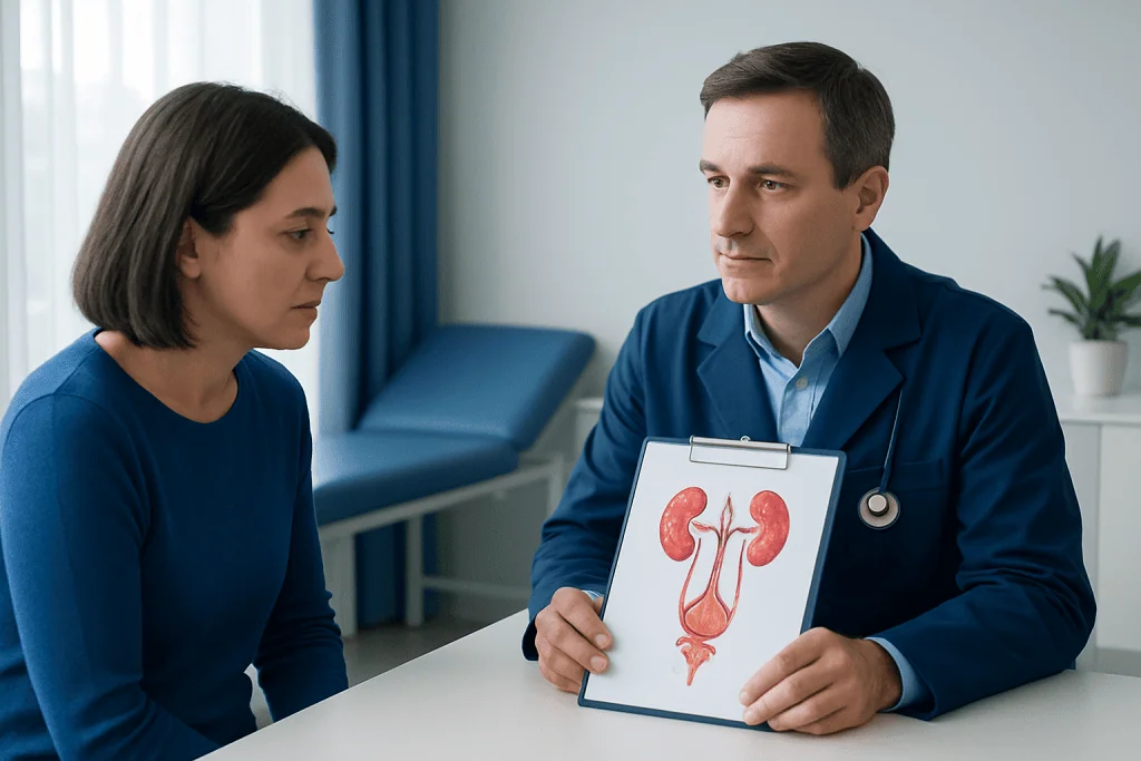
The doctor explains what to expect, possible benefits, and possible risks. This conversation often includes whether a temporary stent may be needed to help urine drain after treatment and whether additional imaging or follow-up visits will be required to confirm that stone fragments have cleared.
Treatment options: shock wave, laser, and related procedures
Shock wave lithotripsy, often called SWL or ESWL, uses focused sound waves from outside the body to break a stone into smaller pieces. It does not require an incision. During the procedure, imaging is used to target the stone accurately. This option can be suitable for selected stones, but success depends on the stone’s location, size, and hardness. Afterward, the patient may pass fragments over days or weeks.
Laser lithotripsy is usually performed during ureteroscopy. A thin scope is passed through the urethra and bladder into the ureter or kidney, allowing the doctor to see the stone directly. A laser fiber then breaks the stone into smaller pieces, which may be removed with tiny baskets or left to pass naturally. In some patients, a temporary ureteral stent is placed to keep urine flowing while swelling settles.
When stones are large, branched, or difficult to reach, the doctor may recommend a more direct stone-removal approach rather than standard lithotripsy alone. This may include kidney stone treatment plans that combine imaging, endoscopic techniques, and follow-up care to achieve complete clearance.
Recovery varies by method. After shock wave treatment, mild flank soreness, bruising, and blood in the urine can occur temporarily. After ureteroscopic laser treatment, urinary urgency, burning, and discomfort from a stent are fairly common for a short period. The care team usually advises on hydration, activity, warning signs, and when normal routines can be resumed.
Prevention, self-care, and long-term follow-up
Even after a successful lithotripsy procedure, new stones can still form. Prevention is therefore an important part of care, especially for people who have had more than one stone episode. The most broadly helpful step is maintaining good hydration so urine stays less concentrated. Many patients are advised to drink enough fluid throughout the day, though the exact amount should be individualized.
Dietary advice depends on the type of stone. In general, reducing excess salt intake may help. Some people also need guidance about oxalate-rich foods, animal protein, or uric acid control. It is important not to make major diet changes based only on internet advice, because prevention works best when matched to the specific stone type and a person’s medical history.
Follow-up may include repeat imaging to make sure fragments have cleared and that there is no persistent blockage. If a stent was placed, it usually needs planned removal. People with recurrent stones may also benefit from metabolic evaluation and a personalized prevention plan from a urologist or nephrologist.
Near the end of the care pathway, some patients seek ongoing support from specialist centers. Acibadem International’s multidisciplinary specialists and JCI-accredited hospitals diagnose and treat urinary stone disease for international patients, including evaluation, minimally invasive procedures, and follow-up planning when appropriate.
When to seek medical care
Medical review is important when symptoms suggest a urinary stone, especially if pain is severe or keeps returning. A person should seek prompt care for strong side or back pain, visible blood in the urine, vomiting that prevents drinking fluids, or trouble passing urine. These symptoms do not always mean lithotripsy is needed, but they do need proper assessment.
Urgent medical care is especially important if stone symptoms are accompanied by fever, chills, marked weakness, or reduced urine output. These can be signs of infection or significant blockage, which may require rapid treatment. Pregnant patients and people with a single kidney should also seek timely medical advice if they suspect a stone.
After a lithotripsy procedure, patients should contact their doctor if pain suddenly worsens, fever develops, urine flow decreases, or bleeding seems heavy or persistent. Passing small fragments and mild discomfort can be expected, but severe or escalating symptoms should never be ignored.
Frequently asked questions
Is a lithotripsy procedure the same as kidney stone surgery?
Not exactly. Lithotripsy is a general term for treatments that break stones into smaller pieces, and some forms are noninvasive while others are done with a scope. It is often considered a minimally invasive way to treat stones, but the exact approach depends on the stone and the patient.
How long does it take to recover from lithotripsy?
Recovery time varies by method. Many people return to light activities within a few days after shock wave treatment, while endoscopic procedures may involve a short recovery period influenced by soreness or a temporary stent. The doctor gives individual advice based on the procedure performed and overall health.
Is lithotripsy painful?
The procedure itself is usually performed with pain control, sedation, or anesthesia depending on the method used. Afterward, some discomfort is common, such as flank soreness, burning with urination, or cramping when stone fragments pass. These symptoms are often temporary, but severe pain should be reviewed by a doctor.
Can stones come back after lithotripsy?
Yes. Lithotripsy treats the existing stone, but it does not remove the tendency to form new stones. That is why hydration, diet advice, and in some cases metabolic testing are important parts of long-term prevention.
Will all stone fragments pass on their own after treatment?
Not always. Many fragments pass naturally, especially after successful shock wave treatment, but some may remain or require further treatment. Follow-up imaging helps show whether the stone has fully cleared.
Who may not be a good candidate for shock wave lithotripsy?
Shock wave lithotripsy may be less suitable for certain large, very hard, or poorly positioned stones. It may also be avoided in situations such as pregnancy, untreated infection, or some bleeding-related concerns. A urologist reviews these factors before recommending the safest option.
References
- National Institute of Diabetes and Digestive and Kidney Diseases
- American Urological Association
- European Association of Urology
- National Kidney Foundation
- Mayo Clinic
This article is for general information only and is not a substitute for professional medical advice. Please consult a qualified doctor about your individual situation.
Explore treatments in Turkey — costs, top hospitals & a free quote
JCI-accredited · board-certified surgeons · reply within 24h
Add us as a Preferred Source to see more of our trusted health content across Google Search, AI Overviews and Discover.