Liver Lesions — Explained by Medical Evidence, Not Myths

A liver lesion is a descriptive imaging finding, not a diagnosis by itself. Many liver lesions are benign, including cysts and hemangiomas.
Key Takeaways
- A liver lesion is a descriptive imaging finding, not a diagnosis by itself.
- Many liver lesions are benign, including cysts and hemangiomas.
- Doctors use imaging features, blood tests, and sometimes biopsy to identify the cause.
- Treatment depends on the type of lesion, symptoms, and overall liver health.
- Urgent assessment is important if liver lesions are linked with pain, jaundice, fever, or known cancer.
Liver lesions are abnormal areas seen in the liver on ultrasound, CT, or MRI. Many liver lesions are harmless, but some need further evaluation to tell whether they are benign, infectious, or cancer-related and to decide if treatment is necessary.
What liver lesions mean
Liver lesions are areas in the liver that look different from the surrounding tissue on an imaging test. In plain terms, the phrase describes what the radiologist sees, not what the lesion definitely is. This matters because a lesion can represent something harmless, such as a simple cyst, or something that needs closer attention, such as an infection, inflammatory change, or tumor.
Many people learn they have liver lesions after an ultrasound, CT scan, or MRI done for another reason. In that situation, the lesion is often incidental, meaning it was found by chance. Incidental findings are common, and in many cases they do not cause symptoms or long-term problems.
What determines the next step is not the word lesion alone, but the lesion’s size, shape, internal appearance, and whether the person has liver disease, symptoms, or a history of cancer. A careful interpretation of imaging usually provides strong clues, and MRI with contrast is often especially useful when a lesion needs to be characterized in more detail.
Common types of liver lesions
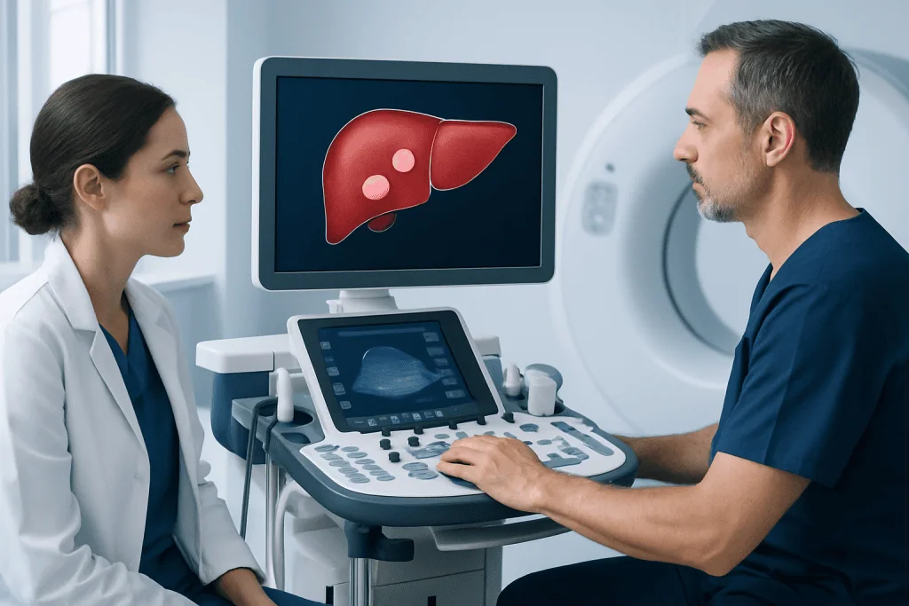
Liver lesions are broadly grouped as benign, malignant, infectious, or less commonly traumatic or vascular. Benign lesions are the most reassuring category and include simple cysts, hemangiomas, focal nodular hyperplasia, and some adenomas. These are not all managed the same way, but many only need monitoring or no treatment at all.
Malignant lesions can start in the liver or spread there from another organ. Primary liver cancers include hepatocellular carcinoma and bile duct cancers, while secondary lesions are metastases from cancers elsewhere in the body. A lesion is not assumed to be cancer based on appearance alone, but certain patterns raise suspicion and lead to further tests. Related conditions such as liver cancer may be considered if imaging and clinical findings point in that direction.
Other liver lesions are caused by infection or inflammation. A liver abscess is a pocket of infection that can cause fever and pain and needs prompt treatment. Some lesions are linked with chronic liver disease, fatty liver, viral hepatitis, or cirrhosis, which change the background risk and influence how doctors interpret imaging findings.
- Benign: simple cyst, hemangioma, focal nodular hyperplasia, hepatic adenoma
- Malignant: primary liver cancer, metastases
- Infectious/inflammatory: liver abscess, granulomatous lesions
- Vascular or other: perfusion changes, regenerative nodules in cirrhosis
Symptoms and how liver lesions are found

Many liver lesions cause no symptoms. They are often discovered incidentally during imaging for abdominal discomfort, routine health checks, or evaluation of another condition. This is especially true for small benign lesions such as simple cysts or hemangiomas.
When symptoms occur, they depend more on the lesion’s size, location, and cause than on the fact that it is in the liver. Some people may feel a dull ache or fullness in the upper right abdomen. Larger lesions can occasionally press on nearby structures and lead to bloating, early fullness after eating, or discomfort.
Symptoms that deserve more urgent attention include jaundice, fever, unexplained weight loss, marked fatigue, or persistent abdominal pain. These do not automatically mean cancer, but they suggest the lesion may be affecting liver function or may be related to infection or another significant liver condition. In some cases, liver lesions are found while assessing possible liver cancer or chronic liver disease, which changes how quickly further evaluation is needed.
Causes and risk factors doctors consider
The causes of liver lesions vary widely. Some develop from congenital or benign changes in liver tissue, while others are linked with hormones, long-standing inflammation, infection, or cancer. A simple cyst may have no clear cause, whereas an adenoma may be associated with hormonal factors, and hepatocellular carcinoma is more often linked with cirrhosis or chronic viral hepatitis.
Doctors look closely at the person’s overall health to estimate risk. Important factors include known liver disease, alcohol-related liver injury, nonalcoholic fatty liver disease, hepatitis B or C, obesity, diabetes, family history, and a personal history of cancer in another organ. Travel history, recent infection, or weakened immunity may increase concern for a liver abscess or parasitic disease in some settings.
Age and sex can also matter because certain lesions are more common in specific groups. The use of contrast-enhanced imaging, along with a person’s medical history, often narrows the possibilities considerably. If there is known chronic liver disease, specialists may also evaluate for related complications and lesions associated with cirrhosis.
How liver lesions are diagnosed
Diagnosis usually begins with imaging. Ultrasound may first detect a lesion, but CT or MRI often provides the detail needed to identify its type. The timing of contrast uptake and washout, the lesion’s borders, and whether it contains fluid, fat, blood vessels, or scar-like tissue all help distinguish a harmless lesion from one that needs more attention.
Blood tests may be used to assess liver function and look for clues such as inflammation, infection, or tumor markers when appropriate. These tests do not diagnose every lesion by themselves, but they add context. If a lesion appears typical for a simple cyst or hemangioma, no further testing may be required. If the finding is indeterminate, follow-up imaging after a set interval may be recommended to see whether it changes over time.
Sometimes a biopsy is needed, particularly if imaging remains uncertain or if the result would change treatment planning. In selected cases, lesion evaluation is part of a broader workup that may include MRI scanning, CT imaging, or image-guided tissue sampling. The goal is to reach a clear diagnosis while avoiding unnecessary procedures whenever possible.
Treatment options and follow-up
Treatment depends entirely on what the liver lesion turns out to be. Many benign lesions need no treatment and only reassurance. A simple cyst or small hemangioma that is not causing symptoms may simply be documented and left alone. Follow-up imaging may be advised if the lesion is large, atypical, or not fully characterized on the first scan.
Some lesions do need active treatment. A liver abscess usually requires antibiotics and sometimes drainage. Symptomatic cysts or adenomas may be monitored more closely or treated if they grow, bleed, or cause pain. If a lesion is malignant, care is tailored to the cancer type, stage, and liver function. This can involve surgery, local ablation, embolization, systemic therapy, or a combination of treatments.
When surgery is appropriate, teams may discuss options such as liver resection for selected tumors or complex benign lesions. Care is often multidisciplinary, involving radiologists, hepatologists, gastroenterologists, surgeons, and oncologists. Near the end of the diagnostic pathway, international patients may also seek evaluation at centers such as Acibadem International, where multidisciplinary specialists in JCI-accredited hospitals assess and treat liver lesions according to the underlying diagnosis.
Self-care, prevention, and living with a liver lesion
There is no single way to prevent all liver lesions because they arise from many different causes. Still, protecting liver health is important. This includes limiting alcohol if advised, maintaining a healthy weight, managing diabetes, discussing hepatitis vaccination where appropriate, and seeking treatment for chronic hepatitis or fatty liver disease.
For people with a known benign lesion, self-care often means keeping follow-up appointments and understanding the plan. It can help to ask whether the lesion is definitely benign, whether another scan is needed, and what symptoms should prompt earlier review. Most stable benign lesions do not interfere with normal daily activities.
People with chronic liver disease should remain under regular medical follow-up because the background liver condition may matter as much as the lesion itself. If symptoms change, the lesion grows, or new abnormalities appear on blood tests, the care plan may need to be updated. A calm, structured follow-up approach usually provides the clearest answers over time.
When to seek medical care
Medical review is appropriate whenever a liver lesion is reported on imaging, even if it was found incidentally and there are no symptoms. Most findings are not emergencies, but they should be interpreted in the context of the person’s age, medical history, and liver health. A clinician can explain whether the lesion appears clearly benign or whether more testing is needed.
Earlier or more urgent assessment is important if there is severe or persistent upper abdominal pain, jaundice, fever, nausea with worsening illness, faintness, or unexplained weight loss. Prompt evaluation is also important for people who already have cirrhosis, hepatitis B or C, a known cancer elsewhere in the body, or abnormal liver blood tests.
If the report uses terms such as indeterminate, suspicious, enhancing, or mass, it does not necessarily mean cancer, but it does mean the lesion deserves proper follow-up. Asking for the exact imaging impression and the recommended next step can help patients move from uncertainty to a clearer, evidence-based diagnosis.
Frequently asked questions
Are liver lesions usually cancer?
No. Many liver lesions are benign, especially when they are found incidentally on imaging. The main question is not whether a lesion exists, but what type it is and whether its imaging features are clearly harmless.
Can liver lesions go away on their own?
Some liver lesions remain stable for years and do not need treatment, while others may change depending on the cause. Simple cysts and hemangiomas often persist without causing problems. Infectious lesions such as abscesses may improve with treatment rather than on their own.
What test is best for evaluating liver lesions?
The best test depends on how the lesion was first found and what needs to be clarified. MRI with contrast is often very helpful for characterizing liver lesions, while CT and ultrasound also play important roles. Doctors choose the test based on the lesion's appearance and the person's medical history.
Do all liver lesions need a biopsy?
No. Many liver lesions can be diagnosed confidently from imaging alone, especially if they have classic benign features. Biopsy is usually reserved for lesions that remain uncertain or when the result would directly affect treatment decisions.
Can liver lesions cause pain?
Yes, some can, but many do not cause any symptoms. Pain is more likely if a lesion is large, stretches the liver capsule, bleeds, or is linked with infection or inflammation. Persistent pain should be assessed by a doctor.
Is a liver lesion the same as cirrhosis or fatty liver disease?
No. A liver lesion is a focal area that looks different on imaging, while cirrhosis and fatty liver disease affect the liver more diffusely. However, chronic liver disease can increase the importance of a lesion and change how it is evaluated.
References
- American College of Radiology
- National Institute of Diabetes and Digestive and Kidney Diseases
- American Association for the Study of Liver Diseases
- European Association for the Study of the Liver
- National Cancer Institute
This article is for general information only and is not a substitute for professional medical advice. Please consult a qualified doctor about your individual situation.
Oncology care in Turkey — second opinion and treatment plan
JCI-accredited · board-certified surgeons · reply within 24h
Add us as a Preferred Source to see more of our trusted health content across Google Search, AI Overviews and Discover.

More from the Health Library

Uncircumcised: An Evidence-Based Guide for Patients

Health — Explained by Medical Evidence, Not Myths

Senile: What Patients Need to Know

Erythematotelangiectatic Rosacea: Redness and Flushing

Throwing Up Bile: A Complete Medical Overview
Shoulder: An Evidence-Based Guide for Patients
Gastroenterology Specialists at Acibadem

Prof. Dr. Çetin Karaca (m)
Gastroenterology
Assoc. Prof. Dr. Diğdem Özer
Gastroenterology
Prof. Dr. Ebubekir Şenateş
Gastroenterology
Assoc. Prof. Dr. Emine Şatır
Gastroenterology