Mammography Procedure: An Evidence-Based Patient Guide
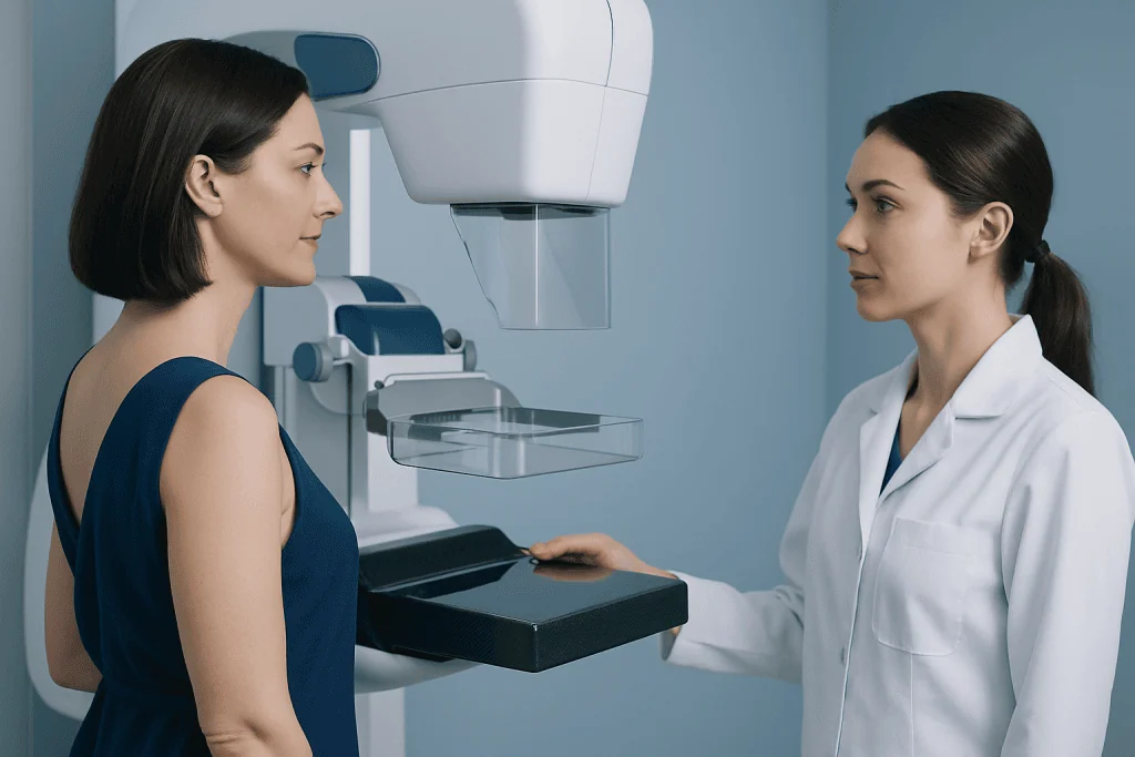
Mammography uses low-dose X-rays and breast compression to create clear images of breast tissue. Screening mammograms are for people without symptoms, while diagnostic mammograms evaluate a symptom or an abnormal screening result.
Key Takeaways
- Mammography uses low-dose X-rays and breast compression to create clear images of breast tissue.
- Screening mammograms are for people without symptoms, while diagnostic mammograms evaluate a symptom or an abnormal screening result.
- Most people can return to normal activities immediately after the examination.
- A callback after mammography is common and does not by itself mean cancer is present.
- Screening decisions should reflect age, overall health, breast density, family history, and personal preferences.
A mammography procedure is a breast X-ray examination that can find changes too small to feel, including some early breast cancers. It is usually brief, and understanding preparation, compression, results, and follow-up can make the experience more manageable.
Mammography Procedure Overview
The mammography procedure is an imaging examination that uses low-dose X-rays to produce detailed pictures of the breast. It is mainly used for screening, meaning checking for breast cancer before symptoms develop, and for diagnosis when a person has a lump, nipple discharge, skin change, breast pain in one area, or an abnormal previous image.
During the examination, each breast is placed briefly between two imaging plates. Gentle but firm compression spreads the tissue apart and reduces movement, allowing the radiologist to see fine details while using the lowest practical radiation dose. The compression can feel uncomfortable for a few seconds, but it should not cause lasting pain.
Evidence-based mammogram practice recognizes that mammography can detect cancers before they can be felt. It also has limits: not every cancer is visible, particularly in dense breast tissue, and some findings require additional images or testing to clarify whether they are harmless or need treatment.
Who May Need a Mammogram and How It Works
Screening mammography is generally offered to people at average risk of breast cancer beginning in midlife, but the right age and interval vary among national guideline groups. People with a strong family history, certain inherited gene changes, prior chest radiation at a young age, previous high-risk breast findings, or a personal history of breast cancer may need an individualized screening plan that can include mammography, breast MRI, or ultrasound.
Screening mammograms usually include standard images of each breast. Diagnostic mammography is more tailored: the radiology team may take additional views, magnify a small area, or use targeted ultrasound during the same visit. Diagnostic imaging helps investigate symptoms and findings, including those associated with breast cancer.
Modern mammography protocols commonly use digital imaging, and many centers use digital breast tomosynthesis, often called 3D mammography. Tomosynthesis obtains images from several angles and reconstructs thin image layers. It may improve visibility in some breasts, although the best approach depends on the facility, equipment, breast tissue, and clinical question.
Mammography Procedure: Step by Step
At check-in, the patient may be asked about breast symptoms, past breast procedures, family history, hormone use, pregnancy possibility, and where earlier mammograms were performed. Comparing new images with prior studies is very valuable, so patients should arrange for previous mammograms to be available whenever possible.
In the changing area, the patient removes clothing and jewelry from the waist up and wears a gown. A mammography technologist positions one breast on the detector and gradually brings a clear compression plate down. The patient is asked to remain still and may need to hold their breath for a moment while the image is taken. This process is repeated in different positions for both breasts.
The imaging portion commonly takes about 10 to 20 minutes, although a diagnostic appointment may take longer because extra images or ultrasound may be needed. The technologist does not usually interpret the images. A radiologist reviews them and sends a report to the referring clinician; timing for results depends on local practice.
When a suspicious area requires tissue sampling, the clinician may recommend a breast biopsy. A biopsy does not mean cancer is confirmed; it is a way to examine cells or tissue and establish an accurate diagnosis.
Mammogram Patient Instructions: Before and After the Visit
When possible, patients may prefer to schedule a routine mammogram when their breasts are least tender. For people who have menstrual cycles, this is often in the week after a period begins. They should tell the imaging center in advance if they are pregnant, might be pregnant, are breastfeeding, have breast implants, or have had breast surgery.
What should you not do the night before a mammogram? There is usually no special diet, fasting requirement, or activity restriction the night before. On the day of the appointment, patients should avoid deodorant, antiperspirant, perfume, lotion, powder, or ointment on the underarms or breasts unless the imaging center says otherwise, because some products can leave particles that appear on X-ray images. Wearing a two-piece outfit can make changing easier.
There is no formal recovery period. Mild temporary tenderness or pressure marks can occur, but normal work, driving, exercise, and daily activities can usually resume immediately. If discomfort is significant or persists, the patient should contact their healthcare professional.
People receiving evaluation or treatment for breast disease may benefit from coordinated input from breast radiology, surgery, oncology, pathology, and genetics. Acibadem International’s multidisciplinary specialists at JCI-accredited hospitals assess and treat breast conditions for international patients, including through breast cancer treatment when indicated.
Benefits, Limits, and Possible Risks
The main benefit of mammography is the ability to identify some cancers at an early stage, when they may be smaller and treatment options may be broader. Mammograms can also reveal noncancerous changes, such as cysts or calcifications, that may need monitoring or further assessment.
Possible drawbacks include temporary discomfort from compression, false-positive findings, and false-negative results. A false positive means an image looks unusual but follow-up testing does not find cancer. A false negative means cancer is present but not identified on the mammogram. Dense breasts can make interpretation more difficult, but density is only one part of overall risk assessment.
Mammography involves a small amount of ionizing radiation. The dose is kept low through quality-controlled mammography protocols, equipment checks, staff training, and radiologist oversight. The potential benefit of screening should be weighed against possible harms such as recalls, unnecessary testing, overdiagnosis, and anxiety, particularly when choosing a screening schedule.
Regulated imaging services follow quality and reporting standards. A mammography protocol for the use of diagnostic facilities includes appropriate image acquisition, documentation, equipment quality assurance, and prompt communication of findings and follow-up recommendations.
What Are the New Mammogram Guidelines for 2026?
What are the new mammogram guidelines for 2026? There is no single worldwide mammogram guideline that applies to every person in 2026. A widely used United States recommendation from the U.S. Preventive Services Task Force advises biennial screening mammography for women aged 40 to 74 years at average risk. Other professional organizations may support starting at age 40 and screening every year or every two years, depending on individual risk and preferences.
For people aged 75 and older, evidence is less certain in some guideline reviews. Decisions are generally individualized according to overall health, life expectancy, previous screening history, breast cancer risk, and willingness to undergo testing and treatment if an abnormality is found.
People at higher-than-average risk should not rely solely on average-risk age ranges. They should ask a clinician about formal risk assessment and whether earlier screening or supplemental imaging is appropriate. Recommendations can evolve as evidence is reviewed, so patients should confirm current guidance with their local health authority and healthcare team.
What Are the New CDC Guidelines for Mammograms?
What are the new CDC guidelines for mammograms? The U.S. Centers for Disease Control and Prevention provides breast cancer screening information and encourages people to discuss the right screening plan with a clinician. The CDC communicates recommendations from expert organizations rather than creating a separate universal mammography schedule for every patient.
In practice, a clinician considers whether the patient has symptoms, age-related risk, breast density, family history, genetic factors, prior breast biopsies, and prior cancer treatment. Screening recommendations apply to people without breast symptoms; anyone with a new breast change needs prompt diagnostic assessment rather than waiting for their next routine screening date.
Good mammogram evidence-based medicine uses both population-level research and the individual patient’s circumstances. Shared decision-making can help a person understand the expected benefits, limitations, possible callbacks, and the type of follow-up that may be recommended after an abnormal result.
When Are Mammograms No Longer Recommended?
When are mammograms no longer recommended? Mammograms are not automatically stopped at one specific age for every person. Some guidelines have clear evidence-based screening age ranges, while beyond those ranges clinicians individualize the decision based on overall health, expected benefit, personal risk, and whether the person would want investigation and treatment if cancer were found.
Screening may be less appropriate when serious illness or frailty substantially limits life expectancy or when a person would not pursue follow-up testing or treatment. This is a personal medical decision and should be discussed respectfully with a trusted clinician. Stopping routine screening does not mean ignoring new symptoms.
After mastectomy, imaging needs depend on whether one or both breasts were removed, whether reconstruction was performed, and the type of cancer treatment received. The treating team can explain the appropriate surveillance plan and whether clinical examinations or imaging are needed.
When to Seek Medical Care
A person should arrange medical assessment promptly for a new breast lump, persistent focal breast pain, swelling, skin dimpling, a change in breast size or shape, a nipple turning inward for the first time, bloody or spontaneous nipple discharge, or redness that does not settle. These symptoms are often caused by noncancerous conditions, but they should be evaluated rather than self-diagnosed.
Patients should also contact their clinician if they receive an abnormal mammogram result, need help understanding breast density information, or are unsure whether their family history changes their screening needs. Most callbacks lead to additional imaging and many do not result in a cancer diagnosis.
Anyone who develops sudden severe breast swelling with fever, rapidly spreading redness, or feels acutely unwell should seek urgent medical advice. Early assessment helps clinicians identify infection or other conditions that may require timely care.
Frequently asked questions
Is a mammography procedure painful?
Compression can cause brief pressure or discomfort, especially if the breasts are tender. It generally lasts only while each image is taken. Patients should tell the technologist if the pressure is painful, as positioning may sometimes be adjusted.
What is the difference between a screening and diagnostic mammogram?
A screening mammogram is performed when there are no breast symptoms and aims to detect early changes. A diagnostic mammogram investigates a symptom, an abnormal screening result, or a specific area of concern. It often includes extra image views and may be combined with ultrasound.
Can mammography detect all breast cancers?
No test detects every breast cancer. Mammography can miss cancers, particularly in dense breast tissue, and it can also show changes that prove harmless after further testing. New breast symptoms should be assessed even if a recent mammogram was normal.
Should a person have a mammogram if they have breast implants?
Yes, people with implants can usually have mammography, but they should notify the facility when booking and on arrival. Special positioning and additional views may be used to visualize as much breast tissue as possible. The imaging team will take care to minimize pressure on the implant.
How soon are mammogram results available?
Result timing depends on the imaging center and local reporting processes. Some centers provide preliminary information on the same day, while others send a report to the referring clinician within several days. Patients should ask how and when they will receive results.
Does an abnormal mammogram mean cancer?
No. An abnormal result means the radiologist saw an area that needs a closer look or comparison with previous images. Many abnormal findings are explained by normal tissue, cysts, benign calcifications, or other noncancerous changes.
References
- U.S. Preventive Services Task Force
- Centers for Disease Control and Prevention
- American Cancer Society
- American College of Radiology
- World Health Organization
This article is for general information only and is not a substitute for professional medical advice. Please consult a qualified doctor about your individual situation.
Oncology care in Turkey — second opinion and treatment plan
JCI-accredited · board-certified surgeons · reply within 24h
Add us as a Preferred Source to see more of our trusted health content across Google Search, AI Overviews and Discover.

More from the Health Library


Related Specialists

Prof. Dr. Zeynep Yezdan Fırat
Otorhinolaryngology
Prof. Dr. Sarper Karaküçük
Ophthalmology
Assoc. Prof. Dr. Saygın Abalı
Pediatric Endocrinology
Prof. Dr. Uygur Er
Neurosurgery