MRI Fusion Prostate Biopsy: Why Targeted Sampling May Improve Diagnosis

MRI fusion prostate biopsy uses previously acquired MRI images together with real-time ultrasound during the biopsy. Targeted sampling may improve detection of clinically significant prostate cancer while reducing unnecessary biopsies of normal tissue.
Key Takeaways
- MRI fusion prostate biopsy uses previously acquired MRI images together with real-time ultrasound during the biopsy.
- Targeted sampling may improve detection of clinically significant prostate cancer while reducing unnecessary biopsies of normal tissue.
- The test is often considered when PSA levels, digital rectal exam findings, or MRI results raise concern.
- Doctors may combine targeted cores with standard systematic biopsy samples for a more complete evaluation.
- Preparation, recovery, and risks are similar to other prostate biopsy methods, including bleeding, infection, and temporary urinary symptoms.
MRI fusion prostate biopsy is an advanced technique that combines MRI and ultrasound to guide targeted tissue sampling from suspicious areas of the prostate. It may improve prostate cancer diagnosis by helping doctors find clinically significant tumors more accurately than standard random sampling alone.
Overview
MRI fusion prostate biopsy is a diagnostic procedure used to investigate possible prostate cancer. It combines the detailed pictures from multiparametric MRI with real-time ultrasound imaging during the biopsy. This “fusion” helps the doctor direct the biopsy needle toward specific areas that look suspicious, rather than relying only on systematic sampling from preset parts of the prostate.
The prostate is a small gland below the bladder that can develop both noncancerous changes and cancer. When prostate-specific antigen (PSA) levels are elevated, a digital rectal exam is abnormal, or MRI shows a concerning lesion, a biopsy may be needed to confirm whether cancer is present. Traditional biopsy methods usually collect samples from several regions of the gland in a pattern, but they may miss some important tumors or detect low-risk cancers that are less likely to cause harm.
MRI fusion prostate biopsy aims to improve accuracy. By focusing on lesions seen on MRI, it may better detect clinically significant cancers, meaning cancers more likely to need treatment or closer follow-up. At the same time, it can reduce unnecessary sampling of areas that appear less concerning.
This approach has become an important example of how imaging technology can support more personalized diagnosis. It does not replace medical judgment, and it is not right for every person, but it can be a valuable tool in modern prostate cancer evaluation.
How MRI Fusion Biopsy Works
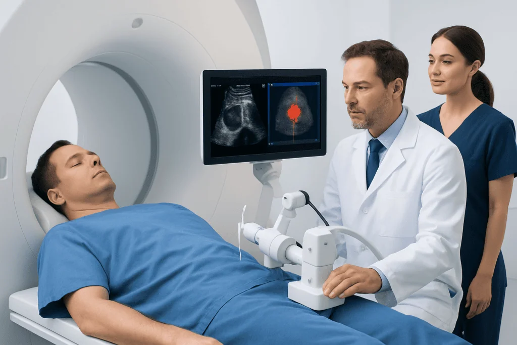
The process usually begins with a multiparametric MRI scan before the biopsy. This MRI provides detailed information about the prostate’s anatomy, blood flow, and tissue characteristics. A radiologist reviews the scan and identifies any suspicious areas, often grading them according to standard reporting systems that help estimate the likelihood of significant cancer.
During the biopsy appointment, the doctor uses ultrasound to see the prostate in real time. Specialized software then overlays, or “fuses,” the MRI images with the ultrasound view. This allows the doctor to track the biopsy needle more precisely and target the lesion seen on MRI. Depending on the center and the clinical situation, the procedure may be performed through the rectum or through the skin between the scrotum and anus, called the perineum.
Most MRI fusion biopsies also include systematic sampling in addition to targeted cores. This is because some cancers may not be visible on MRI, and combining both methods can provide a more complete assessment. The number of samples taken varies based on prostate size, the number of suspicious lesions, and the doctor’s plan.
The biopsy tissue is then examined under a microscope by a pathologist. The results help determine whether cancer is present, how aggressive it appears, and what next steps may be appropriate. When needed, the findings may guide decisions about active surveillance, surgery, radiation therapy, or other prostate cancer treatment options.
Why Targeted Sampling May Improve Diagnosis

The main advantage of MRI fusion prostate biopsy is that it directs sampling toward areas that have already appeared suspicious on MRI. This targeted approach can improve the likelihood of finding clinically significant cancer, especially tumors located in areas that may be harder to sample with standard methods alone. It is particularly useful in men with a prior negative biopsy but ongoing concern based on PSA trends, examination findings, or imaging.
Another benefit is better risk stratification. Detecting a meaningful cancer early can help doctors plan appropriate care, while avoiding overdiagnosis of very low-risk disease may help some patients avoid unnecessary treatment. In this way, MRI fusion biopsy supports more individualized decision-making rather than a one-size-fits-all approach.
Targeted biopsy may also be helpful when MRI identifies a lesion in the front or apex of the prostate, areas that can be challenging to assess with random sampling. In addition, recording the exact biopsy location can make future monitoring more precise, which is important for men on active surveillance.
Even so, no biopsy method is perfect. MRI-visible lesions are not always cancer, and some cancers do not clearly show on MRI. That is why the technique is usually interpreted alongside PSA levels, clinical examination, family history, age, overall health, and sometimes other tests used in prostate check-up and risk assessment.
Who May Need This Procedure
MRI fusion prostate biopsy may be recommended for men whose screening or clinical findings suggest a higher chance of prostate cancer. Common reasons include an elevated or rising PSA level, an abnormal digital rectal exam, or a suspicious lesion seen on prostate MRI. It may also be considered when earlier standard biopsies did not show cancer, but concern remains.
Doctors often use the procedure in people who need a more precise evaluation before deciding on treatment or surveillance. For example, if MRI suggests a focused lesion, targeted biopsy may help determine whether the lesion is likely to be low risk or more aggressive. This can affect whether the next step is close monitoring or active treatment.
Not every patient with prostate concerns will need MRI fusion biopsy. In some cases, a doctor may recommend repeat PSA testing, additional imaging, observation, or a standard biopsy approach. The best choice depends on the person’s symptoms, prostate size, MRI findings, prior biopsy history, medicines, and medical conditions.
Because the procedure is part of a larger diagnostic pathway, patients often benefit from evaluation by specialists in urology and imaging who can explain the options clearly. A doctor can discuss the likely benefits and limitations based on the individual situation.
What to Expect Before, During, and After
Before the biopsy, the care team usually reviews current medicines, allergies, bleeding risk, and any history of infections or urinary problems. Some patients may need to temporarily stop blood-thinning medicines under medical guidance. Antibiotics may be prescribed depending on the biopsy approach and the center’s protocol. The doctor will also explain whether bowel preparation is needed and what kind of anesthesia or local numbing will be used.
During the procedure, the patient is positioned to allow access to the prostate. An ultrasound probe is inserted to visualize the gland, and the MRI images are matched to the ultrasound view with fusion software. The doctor then takes targeted samples from suspicious lesions and may also take systematic cores from standard regions of the prostate. The procedure is usually completed in a relatively short time, though timing can vary.
Afterward, most patients go home the same day. Mild discomfort, blood in the urine, stool, or semen, and temporary urinary irritation can occur for a short period. Patients are usually advised to drink fluids, avoid strenuous activity for a limited time, and follow instructions about medicines and warning signs.
Results are not immediate because the tissue must be analyzed in the laboratory. The pathology report may describe whether cancer is present, how many samples are involved, and how aggressive the cells look. These findings are then reviewed with the patient to decide whether monitoring, repeat testing, or further treatment is needed.
Risks, Limitations, and Accuracy
Like all biopsy procedures, MRI fusion prostate biopsy has risks. The most common are temporary bleeding, bruising, discomfort, and urinary symptoms such as burning or difficulty passing urine. Infection is an important but less common risk, and patients should seek medical advice promptly if they develop fever, chills, worsening pain, or trouble urinating after the biopsy.
Although MRI fusion biopsy can improve detection of clinically significant cancer, it does not guarantee a diagnosis in every case. Some suspicious MRI findings turn out to be inflammation, scarring, or benign prostate enlargement rather than cancer. Conversely, a negative biopsy does not always rule out cancer completely, particularly if suspicion remains high.
Accuracy depends on several factors, including the quality of the MRI, the experience of the radiologist and urologist, the fusion technology used, and whether targeted and systematic samples are both taken. Prostate anatomy, lesion size, and patient movement can also affect the precision of sampling.
For these reasons, biopsy results are best interpreted in context. If the pathology is negative but PSA remains concerning or MRI findings change over time, the doctor may recommend follow-up imaging, repeat biopsy, or evaluation for other prostate conditions such as benign prostate enlargement.
After the Results: Next Steps and Follow-up
If the biopsy shows no cancer, the doctor may recommend ongoing monitoring rather than immediate treatment. Follow-up can include repeat PSA testing, examination, and sometimes another MRI, especially if clinical concern remains. A negative result can be reassuring, but it should always be understood in light of the whole clinical picture.
If the biopsy identifies low-risk cancer, active surveillance may be an option for some men. This approach involves careful monitoring over time instead of immediate treatment, with the goal of avoiding or delaying side effects when the cancer appears unlikely to grow quickly. Repeat MRI and biopsy may be part of this plan.
If clinically significant cancer is found, the care team will discuss the stage, grade, and treatment choices. Options may include surgery, radiation therapy, hormonal treatment, or a combination depending on the case. The decision is guided by cancer features as well as age, overall health, urinary function, and personal preferences.
Near the end of the diagnostic journey, many patients value coordinated care. Acibadem International’s multidisciplinary specialists and JCI-accredited hospitals diagnose and treat prostate conditions for international patients, with planning that may include advanced imaging, biopsy, and follow-up care.
When to Contact a Doctor
Patients should contact their doctor if they have symptoms that suggest a complication after biopsy. These include fever, chills, inability to urinate, heavy bleeding, severe pain, fainting, or symptoms that continue to worsen instead of improving. Early medical advice can help manage problems promptly and safely.
A doctor should also be consulted if biopsy results are unclear, if PSA levels continue to rise, or if symptoms such as urinary difficulty, blood in the urine, pelvic discomfort, or unexplained weight loss persist. These symptoms do not always mean cancer, but they deserve proper evaluation.
In men with a strong family history of prostate cancer or known inherited cancer risk, discussing screening and biopsy timing with a specialist can be especially helpful. A personalized plan may improve both reassurance and early detection.
Regular communication with a qualified healthcare professional is the best way to understand whether MRI fusion prostate biopsy is appropriate, what the results mean, and what follow-up is needed.
Frequently asked questions
What is the difference between MRI fusion biopsy and a standard prostate biopsy?
A standard prostate biopsy usually takes samples from preset areas of the prostate in a systematic pattern. MRI fusion prostate biopsy adds MRI guidance, allowing the doctor to target areas that looked suspicious on imaging. Many doctors use both targeted and systematic samples together for the most complete assessment.
Is MRI fusion prostate biopsy more accurate?
It may be more accurate for detecting clinically significant prostate cancer because it targets suspicious lesions identified on MRI. However, no test is perfect, and some cancers may still be missed. Accuracy also depends on image quality, the technique used, and the experience of the medical team.
Does an MRI fusion biopsy hurt?
Most patients experience some discomfort rather than severe pain. Local anesthesia, sedation, or other pain-control measures are commonly used to make the procedure more comfortable. Mild soreness or urinary irritation may continue for a short time afterward.
How long does it take to recover after the procedure?
Most people go home the same day and return to usual light activities within a short time. Minor bleeding in the urine, stool, or semen can happen temporarily. The care team will explain when to avoid strenuous exercise and what symptoms require medical attention.
Can MRI fusion biopsy replace all other prostate tests?
No. It is one part of prostate cancer evaluation and is usually considered alongside PSA results, digital rectal examination, MRI findings, and medical history. It helps confirm diagnosis, but it does not replace clinical judgment or follow-up.
What happens if the biopsy result is negative but PSA stays high?
A negative biopsy can be reassuring, but it does not always end the evaluation if concern remains. The doctor may recommend repeat PSA testing, another MRI, additional monitoring, or in some cases a repeat biopsy. The next step depends on the overall risk picture and any changes over time.
References
- National Cancer Institute
- American Cancer Society
- European Association of Urology
- American Urological Association
- National Health Service
This article is for general information only and is not a substitute for professional medical advice. Please consult a qualified doctor about your individual situation.
Oncology care in Turkey — second opinion and treatment plan
JCI-accredited · board-certified surgeons · reply within 24h
Add us as a Preferred Source to see more of our trusted health content across Google Search, AI Overviews and Discover.