MRI of Hypothalamus: Preparation, Procedure and Results
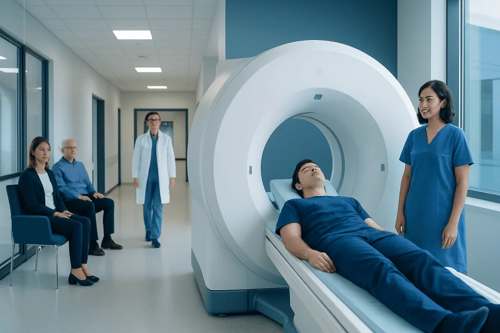
An MRI of hypothalamus uses magnetic fields rather than ionizing radiation. The scan commonly includes the pituitary gland, optic pathways and nearby brain structures.
Key Takeaways
- An MRI of hypothalamus uses magnetic fields rather than ionizing radiation.
- The scan commonly includes the pituitary gland, optic pathways and nearby brain structures.
- Contrast dye may be recommended to improve detection of inflammation, tumors or vascular changes.
- Most people can return to usual activities immediately after a routine MRI.
- MRI findings are interpreted alongside symptoms, examination results and hormone blood tests.
An MRI of hypothalamus is a noninvasive scan that produces detailed images of the small hormone- and temperature-regulating area deep within the brain and the nearby pituitary gland. It can help clinicians investigate certain hormone changes, visual symptoms, unexplained thirst or appetite changes, and suspected abnormalities affecting this region.
Overview: What Is an MRI of Hypothalamus?
An MRI of hypothalamus is a specialized magnetic resonance imaging examination of a very small but important brain region. The hypothalamus sits beneath the thalamus and close to the pituitary gland. It helps coordinate hormone release, body temperature, sleep-wake rhythms, appetite, thirst, stress responses and aspects of reproductive function.
The scan uses a strong magnet and radiofrequency waves to create detailed images of soft tissues. It does not use X-rays or ionizing radiation. Because the hypothalamus is small and close to the pituitary gland, optic nerves and major blood vessels, radiologists usually obtain thin, high-resolution images of the sellar and suprasellar region as well as the brain.
An MRI of hypothalamus may be requested when a clinician needs to assess anatomy, look for structural changes or clarify findings from blood tests and other imaging. It is one part of a wider evaluation; a normal or abnormal scan does not, on its own, explain every hormonal or neurological symptom.
How the Scan Works and Who May Need It
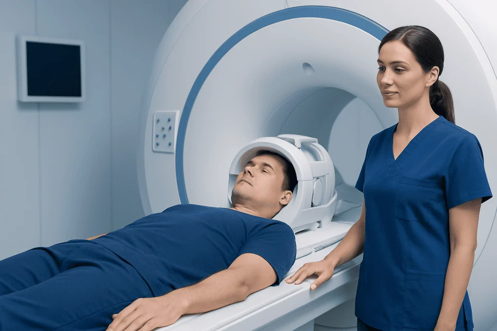
MRI detects signals from hydrogen atoms in the body after they are placed in a powerful magnetic field. Different tissues produce different signals, allowing the scanner to distinguish brain structures in much greater detail than many other imaging methods. Radiologists review images in several planes, including coronal, sagittal and MRI hypothalamus axial views, to understand the relationship between the hypothalamus and surrounding structures.
A clinician may consider an MRI brain hypothalamus protocol for unexplained hormone disturbances, diabetes insipidus, changes in thirst or urination, early or delayed puberty, unexplained weight or appetite changes, visual field symptoms, headaches with endocrine findings, or concern for inflammation, congenital differences or a mass near the pituitary region.
The test may also be appropriate when pituitary hormone testing suggests a problem involving the hypothalamic-pituitary axis. Depending on the concern, an endocrinologist, neurologist, ophthalmologist and neuroradiologist may all contribute to assessment and care.
Most people can have MRI safely. However, the imaging team must know about implanted devices, metal fragments, previous surgery, pregnancy, kidney disease and any past reaction to MRI contrast. Some devices are MRI-compatible under specific conditions, while others may mean that another test is needed.
Preparing for an MRI of Hypothalamus
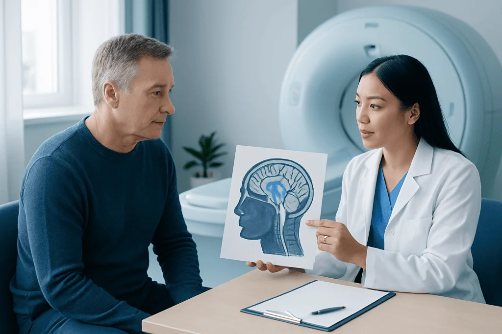
Preparation is usually straightforward. The imaging center will provide instructions based on whether contrast is planned and whether sedation is necessary. Many people can eat, drink and take their usual medicines, but they should confirm this in advance because individual instructions can differ.
Before the scan, the patient completes a safety screening form. It is important to report pacemakers, cochlear implants, neurostimulators, aneurysm clips, insulin pumps, metal fragments from work or injury, dental devices, prior operations and any possibility of pregnancy. Jewelry, hearing aids, watches, removable dental work, hair accessories, bank cards and other metal or electronic items must be removed.
Gadolinium-based contrast may be injected through a small intravenous line during some examinations. It can make certain lesions, inflammation and blood vessels easier to see. The team may ask about kidney function and prior contrast reactions before using it. People who experience claustrophobia should mention this before the appointment; support strategies, a wider-bore scanner or clinician-prescribed medication may be considered.
What Happens During the Procedure
At the appointment, a radiology professional confirms identity, reviews the safety questionnaire and helps the patient change into suitable clothing if needed. The patient lies on a padded table, usually on their back, with the head positioned in a dedicated head coil. Cushions or gentle supports help reduce movement and improve image quality.
The table moves into the MRI scanner. The machine makes repeated loud tapping, knocking or humming sounds while images are acquired, so earplugs or headphones are provided. The patient can communicate with the MRI staff through an intercom and is monitored throughout the examination. Remaining still is especially important for high-detail mri hypothalamus anatomy images.
If contrast is required, it is typically administered partway through the scan. Some people notice a brief cool sensation at the injection site, but most have no significant sensation. The examination itself is painless, although lying still can be uncomfortable for some individuals.
A focused pituitary and hypothalamic MRI often takes about 30 to 60 minutes, although timing varies with the imaging protocol, use of contrast and whether additional brain sequences are needed. The imaging team can give a more specific estimate on the day.
Benefits, Limitations and Possible Risks
The main benefit of an MRI of hypothalamus is detailed visualization of soft tissue without radiation exposure. It can identify or help characterize abnormalities involving the hypothalamus, pituitary stalk, pituitary gland, optic pathways and nearby structures. Images may also support treatment planning and provide a baseline for future comparison when monitoring is appropriate.
MRI is highly informative, but it has limitations. Very subtle tissue changes may not be visible, and some symptoms result from functional, hormonal, genetic, inflammatory or systemic conditions that do not create a distinctive MRI finding. For this reason, clinicians interpret the report together with hormone measurements, medical history, physical examination and sometimes visual field testing or other investigations.
Serious complications are uncommon. MRI safety depends on careful screening for metal and implanted devices. Gadolinium contrast reactions are uncommon and usually mild, but the team should be told about previous reactions. Contrast is used thoughtfully in people with significantly reduced kidney function. Sedation, when needed, has its own considerations and may require arrangements for travel home.
Results and What They May Mean
After the scan, a radiologist reviews the images and prepares a report for the clinician who requested it. The report may describe the hypothalamus, pituitary gland and stalk, optic chiasm, surrounding vessels and any unexpected findings. The referring clinician explains what the findings mean in the context of the person’s health and whether follow-up is needed.
Does hypothalamus damage show on MRI? MRI can show some causes or signs of damage, such as a tumor, inflammation, bleeding, scarring, injury-related changes or a structural abnormality. However, not all hypothalamic dysfunction is visible on MRI, particularly when the problem is mainly functional or microscopic. A normal scan can therefore be reassuring while still leaving a need for further endocrine or neurological assessment in selected cases.
Do you get MRI results quicker if something is wrong? Imaging departments prioritize and communicate urgent findings according to local clinical procedures, and a radiologist may contact the referring team promptly when an immediate concern is identified. However, the timing of a formal report is not a reliable indicator of whether the scan is normal or abnormal. Patients should ask their clinician or imaging center when and how they will receive results.
Acibadem International’s multidisciplinary specialists and JCI-accredited hospitals support international patients needing assessment and treatment for hypothalamic and pituitary-region concerns.
When to Seek Medical Care
People should arrange a medical assessment for persistent or unexplained symptoms that may involve hormones or the nervous system. These can include marked thirst with frequent urination, unexplained changes in menstrual patterns or sexual function, delayed or unusually early puberty, persistent fatigue, major appetite or weight changes, sleep disruption, or new difficulty regulating body temperature. These symptoms have many possible causes, and a clinician can determine whether testing or imaging is appropriate.
Prompt medical attention is important for sudden severe headache, new loss of vision or double vision, confusion, fainting, seizures, severe weakness, or symptoms that develop quickly after a head injury. Emergency care is appropriate for severe or rapidly worsening neurological symptoms.
How to tell if something is wrong with your hypothalamus? Symptoms alone cannot confirm a hypothalamic disorder because this region affects many body systems and similar symptoms are common in other conditions. A careful review by a qualified clinician, often including blood tests for hormone function and targeted imaging when indicated, is the most reliable way to identify the cause.
Frequently asked questions
Does hypothalamus damage show on MRI?
MRI can reveal many structural causes of hypothalamic injury or dysfunction, including masses, inflammation, bleeding, scarring and some effects of trauma. It may not show every problem, especially functional or microscopic changes. Results are interpreted with symptoms, hormone tests and other clinical findings.
How long does a pituitary MRI typically take?
A pituitary MRI commonly takes around 30 to 60 minutes. The exact time depends on the scanner, whether contrast is used and whether the examination also includes a broader brain MRI. Patients should allow extra time for registration, screening and preparation.
Do you get MRI results quicker if something is wrong?
Potentially urgent findings are commonly communicated promptly to the referring clinical team under the imaging provider’s procedures. However, report timing does not reliably indicate whether something is wrong. The imaging center can explain when the formal report is expected.
How to tell if something is wrong with your hypothalamus?
Possible symptoms can involve thirst, urination, appetite, sleep, temperature regulation, puberty, fertility or other hormone-related changes. These symptoms are not specific to the hypothalamus and may have many causes. A medical evaluation is needed to determine whether hypothalamic testing is appropriate.
Is an MRI of hypothalamus painful?
The MRI scan itself is painless. The main challenges are staying still, the loud scanner sounds and, for some people, feeling enclosed in the scanner. Ear protection, communication with staff and advance planning for claustrophobia can make the experience more comfortable.
Can a person eat before an MRI of hypothalamus?
Many people can eat and drink normally before a routine MRI. Instructions may differ if sedation, contrast evaluation or another procedure is planned. The patient should follow the specific directions given by the imaging center or clinician.
References
- Radiological Society of North America
- American College of Radiology
- National Institute of Neurological Disorders and Stroke
- Endocrine Society
This article is for general information only and is not a substitute for professional medical advice. Please consult a qualified doctor about your individual situation.
Explore treatments in Turkey — costs, top hospitals & a free quote
JCI-accredited · board-certified surgeons · reply within 24h
Add us as a Preferred Source to see more of our trusted health content across Google Search, AI Overviews and Discover.