MRI Renal Mass Protocol: Preparation, Procedure and Results

A renal mass MRI is a noninvasive scan that provides detailed images of the kidneys and surrounding structures. Many renal mass protocols include images before and after intravenous contrast to assess enhancement within a lesion.
Key Takeaways
- A renal mass MRI is a noninvasive scan that provides detailed images of the kidneys and surrounding structures.
- Many renal mass protocols include images before and after intravenous contrast to assess enhancement within a lesion.
- Preparation may include screening for implants, kidney function concerns, allergies, pregnancy, and the need for sedation.
- The scan usually takes about 30 to 60 minutes, although timing varies with the protocol and individual needs.
- Results should be interpreted alongside symptoms, blood tests, prior imaging, and, when needed, urology consultation.
An MRI renal mass protocol is a specialized kidney scan that helps doctors characterize a kidney lesion, including whether it contains fat, blood products, fluid, or enhancing tissue. It is commonly performed with and without contrast and provides detailed information to support diagnosis, surveillance, or treatment planning.
Overview: What Is an MRI Renal Mass Protocol?
An MRI renal mass protocol is a tailored magnetic resonance imaging examination used to evaluate a mass, cyst, or other unusual finding in or near a kidney. It creates highly detailed images using a magnetic field and radio waves rather than ionizing radiation. The protocol is designed to help radiologists identify important tissue features and describe whether a lesion appears more consistent with a simple cyst, a complex cyst, a benign growth, an infection-related change, or a lesion that needs further assessment.
A renal mass may first be noticed during an ultrasound, CT scan, or imaging performed for another reason. MRI can be particularly useful when CT findings are uncertain, when a person should avoid radiation where possible, or when detailed soft-tissue characterization is needed. The test does not by itself confirm every diagnosis, but it can meaningfully guide monitoring, referral, biopsy decisions, or kidney cancer treatment planning when appropriate.
The phrase mri renal mass protocol radiology generally refers to the standardized series of image sequences selected by the radiology team. The exact plan may be adjusted for the lesion size and location, kidney function, prior imaging findings, the person’s ability to hold their breath, and whether contrast can be used safely.
How the Scan Works and Who May Need It
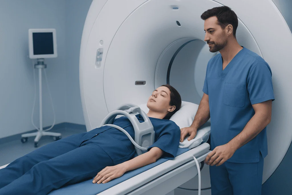
MRI detects signals from water and fat within the body and converts them into detailed cross-sectional images. For renal lesions, images are often obtained in several planes and with different sequence types. Some sequences identify fat or bleeding within a lesion, while others show fluid content, tissue density characteristics, restricted water movement, and blood flow patterns after contrast administration.
People may be referred for a renal mass MRI after an indeterminate kidney finding on ultrasound or CT, during follow-up of a complex kidney cyst, or before treatment for a suspected kidney tumor. It may also be used to assess the opposite kidney, nearby lymph nodes, renal veins, and surrounding tissues. MRI can be valuable for people with a history of kidney surgery or for selected individuals undergoing active surveillance of a small renal lesion.
Not everyone can have MRI in the same way. Some implanted medical devices, metal fragments, or older types of clips may make MRI unsafe or require specialist review. Claustrophobia, difficulty lying still, severe pain, pregnancy, and reduced kidney function do not always prevent imaging, but they should be discussed beforehand so the team can plan the safest and most comfortable approach.
What Should I Do to Prepare for a Kidney MRI?

Preparation instructions can differ between imaging centers, so the person should follow the instructions provided with their appointment. In many cases, normal medicines can be taken with a small amount of water unless the referring doctor advises otherwise. Some centers ask patients not to eat for several hours before a contrast-enhanced abdominal MRI, as this may reduce motion from digestion and improve image quality.
Before the appointment, the imaging team should be told about kidney disease, dialysis, prior reactions to MRI contrast, asthma or significant allergies, pregnancy or possible pregnancy, and any surgery or implanted device. They should also be informed about cochlear implants, pacemakers, neurostimulators, insulin pumps, vascular clips, joint replacements, retained metal fragments, or bullet injuries. The team may request recent blood tests to assess kidney function before administering gadolinium-based contrast.
On arrival, the person will remove metal-containing items such as jewelry, watches, hearing aids, credit cards, hairpins, and some clothing with metallic fasteners. Comfortable clothing without metal may be helpful. Anyone who feels anxious in enclosed spaces can ask in advance about relaxation measures, an accompanying support person where permitted, or sedation options; if sedation is planned, transport home will usually be needed.
What Is the Typical MRI Protocol for Renal Masses?
A typical MRI renal mass protocol begins with localizer images that help the radiologist position the detailed scan. It commonly includes T1-weighted and T2-weighted sequences, images that compare fat and water signals, and diffusion-weighted imaging. These components can help evaluate whether a lesion contains fluid, fat, blood products, scar-like tissue, or solid areas.
For many patients, the MRI renal mass protocol is performed with and without contrast. Images are acquired before contrast and then at timed phases after a gadolinium-based contrast agent is injected into a vein. Early and later contrast phases help show whether and how a lesion enhances, which is an important feature in distinguishing simple fluid-filled cysts from lesions containing vascularized tissue. The scan may also include delayed images to assess the kidneys’ collecting systems when clinically relevant.
The specific sequences and timing are not identical for every person. A radiologist may tailor the examination after reviewing earlier ultrasound or CT images. When contrast is not suitable, a non-contrast MRI may still provide useful information, although it may not answer every question. The term mri renal mass protocol cpt code concerns medical billing and coding rather than the clinical content of the scan; the appropriate code depends on the country, facility, and whether contrast is used.
- Non-contrast sequences assess anatomy, fluid, fat, blood products, and tissue characteristics.
- Diffusion sequences may provide additional clues about cellular tissue and lesion behavior.
- Dynamic contrast images evaluate enhancement and blood supply patterns.
- Additional images may assess renal veins, lymph nodes, collecting systems, or nearby organs.
What Should You Not Do the Day Before an MRI?
Most people do not need major lifestyle changes the day before a kidney MRI. They should not stop prescribed medicines, alter diabetes treatment, or restrict fluids unless their doctor or imaging department gives specific instructions. Following individualized instructions is especially important for people with diabetes, kidney disease, heart failure, or a planned sedative medication.
It is sensible to avoid arriving dehydrated unless a clinician has recommended fluid restriction. The person should also avoid wearing or bringing unnecessary metal items to the appointment. If the center requests fasting, it is important not to eat or drink outside the stated time frame, while still taking essential medicines as instructed.
People should not overlook new or relevant health information. For example, a recent surgery, a newly implanted device, possible pregnancy, worsening kidney problems, or a prior contrast reaction should be reported before the scan rather than on the day if possible. Alcohol or recreational substances should be avoided if sedation may be used, as they can interact with sedative medicines and affect safety.
Step-by-Step Procedure, Scan Duration and Recovery
At the imaging center, a radiographer reviews safety questions and confirms the reason for the scan. If contrast is planned, an intravenous cannula is placed in the arm or hand. The person lies on a padded table, usually on their back, and a receiving coil is positioned around the abdomen. Ear protection is provided because the scanner produces loud tapping and thumping sounds.
The table moves slowly into the MRI scanner. During the examination, the radiographer can communicate through an intercom and can see and hear the patient. Several image sets are obtained, and the person may be asked to hold their breath for short periods. Remaining as still as possible improves image clarity. Contrast, if used, is injected partway through the examination; some people notice a cool sensation at the injection site, but many feel no significant change.
How long does a renal MRI take? A renal mass MRI commonly takes about 30 to 60 minutes. More complex studies, additional delayed images, the use of sedation, movement during scanning, or the need to repeat sequences can make the visit longer. Once the images are complete, most people can resume usual activities immediately unless they received sedation or were given different instructions.
There is generally no recovery period after a routine MRI. Drinking fluids as normally advised may be reasonable after contrast, unless fluid intake is restricted for a medical reason. If sedation was used, the person should avoid driving, operating machinery, making important decisions, or drinking alcohol until the effects have fully worn off and according to the care team’s guidance.
Benefits, Risks and Understanding the Results
The main benefit of MRI is detailed tissue characterization without radiation exposure. It may help distinguish a simple benign cyst from a complex cyst or solid lesion and can provide information about lesion size, location, enhancement, possible fat content, and involvement of nearby structures. These details support a more informed discussion with a urologist, nephrologist, oncologist, or other specialist.
MRI is generally considered safe when appropriate screening is completed. However, the confined space and noise can be uncomfortable for some people. Contrast reactions are uncommon and are usually mild, but serious reactions can occur rarely. Gadolinium-based contrast requires particular consideration in people with severely reduced kidney function, so the imaging team assesses individual risks and benefits beforehand.
A radiologist reviews the images and sends a report to the referring clinician, often within several working days depending on local processes. The report may describe a lesion as a simple cyst, a complex cyst using a recognized classification system, a likely benign lesion, an indeterminate lesion, or a finding requiring further assessment. A recommendation for follow-up imaging, specialist evaluation, biopsy, or treatment does not automatically mean cancer; it means the finding needs appropriate clarification.
At Acibadem International, multidisciplinary specialists at JCI-accredited hospitals can coordinate imaging interpretation and care for international patients with kidney lesions, including referral for kidney cancer evaluation when indicated.
When to Seek Medical Care
A person should contact the clinician who ordered the scan to discuss results and the recommended next step, even if symptoms have improved. Timely follow-up is especially important when a report describes a solid lesion, a complex cyst, growth compared with previous imaging, or a finding that needs urology review. Many kidney findings are not emergencies, and clear follow-up helps ensure they are assessed appropriately.
More urgent medical assessment is appropriate for visible blood in the urine, severe or persistent flank or abdominal pain, fever with urinary symptoms, inability to pass urine, unexplained marked weakness, or new swelling. These symptoms can have several causes and should not be assumed to be caused by a renal mass, but they warrant professional evaluation.
Emergency care is needed for signs of a severe allergic reaction after contrast, such as trouble breathing, swelling of the face or throat, widespread hives, fainting, or severe dizziness. Imaging staff usually monitor patients briefly after contrast when necessary and can provide immediate care if a reaction occurs.
Frequently asked questions
Is contrast always needed for an MRI renal mass protocol?
Contrast is often used because it helps show whether a kidney lesion enhances after injection, which is important for characterization. However, contrast is not appropriate for every person, particularly in some cases of significant kidney impairment or prior contrast reaction. The radiologist and referring clinician decide whether contrast is needed based on the clinical question and individual safety factors.
Can a renal MRI tell if a kidney mass is cancer?
MRI can identify imaging features that make a lesion more likely to be benign or more concerning for cancer. However, imaging cannot definitively classify every renal mass. Depending on the findings, doctors may recommend follow-up imaging, urology review, biopsy, surgery, or another management approach.
Do I need to fast before a kidney MRI?
Some imaging centers request fasting for several hours before an abdominal MRI, especially when contrast is planned, while others may not. The patient should follow the instructions from the imaging center rather than making changes independently. Essential medicines are often continued with a small sip of water, but this should be confirmed in advance.
Is a renal MRI painful?
The scan itself is not painful, and MRI does not involve needles unless intravenous contrast is used. Lying still and holding the breath briefly can be uncomfortable for some people, and the scanner is loud. The radiographer can provide ear protection and communicate throughout the examination.
What happens if I am claustrophobic?
Claustrophobia is common and should be mentioned when booking the scan. The imaging team may offer practical support, relaxation strategies, a wider-bore scanner where available, or clinician-prescribed sedation in selected cases. If sedation is used, a responsible adult is usually needed to take the person home.
How soon will renal MRI results be available?
A radiologist must review the images and prepare a formal report, so timing varies by facility and clinical urgency. The referring clinician commonly receives the report within several working days. Urgent or unexpected findings may be communicated more quickly when appropriate.
References
- American College of Radiology
- Radiological Society of North America
- National Institute of Diabetes and Digestive and Kidney Diseases
- European Association of Urology
- U.S. Food and Drug Administration
This article is for general information only and is not a substitute for professional medical advice. Please consult a qualified doctor about your individual situation.
Explore treatments in Turkey — costs, top hospitals & a free quote
JCI-accredited · board-certified surgeons · reply within 24h
Add us as a Preferred Source to see more of our trusted health content across Google Search, AI Overviews and Discover.

More from the Health Library



Related Specialists

Prof. Dr. Burak Pamukçu
Cardiology
Dr. Haluk Mumcuoğlu
Internal Medicine
Dr. Ersin Adem Hadzhiyusein
Inpatient Clinic Physicians Clinical Service
Dr. Aytekin Ünlükaplan
Anesthesiology