MRI Syrinx: Preparation, Procedure and Results

MRI is the preferred test for detecting and measuring a spinal syrinx. A syrinx may be linked with conditions that affect cerebrospinal fluid flow, including Chiari malformation, injury, tumors or inflammation.
Key Takeaways
- MRI is the preferred test for detecting and measuring a spinal syrinx.
- A syrinx may be linked with conditions that affect cerebrospinal fluid flow, including Chiari malformation, injury, tumors or inflammation.
- Not every syrinx requires surgery; symptoms, progression and the underlying cause guide care.
- MRI results show anatomy but may not always establish exactly why a syrinx formed.
- New weakness, walking difficulty, bladder changes or rapidly worsening symptoms need prompt medical assessment.
An MRI syrinx examination is the main imaging test used to identify and assess a syrinx, a fluid-filled cavity within the spinal cord. It can show the cavity’s location and extent and may help doctors look for an underlying cause, guiding monitoring or treatment decisions.
MRI Syrinx Overview
An MRI syrinx scan uses magnetic resonance imaging to look for a syrinx: a fluid-filled cavity, also called a cyst, that develops inside the spinal cord. The condition is often called syringomyelia. MRI produces detailed images of the spinal cord and surrounding structures without using ionizing radiation, making it the most useful test for confirming a syrinx and following it over time.
Some syrinxes are found while investigating neck or back pain, numbness, weakness, scoliosis, or a previous spinal injury. Others cause no symptoms and are discovered incidentally. The finding itself does not automatically mean that treatment is needed. A neurologist or neurosurgeon considers the MRI appearance together with symptoms, examination findings and whether the cavity is changing.
A syrinx can occur in the neck, chest or lower spinal cord, although the cervical and upper thoracic regions are common locations. It may be associated with Chiari malformation, trauma, spinal cord tumors, inflammation, scarring around the spinal cord, or other conditions that interfere with the normal circulation of cerebrospinal fluid.
How MRI Detects a Syrinx and What It Can Show
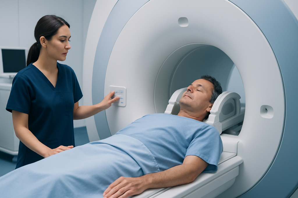
MRI uses a powerful magnet and radiofrequency waves to create cross-sectional images of soft tissues. Cerebrospinal fluid appears differently from spinal cord tissue on standard MRI sequences, allowing the radiologist to identify a syrinx, estimate its length and width, and see whether it affects nearby spinal cord structures.
The scan commonly includes several areas of the spine. If a syrinx is identified, imaging of the brain and the junction between the skull and upper spine may also be recommended, particularly when a doctor is assessing for Chiari malformation. In some situations, contrast material given through a vein helps evaluate a possible tumor, inflammation, infection or prior surgical change.
MRI can document important features for future comparison, including the syrinx level, size, shape and any associated spinal cord swelling. It can also reveal possible contributors, such as crowding at the base of the skull, a mass, tethering, scar tissue or changes after injury. A radiology report is interpreted alongside the clinical evaluation rather than used in isolation.
Who May Need an MRI Syrinx Examination

Doctors may request an MRI when symptoms suggest an issue affecting the spinal cord. These may include altered sensitivity to pain or temperature, persistent numbness or tingling, weakness in an arm or leg, muscle wasting, stiffness, balance changes, or scoliosis that requires further assessment. Symptoms vary greatly, and similar symptoms can have many other causes.
An MRI may also be appropriate after a significant spinal cord injury, when there is concern about a spinal tumor or inflammatory condition, or when an existing syrinx needs monitoring. People with a known Chiari malformation may be advised to have spinal MRI if symptoms or examination findings raise concern for syringomyelia.
MRI is safe for many people, but screening is essential. Certain implanted devices, metallic fragments, cochlear implants, clips used in past surgery, and some stimulators may affect whether MRI can be performed or require a specialized protocol. Pregnancy, kidney disease and previous reactions to contrast should also be discussed before a contrast-enhanced scan.
Preparing for and Having an MRI Syrinx Scan
Preparation is usually straightforward. The imaging center will ask about implants, previous operations, allergies, kidney problems and possible pregnancy. Patients may normally eat and drink unless the facility gives different instructions, such as when sedation or contrast is planned. Jewelry, watches, hairpins, hearing aids, removable dental devices and clothing with metal parts must be removed before the examination.
During the scan, the patient lies on a padded table that moves slowly into the MRI scanner. A coil is placed around the area being imaged to improve image quality. The procedure is painless, though the machine makes repeated loud tapping and knocking sounds; ear protection is provided. It is important to remain as still as possible so the images are clear.
A spinal MRI often takes about 30 to 60 minutes, depending on the areas being scanned and whether contrast is used. The radiographer can see and speak with the patient throughout the exam. People with claustrophobia, pain that makes lying still difficult, or young children may benefit from planning ahead with the care team; options can include reassurance measures, medication or sedation when clinically appropriate.
If gadolinium contrast is needed, it is injected through a small intravenous line during the examination. Most people tolerate it well. Serious reactions are uncommon, but staff are trained to respond if they occur. The team may check kidney function before contrast in people with relevant health concerns.
MRI Syrinx Results, Benefits and Limitations
After the scan, a radiologist reviews the images and sends a report to the referring clinician. The report may describe whether a syrinx is present, its dimensions and spinal levels, and any findings that may be related. The clinician then explains what the result means in the context of the person’s symptoms and medical history.
The central benefit of MRI is its detailed view of the spinal cord and cerebrospinal fluid spaces. It helps establish a baseline, supports decisions about observation or further investigation, and can be repeated to monitor changes. Unlike CT scanning, conventional MRI does not expose the patient to radiation.
Does an MRI tell you how a syrinx showed up? MRI can often identify a likely associated condition, such as Chiari malformation, prior injury, a tumor or obstruction to fluid flow. However, it cannot always prove the exact reason a syrinx developed. Additional imaging, medical history, neurological examination and sometimes repeat scans are needed to build the most complete picture.
Most people can return to normal activities immediately after a non-sedated MRI. If sedation was used, someone else should accompany the patient home and driving or important decisions should be avoided for the rest of that day. The timing of results varies by service, but urgent findings are usually communicated promptly to the referring team.
Treatment, Monitoring and Recovery Expectations
Management depends on symptoms, MRI changes and the cause of the syrinx. A small, stable syrinx without symptoms may be monitored with clinical reviews and follow-up MRI. Treating the underlying problem is often the priority when symptoms are progressive or when the syrinx is enlarging.
For example, surgery may be considered to improve cerebrospinal fluid flow in people whose syrinx is related to Chiari malformation. If a tumor, tethering, scar-related blockage or another structural issue is involved, treatment is tailored to that cause. In selected situations, surgeons may drain the syrinx or place a shunt, although the approach depends on individual anatomy and the likely source of fluid-flow disruption.
How long does it take to recover from a syrinx spinal cord surgery? Recovery varies with the procedure, the underlying cause, overall health and any spinal cord symptoms present before surgery. Hospital recovery may take several days for some operations, while full return to usual activity can take weeks or longer. The surgical team provides individualized restrictions, wound care guidance, rehabilitation planning and follow-up MRI recommendations.
Benefits of surgery may include preventing further neurological decline and improving fluid flow or reducing the syrinx size. Improvement in long-standing numbness, weakness or pain is less predictable because spinal cord nerves may have been affected before treatment. Potential risks, which vary by procedure, include infection, bleeding, cerebrospinal fluid leakage, neurological symptoms, recurrence or need for additional treatment; these should be discussed carefully with a neurosurgeon.
What to Avoid if You Have a Syrinx
What to avoid if you have a syrinx? Until a clinician has assessed the condition, it is sensible to avoid activities that reliably trigger symptoms or place unusual strain on the neck or spine. This may include heavy lifting, high-impact contact sports, forceful neck movements, and activities that involve repeated straining or breath-holding. Recommendations should be individualized, since activity limits depend on the syrinx location, symptoms and underlying cause.
People should not ignore changes in sensation simply because they are mild. Reduced ability to feel pain or temperature can increase the chance of accidental burns, cuts or injuries, particularly to the hands. Using protective gloves when appropriate, checking the skin regularly, and taking care with hot water, cooking surfaces and heating devices can be helpful.
Healthy routines can support overall wellbeing: regular gentle activity approved by the clinical team, adequate sleep, balanced nutrition and avoiding tobacco are reasonable goals. Physical therapy may be recommended for some people, but exercises should be selected by professionals who understand the diagnosis. Avoid starting intensive strengthening, manipulation or traction programs without medical advice.
How Serious Is a Spinal Syrinx and When to Seek Medical Care
How serious is a spinal syrinx? The seriousness ranges from an incidental, stable finding that needs observation to a condition that can gradually affect spinal cord function. A syrinx is more concerning when it is enlarging, associated with progressive symptoms, or caused by a condition that requires treatment. Early specialist evaluation helps clarify the outlook and creates an appropriate monitoring plan.
Medical assessment should be arranged for persistent or worsening neck, shoulder, arm or back symptoms; unexplained numbness; weakness; reduced temperature sensation; balance difficulties; or new curvature of the spine. Anyone with a known syrinx should attend recommended follow-up appointments, even when symptoms are stable, because MRI comparisons can help identify meaningful changes.
Urgent medical care is appropriate for sudden or rapidly worsening weakness, new difficulty walking, loss of bladder or bowel control, marked loss of sensation, severe new pain after injury, or symptoms suggesting infection following surgery. These symptoms do not always indicate a syrinx-related emergency, but they require timely assessment.
Acibadem International’s multidisciplinary neurology, radiology and neurosurgery specialists at JCI-accredited hospitals assess spinal cord conditions and discuss monitoring or treatment options for international patients. A referral to a qualified specialist is appropriate whenever MRI findings or symptoms raise concerns about a syrinx.
Frequently asked questions
What is an MRI syrinx scan?
An MRI syrinx scan is an MRI examination used to identify a fluid-filled cavity within the spinal cord. It shows the syrinx location, size and relationship to surrounding spinal cord tissue. It may also help doctors investigate conditions that can affect cerebrospinal fluid flow.
Is contrast always needed for an MRI syrinx?
No. Many syrinxes can be identified clearly on standard non-contrast MRI sequences. Contrast may be added when doctors need to assess for a tumor, inflammation, infection, prior surgery-related changes or another possible underlying cause.
How serious is a spinal syrinx?
A spinal syrinx can range from a stable finding with no symptoms to a condition that gradually affects sensation, strength or walking. Its significance depends on symptoms, whether it changes over time and what caused it. A neurological or neurosurgical assessment helps determine the appropriate follow-up.
What should someone avoid if they have a syrinx?
People should avoid activities that cause symptoms or involve major strain, forceful neck movement or high-impact injury risk until they have personalized medical advice. They should also take precautions against burns and cuts if pain or temperature sensation is reduced. The right activity plan differs from person to person.
Does an MRI tell you how a syrinx showed up?
MRI can show features that suggest a cause, including Chiari malformation, spinal injury, a tumor, inflammation or an obstruction to cerebrospinal fluid flow. However, it may not identify an exact cause in every case. Doctors combine MRI findings with symptoms, examination and medical history.
How long does it take to recover from syrinx spinal cord surgery?
Recovery depends on the operation performed, the underlying condition and the person’s neurological function before surgery. Initial hospital recovery may take days, while return to usual activities can take several weeks or longer. Follow-up imaging and rehabilitation may be part of recovery.
References
- National Institute of Neurological Disorders and Stroke
- American Association of Neurological Surgeons
- Radiological Society of North America
- Mayo Clinic
- Merck Manual Consumer Version
This article is for general information only and is not a substitute for professional medical advice. Please consult a qualified doctor about your individual situation.
Joint and spine care in Turkey — expert assessment & treatment
JCI-accredited · board-certified surgeons · reply within 24h
Add us as a Preferred Source to see more of our trusted health content across Google Search, AI Overviews and Discover.

More from the Health Library



Related Specialists

Prof. Dr. Ali Kurtsoy
Neurosurgery
Prof. Dr. Deniz Tuna Edizer
Otorhinolaryngology
Dr. M. Mümtaz Özarar
Radiology
Dr. Hayati Yavuz
Internal Medicine