

Neuro-Ophthalmology Tests Explained: MRI, Visual Fields, and OCT

MRI, visual field testing, and OCT look at different parts of the visual system and are often used together. These tests can help evaluate blurred vision, double vision, vision loss, and suspected optic nerve disease.
Key Takeaways
- MRI, visual field testing, and OCT look at different parts of the visual system and are often used together.
- These tests can help evaluate blurred vision, double vision, vision loss, and suspected optic nerve disease.
- Most neuro-ophthalmology tests are noninvasive and completed as outpatient procedures.
- Test results are interpreted alongside symptoms, eye examination findings, and medical history.
- Early assessment can help identify treatable causes and guide follow-up care.
Neuro-ophthalmology tests help doctors understand how the eyes, optic nerves, and brain work together. MRI, visual field testing, and OCT are commonly used to investigate vision changes, eye movement problems, and optic nerve disorders.
Overview of Neuro-Ophthalmology Tests
Neuro-ophthalmology is the area of medicine that connects eye care with neurology. It focuses on conditions that affect vision because of problems in the optic nerves, eye movement nerves, brain pathways, or the way the brain processes visual information. When a person has symptoms such as sudden or gradual vision loss, double vision, drooping eyelids, blind spots, or unexplained headaches with visual changes, a neuro-ophthalmologist may recommend specialized testing.
Three of the most commonly discussed tests are magnetic resonance imaging (MRI), visual field testing, and optical coherence tomography (OCT). Each one provides different information. MRI shows detailed images of the brain, eye sockets, optic nerves, and surrounding structures. Visual field testing measures how well a person sees in central and peripheral vision. OCT creates detailed cross-sectional images of the retina and optic nerve head.
These tests do not replace a clinical examination. Instead, they add important pieces to the overall picture. The doctor combines the patient’s symptoms, medical history, eye examination, and test results to look for causes such as optic neuritis, compression of the optic nerve, inflammation, stroke, increased pressure around the brain, or other neurological and eye-related conditions.
When These Tests May Be Recommended
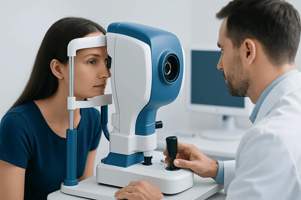
Neuro-ophthalmology tests are ordered based on symptoms and examination findings rather than as routine screening for everyone. A doctor may suggest them when a person reports blurred vision that cannot be explained by a standard eye exam, episodes of temporary vision loss, side vision loss, color vision changes, eye pain with movement, or double vision. They may also be useful when the optic nerve appears swollen or pale, or when eye movements are not normal.
Sometimes these tests help evaluate conditions that begin outside the eye itself. For example, a patient with headaches and optic nerve swelling may need assessment for raised pressure in or around the brain. Someone with one-sided vision loss may need testing for optic nerve inflammation or compression. A person with difficulty seeing on one side of space may need evaluation of the visual pathways in the brain.
Neuro-ophthalmology testing may also be part of monitoring after a diagnosis has already been made. Doctors may repeat visual fields or OCT over time to check whether a condition is stable, improving, or progressing. This can be important in disorders such as glaucoma when the optic nerve is being monitored, or in optic nerve diseases that require regular follow-up.
MRI: What It Shows and What to Expect
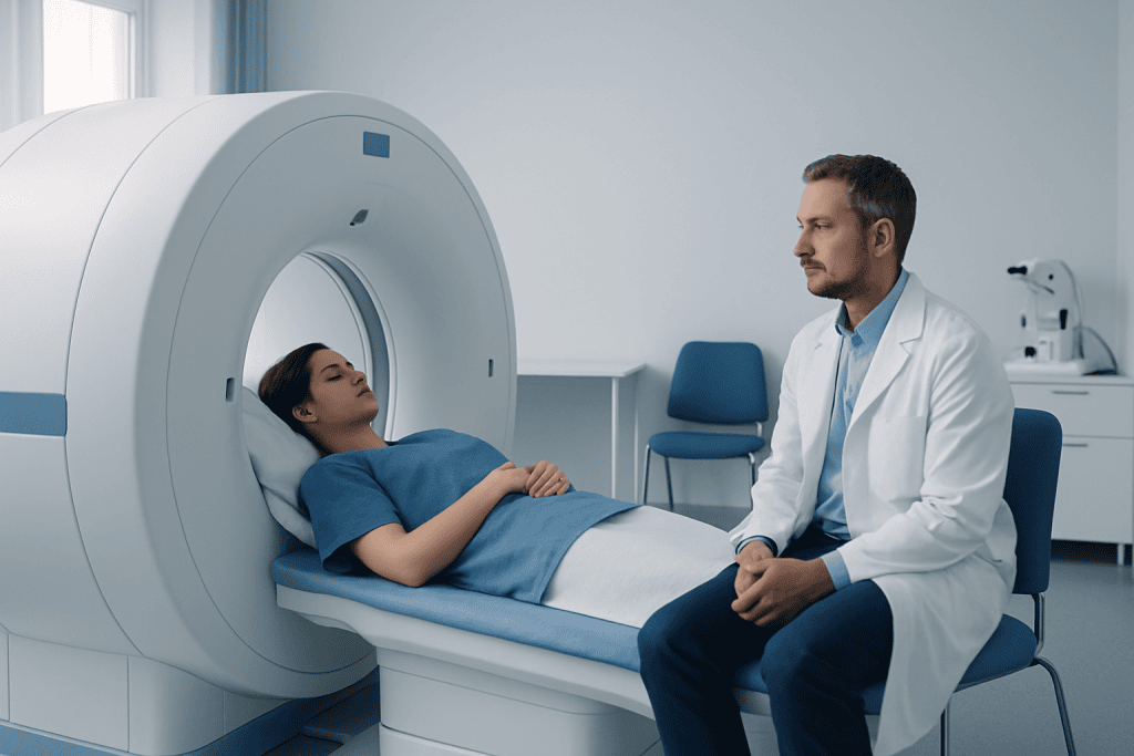
MRI uses a strong magnetic field and radio waves to create detailed images of internal structures. In neuro-ophthalmology, MRI can help examine the brain, optic nerves, eye sockets, pituitary region, and pathways that carry visual information from the eyes to the brain. It is especially helpful when doctors suspect inflammation, demyelination, tumors, compression, stroke, structural abnormalities, or other neurological causes of visual symptoms.
For some patients, contrast material may be used to improve the visibility of certain tissues and areas of inflammation. The scan itself is painless, but it requires lying still inside the MRI machine for a period of time. Because the machine can be noisy and enclosed, some people feel uncomfortable or claustrophobic. The imaging team can explain what to expect and discuss options if anxiety is a concern. If needed, a specialist may arrange MRI scanning as part of a broader evaluation.
MRI is not always the first or only test. It is usually chosen when the pattern of symptoms suggests that the problem may involve the optic nerves, brain, or eye movement pathways. MRI findings are interpreted carefully because some changes can be subtle, and not every MRI abnormality explains a person’s symptoms. This is why imaging works best when paired with a neuro-ophthalmic examination and other tests.
Before an MRI, patients should tell the medical team about implanted devices, metal fragments, kidney problems, pregnancy, or previous reactions to contrast. In many cases, MRI is very safe, but these details help the team plan the study appropriately and choose the best approach.
Visual Field Testing: Measuring Side Vision and Blind Spots
Visual field testing checks how well a person sees across the entire field of vision, including central and peripheral areas. It is one of the most useful tools for finding blind spots, reduced sensitivity, or characteristic patterns of vision loss. In neuro-ophthalmology, this can help doctors decide whether a problem is likely affecting the optic nerve, optic chiasm, visual pathways in the brain, or the retina.
The most common type is automated perimetry. During the test, the person looks into a bowl-shaped instrument and presses a button when lights appear in different locations. The test is noninvasive, but it does require concentration. It is common for the first test to feel tiring or unfamiliar, and sometimes a repeat test is needed to confirm reliable results. A poor night’s sleep, dry eyes, droopy eyelids, or difficulty understanding the instructions can affect performance.
Doctors look not only at whether the test is normal or abnormal, but also at the specific pattern of loss. For example, some patterns may suggest optic nerve disease, while others may point to a problem where the optic nerves cross or farther back in the brain. Visual field testing can therefore guide the need for further imaging or neurological investigation.
This test is also valuable for follow-up. Repeating visual fields over time helps show whether vision is stable or changing. It may be used alongside a detailed eye examination and other studies to track diseases that affect the optic nerve and visual pathways.
OCT: A Detailed Look at the Retina and Optic Nerve
Optical coherence tomography, or OCT, is a quick and noninvasive imaging test that uses light waves to create highly detailed cross-sectional pictures of the retina. In neuro-ophthalmology, OCT is especially useful for examining the retinal nerve fiber layer and the optic nerve head. These structures can become swollen, thinned, or otherwise changed in diseases involving the optic nerve.
OCT can help distinguish between true swelling of the optic nerve and conditions that can look similar on examination. It may also show thinning after past optic nerve injury, which can support the diagnosis of previous damage. Because the images are measured precisely, OCT can be repeated over time to monitor changes and compare one visit with another.
The test is usually simple and comfortable. The patient places the chin on a support and looks at a target while the scanner captures images. There is no contact with the eye, and the scan takes only a short time. In some settings, OCT is performed after pupil dilation, depending on the equipment and the doctor’s needs.
OCT is often used together with visual field testing because the two tests complement each other. OCT shows structure, while visual fields show function. In some patients, structural changes appear first; in others, symptoms or field changes may come before clear OCT abnormalities. When appropriate, a specialist may include OCT imaging in a comprehensive neuro-ophthalmology assessment.
How Doctors Combine Test Results
No single test can answer every question in neuro-ophthalmology. A normal MRI does not always rule out disease, and an abnormal OCT or visual field does not automatically identify the cause. Doctors combine all available information to understand whether the main issue is in the eye, optic nerve, eye movement system, or brain.
For example, a person with painful vision loss and reduced color vision may have examination findings that raise concern for optic neuritis. MRI may help look for inflammation or related brain changes, while OCT and visual fields can document optic nerve function and structure over time. In another case, a patient with gradual side vision loss may have a visual field pattern that suggests compression near the optic chiasm, leading to targeted MRI imaging.
Sometimes these tests also help distinguish neuro-ophthalmic disorders from other eye diseases. A person with blurred vision may need evaluation to determine whether the problem is retinal, optic nerve-related, or neurological. OCT can reveal retinal disease, while visual fields and MRI may help clarify the rest of the picture. This may be relevant in patients being assessed for disorders such as retinal detachment or other conditions that can mimic neuro-ophthalmic symptoms.
Because interpretation can be complex, patients may benefit from care that includes ophthalmologists, neurologists, neuroradiologists, and other specialists when needed. Near the end of the diagnostic process, centers such as Acibadem International may offer this multidisciplinary evaluation in JCI-accredited hospitals for international patients.
Other Tests That May Be Used
Although MRI, visual fields, and OCT are central tools, some patients need additional tests. Blood tests may be ordered if the doctor suspects inflammation, infection, autoimmune disease, nutritional deficiency, or other systemic causes. In selected cases, a lumbar puncture may be recommended to assess pressure around the brain or to analyze cerebrospinal fluid.
Eye movement testing, color vision assessment, pupil evaluation, and photographs of the optic nerve may also be useful. Some patients may need CT imaging rather than MRI, especially if MRI is not possible. Others may be referred for more specialized retinal imaging, ultrasound, or electrophysiological testing depending on the clinical question.
The choice of tests depends on the person’s age, symptoms, examination findings, and general health. A careful, step-by-step approach helps avoid unnecessary procedures while ensuring that important causes are not missed. If treatment is needed, the next steps may involve neurology care, eye care, or targeted interventions such as treatment for a brain lesion when imaging identifies a structural cause.
Preparing for Testing and When to Seek Medical Care
Patients can prepare for neuro-ophthalmology tests by bringing a list of symptoms, current medications, previous scan results, and eyeglasses or contact lens information. It helps to mention when symptoms started, whether they are constant or intermittent, and whether headaches, pain, or neurological symptoms are present. For MRI, patients should inform the team about implants, pacemakers, metal exposure, or pregnancy. For visual field testing, being rested and comfortable can improve reliability.
After the tests, results may not be fully explained by one report alone. Doctors often review the images and measurements together with the eye examination before discussing what they mean. Follow-up may include repeating one or more tests after weeks or months to see whether changes persist, improve, or progress.
Urgent medical attention is important for sudden vision loss, sudden double vision, severe new headache with visual symptoms, new drooping of the eyelid, eye pain with vision changes, or weakness, numbness, or speech difficulty. These symptoms do not always indicate an emergency, but they deserve prompt medical assessment. Seeking timely care gives the best chance of identifying the cause and starting appropriate treatment.
Frequently asked questions
What is the difference between MRI, visual field testing, and OCT?
These tests examine different parts of the visual system. MRI shows detailed images of the brain, optic nerves, and surrounding structures; visual field testing measures how well a person sees in central and peripheral vision; and OCT shows the structure of the retina and optic nerve in fine detail. Doctors often use them together because each provides different information.
Are neuro-ophthalmology tests painful?
Most of these tests are not painful. Visual field testing and OCT are noninvasive and usually well tolerated, while MRI is also painless but may feel uncomfortable for people who dislike enclosed spaces or loud sounds. The care team can explain each step and help make the experience easier.
Why would a doctor order an MRI for vision problems?
An MRI may be ordered when the doctor suspects that visual symptoms are related to the optic nerves, brain, eye movement pathways, or structures around the eyes. It can help look for inflammation, compression, stroke, demyelination, or other structural causes. The decision depends on the pattern of symptoms and examination findings.
How long do visual field and OCT tests take?
OCT is usually very quick and often takes only a few minutes per eye. Visual field testing usually takes longer because it depends on concentration and careful responses to light signals. The exact timing varies by clinic and by whether repeat testing is needed for accuracy.
Can OCT detect optic nerve problems?
OCT can show structural changes in and around the optic nerve, including swelling or thinning of nerve fiber layers. This makes it very useful in many optic nerve disorders. However, OCT does not replace a full examination, and its findings are most meaningful when interpreted with symptoms and other tests.
If one test is normal, does that mean nothing is wrong?
Not necessarily. Some neuro-ophthalmic conditions may not appear clearly on one test, especially early on or if the abnormality is subtle. Doctors usually consider the full clinical picture, and they may recommend repeat testing or additional studies if symptoms continue.
References
- American Academy of Ophthalmology
- North American Neuro-Ophthalmology Society
- National Eye Institute
- National Institute of Neurological Disorders and Stroke
- American Academy of Neurology
This article is for general information only and is not a substitute for professional medical advice. Please consult a qualified doctor about your individual situation.
Explore treatments in Turkey — costs, top hospitals & a free quote
JCI-accredited · board-certified surgeons · reply within 24h
Add us as a Preferred Source to see more of our trusted health content across Google Search, AI Overviews and Discover.






