

Neuro Scans for Headache, Dizziness, and Weakness: Which Test Might Be Used?
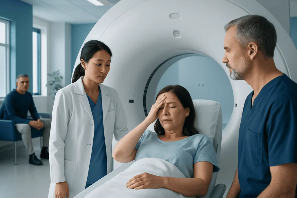
Not every headache, dizzy spell, or episode of weakness needs a brain scan. CT is often used in emergencies because it is fast and good at detecting bleeding.
Key Takeaways
- Not every headache, dizzy spell, or episode of weakness needs a brain scan.
- CT is often used in emergencies because it is fast and good at detecting bleeding.
- MRI provides more detail and is often preferred for many non-emergency neurological concerns.
- MRA, MRV, and CTA focus on blood vessels and may be used when stroke, aneurysm, or clot is suspected.
- A doctor chooses imaging based on symptoms, examination findings, medical history, and timing.
Neuro scans can help doctors look for causes of headache, dizziness, and weakness when symptoms suggest a problem in the brain, nerves, or blood vessels. The right test depends on how symptoms started, how severe they are, and whether there are warning signs that need urgent evaluation.
Overview: Why neuro scans may be used
Headache, dizziness, and weakness are common symptoms, and they can happen for many different reasons. In many cases, they are related to conditions that do not require brain imaging, such as tension headaches, inner ear disorders, dehydration, medication side effects, or temporary viral illness. However, when symptoms are severe, sudden, unusual, or accompanied by neurological changes, doctors may recommend neuro imaging to look more closely.
Neuro scans create pictures of the brain and, in some cases, the blood vessels of the head and neck. These tests can help identify bleeding, stroke, tumors, inflammation, structural changes, infections, or blood vessel problems. Imaging does not always give the diagnosis by itself, but it can provide important clues and help guide treatment decisions.
The main tests used are computed tomography (CT), magnetic resonance imaging (MRI), and specialized vessel studies such as CT angiography (CTA), magnetic resonance angiography (MRA), and magnetic resonance venography (MRV). Each test has strengths and limitations. The most suitable scan depends on the clinical situation, how quickly symptoms developed, and whether the doctor is looking for bleeding, stroke, a mass, or another cause.
Symptoms and situations that may lead to imaging
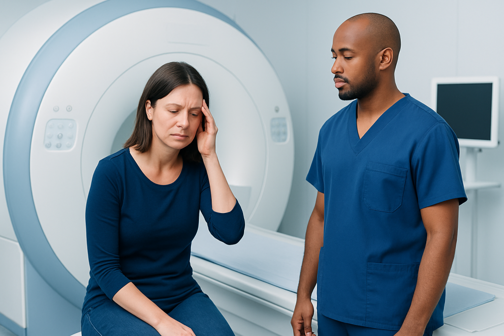
Doctors are more likely to order a scan when symptoms suggest a possible problem involving the brain or its blood supply. For headaches, this may include a sudden “worst headache” of life, a new headache in an older adult, headache with fever or neck stiffness, headache after head injury, or headache with weakness, confusion, seizures, or vision changes. In these settings, imaging helps rule out urgent causes.
Dizziness is a broad symptom and can describe spinning, imbalance, lightheadedness, or a floating sensation. Imaging may be considered when dizziness happens together with difficulty walking, double vision, slurred speech, one-sided weakness, numbness, severe headache, or a sudden change in hearing. These combinations can raise concern for a neurological cause rather than a simple inner ear problem.
Weakness is especially important when it affects one side of the body, appears suddenly, or comes with facial drooping, difficulty speaking, or confusion. These features can point to stroke or another urgent brain condition. A doctor will also consider how long symptoms have lasted, whether they come and go, and whether they affect an arm, a leg, the face, or the whole body.
- Sudden severe headache
- New neurological symptoms such as weakness or numbness
- Problems with speech, vision, or balance
- Symptoms after trauma
- Possible seizure, infection, or altered consciousness
Which scans might be used
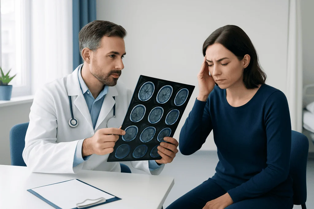
A non-contrast head CT is often the first scan used in emergency settings. It is quick, widely available, and very useful for detecting fresh bleeding, some fractures, and major structural problems. When someone arrives with sudden severe headache, new weakness, or suspected stroke, CT can often be performed within minutes and can help doctors make urgent decisions.
MRI of the brain gives more detailed images of brain tissue than CT. It is often used when symptoms are less time-critical, when CT is normal but symptoms continue, or when a doctor needs a closer look at areas such as the brainstem, small strokes, inflammation, demyelination, or certain tumors. Depending on the question, MRI may be done with or without contrast.
Specialized vessel imaging may also be needed. CTA and MRA look at arteries and can help detect narrowing, aneurysms, dissections, or blockages. MRV focuses on veins and may be used when a doctor suspects a venous clot. In some cases, neck vessel imaging is added, especially if symptoms suggest reduced blood flow to the brain. A neuroradiology team may recommend MRI or CT scanning based on the urgency and the likely diagnosis.
Less commonly, doctors may use advanced studies such as perfusion imaging, contrast-enhanced MRI, or targeted imaging of the inner ear, pituitary region, or spine. The choice depends on the pattern of symptoms and what the examination suggests.
How doctors choose the right test
The choice of scan is guided by the story the symptoms tell. A sudden collapse, a thunderclap headache, or one-sided weakness usually calls for rapid emergency imaging, often starting with CT. A headache that has gradually changed over weeks, or dizziness with persistent neurological signs, may lead to MRI because it shows more detail in soft tissues and certain areas of the brain.
Doctors also look at age, medical history, medications, and risk factors. For example, a person taking blood thinners may need prompt imaging after a fall or severe headache because of bleeding risk. Someone with cancer, infection risk, immune suppression, or a history of migraine may need a different imaging approach based on the clinical question.
Safety and practicality matter too. CT uses ionizing radiation, while MRI does not. MRI takes longer and may not be suitable for everyone, such as some patients with non-compatible metal implants or severe claustrophobia. Contrast dye may be needed in some studies, so doctors check kidney function, allergy history, and whether contrast is truly necessary. When blood vessel disease is a concern, MRA or MRV may be particularly helpful.
What the scans can show
Imaging may help reveal conditions that need urgent treatment, including bleeding in or around the brain, stroke, swelling, hydrocephalus, infection, or a mass. In people with weakness, scans can identify small or large areas of brain injury, pressure on nearby structures, or problems in the blood vessels that supply the brain. In headache evaluation, imaging can also look for causes such as sinus-related complications, tumors, or changes in pressure around the brain.
For dizziness, MRI can be useful when doctors suspect problems in the brainstem or cerebellum, where balance is coordinated. Vessel imaging may show reduced blood flow or narrowing of arteries. MRV can help detect venous sinus thrombosis, a less common but important cause of headache and neurological symptoms.
At the same time, a normal scan does not mean symptoms are not real. Many conditions, including vestibular disorders, some migraines, medication-related dizziness, and functional neurological symptoms, may not show visible abnormalities on standard imaging. That is why scan results are always interpreted alongside the medical history and neurological examination.
What to expect during CT, MRI, MRA, or MRV
A head CT is usually quick and painless. The patient lies on a table that slides through a donut-shaped scanner, and the test often takes just a few minutes. In urgent situations, this speed is one of CT’s main advantages. Some CT studies require contrast dye through a vein, especially when blood vessels need to be seen clearly.
MRI, MRA, and MRV use a powerful magnet rather than radiation. The patient lies still on a table that moves into the scanner. These tests take longer than CT and can be noisy, but ear protection is provided. Some people receive contrast, depending on what the doctor needs to evaluate. It is important to inform the imaging team about implants, pacemakers, prior surgeries, kidney disease, pregnancy, or anxiety in enclosed spaces.
After the scan, a radiologist or neuroradiologist reviews the images and sends a report to the treating doctor. In emergencies, important findings are communicated quickly. For non-urgent problems, results are usually discussed at a follow-up visit, where the doctor explains what the scan means and whether further testing is needed.
Limits of imaging and next steps after results
Neuro imaging is valuable, but it is only one part of diagnosis. Some causes of headache, dizziness, and weakness are diagnosed mainly through the history, physical examination, blood tests, heart rhythm testing, or inner ear assessment. A scan may answer one question but not every question. Sometimes it helps by ruling out dangerous causes, which allows the doctor to focus on more likely explanations.
Follow-up depends on the results. If imaging shows a stroke, bleeding, or another urgent condition, treatment may begin immediately. If the scan is normal but symptoms continue, the doctor may recommend additional evaluation, such as blood pressure review, vestibular testing, migraine management, nerve studies, or referral to a neurologist, ENT specialist, or another expert.
When symptoms are complex, care often involves more than one specialty. Near the end of the diagnostic journey, some patients benefit from centers with coordinated imaging and neurological care. Acibadem International’s multidisciplinary specialists and JCI-accredited hospitals diagnose and treat neurological conditions for international patients when a more comprehensive assessment is needed.
When to seek urgent medical care
Immediate medical attention is important for symptoms that could signal a neurological emergency. This includes sudden one-sided weakness, facial drooping, difficulty speaking, severe confusion, new seizures, fainting, loss of consciousness, or a very abrupt severe headache. These symptoms should not be watched at home to see if they improve.
Urgent care is also needed for dizziness with inability to walk, double vision, new hearing loss, chest pain, or persistent vomiting, as well as headache after injury or headache with fever and neck stiffness. Even if symptoms come and go, transient neurological events can still be serious and deserve prompt evaluation.
For non-urgent but ongoing symptoms, a planned medical visit is the best next step. A doctor can decide whether imaging is necessary and which type will be most useful. This helps avoid unnecessary scans while making sure important conditions are not missed.
Frequently asked questions
Do all headaches require a brain scan?
No. Many headaches, including common tension-type headaches and many migraines, do not need imaging when the history and examination are typical and there are no warning signs. Doctors usually recommend a scan when the headache is sudden, unusually severe, new, changing, or associated with neurological symptoms.
Is CT or MRI better for headache, dizziness, or weakness?
Neither test is always better; each has a different role. CT is often preferred in emergencies because it is fast and very good at detecting acute bleeding, while MRI gives more detailed information about brain tissue and is often used for less urgent or more complex questions.
What is the difference between MRA and MRV?
MRA looks mainly at arteries, the blood vessels that carry blood to the brain. MRV looks at veins, which carry blood away from the brain, and may be used when a venous clot is suspected.
If the scan is normal, does that mean nothing is wrong?
Not necessarily. A normal scan can be reassuring because it may rule out bleeding, large stroke, mass lesions, or other structural causes, but some conditions do not appear on standard imaging. Symptoms still need to be interpreted in the context of the examination and medical history.
Are neuro scans safe?
In general, yes, when they are used appropriately. CT involves radiation, so doctors order it when the expected benefit outweighs that exposure, while MRI does not use radiation but may not be suitable for some people with certain implants or severe claustrophobia.
Will contrast dye always be needed?
No. Many scans are performed without contrast, especially an initial non-contrast CT for suspected bleeding. Contrast is used only when it is likely to add important information, such as when evaluating blood vessels, inflammation, infection, or certain masses.
References
- American College of Radiology
- National Institute of Neurological Disorders and Stroke
- National Institute for Health and Care Excellence
- Radiological Society of North America
- World Health Organization
This article is for general information only and is not a substitute for professional medical advice. Please consult a qualified doctor about your individual situation.
Explore treatments in Turkey — costs, top hospitals & a free quote
JCI-accredited · board-certified surgeons · reply within 24h
Add us as a Preferred Source to see more of our trusted health content across Google Search, AI Overviews and Discover.

More from the Health Library



Related Specialists

Fzt. Sevgi Nama
Physical Medicine and Rehabilitation
Dt. Gülçin Doğusal İşgüder
Dentistry
Prof. Dr. Koray Güven
Interventional Radiology
Dr. Nihat Cem Çelebi
Pediatrics