Neurointerventional Radiology: Conditions Commonly Treated

Neurointerventional radiology uses imaging guidance to treat conditions affecting the brain, spine, and blood vessels. Commonly treated problems include stroke, brain aneurysms, arteriovenous malformations, carotid artery narrowing, and some spinal fractures.
Key Takeaways
- Neurointerventional radiology uses imaging guidance to treat conditions affecting the brain, spine, and blood vessels.
- Commonly treated problems include stroke, brain aneurysms, arteriovenous malformations, carotid artery narrowing, and some spinal fractures.
- Many procedures are performed through a small catheter inserted into an artery, usually in the wrist or groin.
- Treatment choice depends on the condition, urgency, anatomy, overall health, and specialist assessment.
- Early evaluation is especially important for stroke symptoms and suspected bleeding in the brain.
Neurointerventional radiology is a medical specialty that diagnoses and treats certain brain, spine, head, and neck conditions from inside the blood vessels or through very small skin openings. It often offers a minimally invasive alternative to open surgery for carefully selected patients.
Overview of Neurointerventional Radiology
Neurointerventional radiology, also called interventional neuroradiology or endovascular neurosurgery in some settings, is a highly specialized field that treats disorders of the brain, spine, head, neck, and blood vessels using advanced imaging guidance. Instead of making a large surgical opening, specialists often work through a thin tube called a catheter, which is guided through the blood vessels to the treatment area.
This approach can be used to diagnose a problem, deliver treatment, restore blood flow, block abnormal vessels, or stabilize damaged structures. Because the procedures are minimally invasive, they may reduce recovery time, discomfort, and hospital stay for some patients. However, they are not suitable for every condition, and open surgery or medical therapy may still be the best option in some cases.
Neurointerventional radiologists work closely with neurologists, neurosurgeons, stroke specialists, vascular surgeons, and critical care teams. This team-based approach helps patients receive the most appropriate treatment plan based on the exact diagnosis and the urgency of the condition.
Conditions Commonly Treated
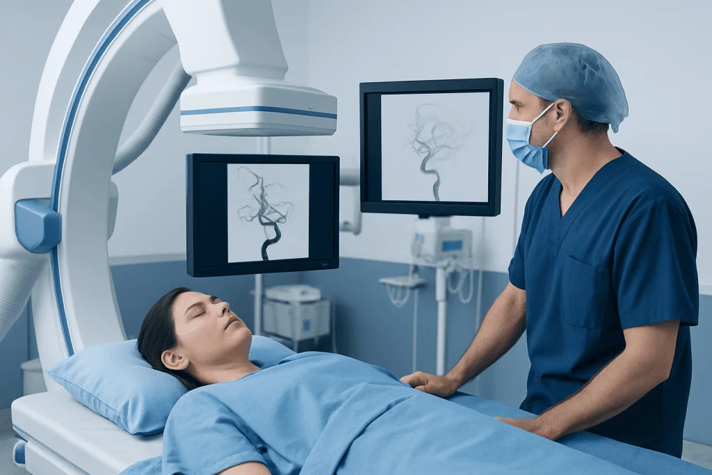
One of the best-known uses of neurointerventional radiology is emergency treatment for certain types of stroke. In ischemic stroke, a blood clot blocks an artery supplying the brain. In selected patients, specialists may perform a catheter-based clot removal procedure to reopen the artery quickly and limit brain injury. Neurointerventional techniques are also used in some cases of bleeding in or around the brain, depending on the cause.
Another common group of conditions involves abnormalities of blood vessels. These include brain aneurysms, arteriovenous malformations, dural arteriovenous fistulas, and narrowing of the carotid or intracranial arteries. Treatments may aim to seal an aneurysm, reduce the risk of rupture, block abnormal vessel connections, or improve blood flow through narrowed arteries. Patients looking for more background may also come across related topics such as aneurysm and stroke.
Neurointerventional radiology can also help with selected spinal conditions. A common example is a painful vertebral compression fracture, often related to osteoporosis or cancer affecting the spine. In suitable patients, image-guided procedures may stabilize the fractured bone and help relieve pain. Some tumors or vascular lesions involving the spine and head-and-neck region may also be evaluated and treated using these techniques.
- Acute ischemic stroke caused by large-vessel blockage
- Brain aneurysms
- Arteriovenous malformations and dural fistulas
- Carotid artery stenosis and some intracranial arterial narrowings
- Vertebral compression fractures
- Certain vascular tumors or bleeding-related conditions
Symptoms That May Lead to Evaluation

The symptoms that lead to neurointerventional evaluation depend on the underlying condition. Stroke symptoms usually start suddenly and may include facial drooping, weakness or numbness on one side of the body, trouble speaking, vision changes, severe dizziness, loss of balance, or confusion. These symptoms need immediate emergency care because treatment is most effective when delivered quickly.
Brain aneurysms and vascular malformations may not cause symptoms until they are found on imaging for another reason. When symptoms do occur, they can include severe headache, double vision, eyelid drooping, seizures, weakness, or bleeding into the brain. A sudden, unusually severe headache can be a medical emergency.
Spinal compression fractures often cause sudden or gradually worsening back pain, reduced mobility, and pain that increases when standing or walking. In some cases, symptoms such as limb weakness, numbness, or bowel and bladder changes suggest pressure on nerves or the spinal cord and require urgent assessment.
Causes and Risk Factors
Different conditions treated by neurointerventional radiology have different causes. Ischemic stroke is commonly linked to blood clots that form in the heart or arteries, while brain aneurysms may be associated with weakness in the blood vessel wall. Arteriovenous malformations are often congenital, meaning they develop before birth, although they may not be discovered until later in life.
Several vascular risk factors increase the chance of stroke and arterial narrowing. These include high blood pressure, smoking, diabetes, high cholesterol, obesity, atrial fibrillation, lack of physical activity, and advancing age. Family history may also play a role for some blood vessel conditions, including certain aneurysms.
Vertebral compression fractures are more likely in people with osteoporosis, older age, long-term steroid use, trauma, or cancers that spread to bone. Understanding the risk factors helps doctors recommend not only treatment, but also ways to lower the chance of future complications or recurrence.
How Diagnosis and Planning Are Done
Diagnosis usually begins with a careful history, physical examination, and imaging tests. Depending on the suspected condition, doctors may use CT, MRI, CT angiography, MR angiography, ultrasound, or a diagnostic cerebral angiogram. These tests help show the shape of the blood vessels, the location of a blockage or aneurysm, and whether there is active bleeding or reduced blood flow.
In emergencies such as stroke, evaluation happens rapidly to determine whether the patient is eligible for urgent treatment. Timing, the size and location of the blocked artery, brain tissue at risk, bleeding risk, and overall medical stability all influence the decision. For aneurysms, malformations, and carotid disease, planning may be more elective and involves detailed review by a multidisciplinary team.
The doctor will also discuss possible benefits, alternatives, and risks. As with any invasive procedure, potential complications can include bleeding, infection, blood vessel injury, allergic reaction to contrast material, kidney effects from contrast in some patients, or neurological complications. The exact risk varies greatly depending on the condition and the type of procedure.
Treatment Options Used in Neurointerventional Radiology
Treatment is tailored to the condition. For acute ischemic stroke, one major procedure is mechanical thrombectomy, in which a catheter-based device removes a clot from a blocked brain artery. In some patients, clot-dissolving medication may also be part of care, depending on timing and other medical factors. This is often coordinated with stroke treatment pathways in specialized centers.
For brain aneurysms, specialists may use endovascular coiling, flow-diverting stents, or other embolization techniques to seal the aneurysm from inside the vessel and lower the risk of bleeding. Vascular malformations and fistulas may be treated with embolization, which blocks abnormal vessels using coils, liquid embolic agents, or other materials. Some patients may need a combination of embolization, surgery, and radiosurgery. This can be part of broader brain aneurysm treatment planning.
In narrowed arteries, angioplasty and stenting may be considered in selected situations, particularly when symptoms continue despite medical treatment or when anatomy is suitable. For painful vertebral compression fractures, image-guided procedures such as kyphoplasty and vertebroplasty may stabilize the bone. If a brain or spinal lesion ultimately requires open surgery, patients may also be referred within a wider neurosurgery program.
Most procedures are performed under local anesthesia with sedation or under general anesthesia, depending on complexity and urgency. After treatment, patients are monitored closely, sometimes in an intensive care or stroke unit. Follow-up imaging may be needed to confirm the result and watch for recurrence or new changes over time.
Recovery, Prevention, and Self-care
Recovery depends on the treated condition and how urgently treatment was needed. After an endovascular procedure, the small access site in the wrist or groin usually heals within days, but recovery of neurological function after stroke or bleeding may take much longer. Some patients need rehabilitation for speech, movement, balance, or daily activities.
Long-term prevention is especially important for vascular conditions. Doctors often recommend controlling blood pressure, cholesterol, and blood sugar; stopping smoking; maintaining a healthy weight; being physically active; and taking prescribed medicines exactly as directed. In some patients, antiplatelet or anticoagulant medicines are part of preventing future strokes, but these should only be used under medical supervision.
For spinal compression fractures, self-care may include osteoporosis evaluation, bone-strengthening treatment, safe movement strategies, and physical therapy. Falls prevention, good nutrition, and vitamin and mineral advice from a clinician may also be helpful. Regular follow-up helps ensure healing is progressing and that any underlying cause is being addressed.
When to See a Doctor
Immediate medical attention is essential for possible stroke symptoms, sudden severe headache, new seizures, sudden vision loss, sudden confusion, or weakness on one side of the body. These symptoms can be caused by serious brain or blood vessel conditions, and early treatment can make an important difference.
People should also arrange medical evaluation for repeated transient neurological symptoms, such as brief speech difficulty, temporary vision changes, or short episodes of weakness, because these may be warning signs of a transient ischemic attack or evolving vascular disease. Persistent or worsening back pain after a fall, in older adults, or in people with osteoporosis or cancer should also be assessed.
For international patients seeking coordinated care, Acibadem International’s multidisciplinary specialists and JCI-accredited hospitals diagnose and treat conditions managed with neurointerventional radiology using individualized evaluation and follow-up plans. A qualified doctor can explain whether a minimally invasive approach is appropriate and what to expect before and after treatment.
Frequently asked questions
What is neurointerventional radiology?
Neurointerventional radiology is a specialty that treats certain disorders of the brain, spine, and blood vessels using imaging-guided, minimally invasive techniques. Doctors usually work through a thin catheter inserted into a blood vessel rather than through a large surgical incision.
Which conditions are most commonly treated with neurointerventional radiology?
Commonly treated conditions include acute ischemic stroke, brain aneurysms, arteriovenous malformations, dural fistulas, and some cases of carotid artery narrowing. Certain spinal compression fractures and selected bleeding-related or vascular tumors may also be treated this way.
Is neurointerventional radiology the same as brain surgery?
Not exactly. It is a minimally invasive approach that may be used instead of, before, or together with traditional brain or spine surgery. The best option depends on the diagnosis, the patient's anatomy, and the urgency of treatment.
How is a stroke treated with neurointerventional radiology?
In selected patients with an artery blocked by a large clot, doctors may perform mechanical thrombectomy to remove the clot and restore blood flow. This is an emergency treatment, so getting to a hospital quickly is very important.
Are these procedures safe?
These procedures are widely used and can be very effective when performed in appropriate patients by experienced teams. Like all invasive treatments, they carry risks such as bleeding, blood vessel injury, contrast reactions, or neurological complications, so doctors carefully assess the balance of benefit and risk.
What is recovery like after a neurointerventional procedure?
Recovery varies based on the condition being treated and the person's overall health. Some patients go home within a short time, while others need longer hospital care, rehabilitation, and follow-up imaging, especially after stroke or brain bleeding.
References
- World Stroke Organization
- American Heart Association
- National Institute of Neurological Disorders and Stroke
- Radiological Society of North America
- Society of NeuroInterventional Surgery
This article is for general information only and is not a substitute for professional medical advice. Please consult a qualified doctor about your individual situation.
Joint and spine care in Turkey — expert assessment & treatment
JCI-accredited · board-certified surgeons · reply within 24h
Add us as a Preferred Source to see more of our trusted health content across Google Search, AI Overviews and Discover.
Check your numbers in seconds
BMI, calories, due date, blood pressure and 30+ more clinical calculators — free, instant, doctor-reviewed ranges.