Neurointerventional Radiology: Conditions Treated and Common Procedures

Neurointerventional radiology uses thin catheters and real-time imaging to reach blood vessels in the brain, neck, and spine. It is often used to treat stroke, brain aneurysms, arteriovenous malformations, and narrowed blood vessels.
Key Takeaways
- Neurointerventional radiology uses thin catheters and real-time imaging to reach blood vessels in the brain, neck, and spine.
- It is often used to treat stroke, brain aneurysms, arteriovenous malformations, and narrowed blood vessels.
- Many procedures are less invasive than open surgery and may shorten recovery time for selected patients.
- Treatment decisions depend on the exact condition, symptoms, anatomy, overall health, and urgency.
- Early medical assessment is especially important for sudden neurological symptoms such as weakness, trouble speaking, or severe headache.
Neurointerventional radiology is a medical specialty that treats certain brain, spine, head, and neck conditions from inside the blood vessels using image-guided, minimally invasive techniques. It can play an important role in emergencies such as stroke as well as in planned treatment for aneurysms, vascular malformations, and other complex conditions.
Overview
Neurointerventional radiology is a highly specialized field focused on diagnosing and treating disorders of the blood vessels and related structures of the brain, spinal cord, head, and neck. It is also called endovascular surgical neuroradiology or interventional neurology in some settings. Instead of making a large surgical opening, specialists guide very thin tubes called catheters through the blood vessels, usually starting from the wrist or groin, while watching detailed imaging in real time.
This approach allows doctors to reach areas deep inside the body with precision. Once the catheter is in place, the specialist may remove a clot, open a narrowed artery, place a stent, seal an aneurysm, or block abnormal blood vessels. Because these procedures are minimally invasive, they may reduce tissue disruption and support faster recovery in appropriate cases.
Neurointerventional radiology is often used in urgent situations, especially when a person has signs of acute stroke. It is also used in planned care for conditions such as aneurysms, vascular malformations, some tumors, and certain causes of head or spine bleeding. Care is usually delivered by a multidisciplinary team that may include neurologists, neurosurgeons, neuroradiologists, anesthesiologists, and intensive care specialists.
Conditions Commonly Treated
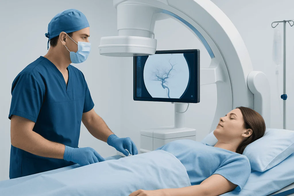
One of the best-known uses of neurointerventional radiology is emergency treatment for ischemic stroke, which happens when a blood clot blocks blood flow to part of the brain. In selected patients, doctors may perform mechanical thrombectomy, a procedure that removes the clot to restore circulation. This can be life-changing when done quickly and in the right clinical setting.
Another major area is treatment of brain aneurysms, which are weakened, bulging areas in a blood vessel wall. Some aneurysms can be treated from inside the vessel using coils, flow-diverting stents, or other devices rather than open surgery. Neurointerventional specialists also treat arteriovenous malformations and dural arteriovenous fistulas, which are abnormal tangles or connections between blood vessels that can raise the risk of bleeding or neurological symptoms.
Additional conditions include narrowing of the carotid or intracranial arteries, certain types of spinal vascular malformations, and some causes of bleeding in or around the brain. In some cases, neurointerventional techniques are used to reduce blood flow to tumors before surgery or to manage complications after trauma. Patients may also be referred for evaluation of conditions such as brain aneurysm or stroke when a minimally invasive treatment option is being considered.
- Acute ischemic stroke
- Brain aneurysms
- Arteriovenous malformations (AVMs)
- Dural arteriovenous fistulas
- Carotid or intracranial artery narrowing
- Some spinal vascular disorders and selected tumor-related vessel problems
Common Procedures Used in Neurointerventional Radiology

Procedures vary depending on the condition being treated. In acute stroke, a specialist may perform mechanical thrombectomy to remove a clot from a blocked artery in the brain. This is usually done by advancing a catheter through the blood vessels and using tiny devices to retrieve or suction the clot. In some situations, medicine to dissolve clots may also be part of treatment, depending on timing and individual factors.
For aneurysms, common procedures include endovascular coiling and flow diversion. In coiling, very small coils are placed inside the aneurysm to help prevent blood from entering it. Flow-diverting stents are placed in the parent artery to redirect blood flow away from the aneurysm and support healing of the vessel wall. Some patients may instead need clipping or another surgical approach, so the best treatment depends on aneurysm size, shape, location, and rupture status.
Other procedures include embolization, which blocks abnormal or unwanted blood flow. Embolization may be used for AVMs, fistulas, some tumors, or sources of bleeding. Balloon angioplasty and stenting can be used to widen narrowed arteries in carefully selected patients. These options may be discussed alongside aneurysm treatment or stroke treatment plans when a team is choosing the safest and most effective strategy.
Although these procedures are less invasive than open surgery, they are still advanced medical treatments and require careful monitoring before, during, and after the intervention. Most are performed in a specialized angiography suite with continuous imaging and support from a trained team.
Symptoms and Situations That May Need Urgent Assessment
Symptoms depend on the underlying condition. Stroke often causes sudden weakness or numbness on one side of the body, facial drooping, trouble speaking, confusion, vision loss, dizziness, or difficulty walking. These symptoms need immediate emergency attention because treatment is highly time-sensitive.
A ruptured aneurysm may cause a sudden, extremely severe headache that is different from usual headaches and may be accompanied by nausea, vomiting, neck stiffness, fainting, or confusion. Unruptured aneurysms and vascular malformations may sometimes cause no symptoms at all, but in other cases they can lead to headaches, seizures, visual changes, or nerve-related symptoms.
Narrowed arteries in the neck or brain may contribute to transient ischemic attacks, sometimes called mini-strokes, with brief episodes of weakness, speech problems, or vision changes. Bleeding, trauma-related vessel injury, or spinal vascular disorders can cause severe pain, new neurological symptoms, or changes in strength and sensation. Any sudden neurological symptom should be treated as urgent until a doctor says otherwise.
How Diagnosis and Treatment Planning Work
Diagnosis usually begins with a detailed medical history, symptom review, and neurological examination. Doctors look at when symptoms began, how they progressed, and whether there are risk factors such as high blood pressure, smoking, high cholesterol, diabetes, or a family history of vascular disease. In emergency situations, this evaluation happens very quickly so treatment decisions can be made without delay.
Imaging is central to neurointerventional care. Depending on the situation, a patient may need CT, CT angiography, MRI, MR angiography, ultrasound, or catheter angiography. Catheter angiography is often considered the most detailed way to view blood vessels and may be used both to confirm the diagnosis and to guide treatment during the same session.
Treatment planning is individualized. The team considers the exact diagnosis, the anatomy of the blood vessels, the risk of bleeding or clotting, the patient’s age and general health, and whether symptoms are stable or rapidly worsening. In some cases, careful monitoring or medication is the best choice; in others, endovascular treatment or surgery is recommended. Shared decision-making is important whenever the situation is not an emergency.
Benefits, Risks, and Recovery
A major benefit of neurointerventional radiology is that many conditions can be treated without large incisions. This may mean less pain, shorter hospital stays, and faster return to daily activities for selected patients. It can also make treatment possible in areas that are difficult to reach with traditional surgery.
Still, every procedure carries risk. Possible complications may include bleeding, infection, damage to a blood vessel, allergic reaction to contrast dye, kidney strain in susceptible patients, stroke, or recurrence of the treated problem. The exact risks vary by procedure and by the person’s overall health, medications, and anatomy.
Recovery depends on what was treated and whether the procedure was planned or done in an emergency. Some people go home within a day or two after a straightforward intervention, while others need intensive monitoring, rehabilitation, or longer hospitalization. Follow-up imaging is often important to confirm that the blood vessel remains open, the aneurysm stays sealed, or the abnormal connection has been fully treated.
After discharge, patients are usually advised to follow medication instructions carefully, keep follow-up appointments, and watch for warning signs such as new weakness, worsening headache, trouble speaking, fever, or bleeding at the catheter site. If these occur, prompt medical review is important.
Prevention, Self-care, and When to See a Doctor
Not all conditions treated by neurointerventional radiology can be prevented, but reducing vascular risk factors can lower the chance of stroke and some blood vessel problems. Helpful steps include controlling blood pressure, managing cholesterol and diabetes, avoiding smoking, staying physically active, and following a balanced diet. Taking prescribed medications as directed is also important, especially after a prior stroke or vascular procedure.
People with a known aneurysm or vascular malformation should keep regular appointments and ask their doctor which symptoms should prompt urgent review. Family history may matter in some cases, so it is useful to share information about relatives with aneurysms, stroke, or inherited vascular disorders. Patients should never stop blood thinners, antiplatelet medicines, or blood pressure medicines without professional advice.
Immediate emergency care is needed for signs of stroke, sudden severe headache, loss of consciousness, seizures, or sudden vision or speech changes. Early treatment can improve outcomes and may open the door to time-sensitive neurointerventional procedures. For planned care, a neurologist, neurosurgeon, or neurointerventional specialist can explain whether a minimally invasive approach is suitable.
In complex cases, care may be coordinated across several specialties. Acibadem International’s multidisciplinary specialists and JCI-accredited hospitals evaluate and treat neurovascular conditions for international patients, including advanced endovascular options when appropriate.
Frequently asked questions
What is neurointerventional radiology?
Neurointerventional radiology is a specialty that treats certain brain, spine, head, and neck conditions using catheters and imaging guidance. It is commonly used for blood vessel disorders such as stroke, aneurysms, and vascular malformations.
Is neurointerventional radiology the same as brain surgery?
It is different from traditional open brain surgery because it usually works from inside the blood vessels through a small entry point in the wrist or groin. However, it is still a highly specialized procedure and may be used alongside neurosurgery or other treatments when needed.
Which conditions are most often treated with these procedures?
Common examples include acute ischemic stroke, brain aneurysms, arteriovenous malformations, dural fistulas, and some narrowed arteries in the brain or neck. The right treatment depends on the exact diagnosis, symptoms, and blood vessel anatomy.
Are neurointerventional procedures safe?
These procedures are widely used and can be very effective in the right setting, but no medical procedure is risk-free. Doctors weigh the expected benefit against possible risks such as bleeding, vessel injury, or stroke before recommending treatment.
How long does recovery usually take?
Recovery varies depending on the procedure and the condition being treated. Some patients recover quickly after a planned intervention, while others need a longer hospital stay and rehabilitation, especially after stroke or bleeding.
When should someone seek emergency help?
Emergency care is needed for sudden weakness, facial drooping, trouble speaking, severe new headache, seizures, fainting, or sudden vision changes. These may be signs of stroke or bleeding and should be assessed immediately.
References
- World Health Organization
- American Stroke Association
- National Institute of Neurological Disorders and Stroke
- Society of NeuroInterventional Surgery
- Radiological Society of North America
This article is for general information only and is not a substitute for professional medical advice. Please consult a qualified doctor about your individual situation.
Joint and spine care in Turkey — expert assessment & treatment
JCI-accredited · board-certified surgeons · reply within 24h
Add us as a Preferred Source to see more of our trusted health content across Google Search, AI Overviews and Discover.

More from the Health Library



Related Specialists

Assoc. Prof. Dr. Ayça Erşen Danyeli
Pathology
Dr. Ercan Biçakci
Gastroenterology
Prof. Dr. Abdullah Büyükçelik
Medical Oncology
Prof. Dr. Remzi Tözün
Orthopedic Surgery & Traumatology