Neurointerventional Radiology for Brain Aneurysms: Coiling, Flow Diversion, and Stents

Brain aneurysms can often be treated from inside the blood vessel using catheters rather than open surgery. Coiling, flow diverters, and stents are different endovascular tools used for different aneurysm shapes and locations.
Key Takeaways
- Brain aneurysms can often be treated from inside the blood vessel using catheters rather than open surgery.
- Coiling, flow diverters, and stents are different endovascular tools used for different aneurysm shapes and locations.
- Treatment choice depends on whether the aneurysm has ruptured, its anatomy, and the patient’s overall health.
- Imaging tests such as CT, MRI, and cerebral angiography help diagnose aneurysms and guide treatment planning.
- Regular follow-up imaging is important after treatment to confirm the aneurysm remains safely sealed.
Neurointerventional radiology for brain aneurysms uses minimally invasive, catheter-based techniques to treat weakened blood vessels in the brain. Common options include coiling, flow diversion, and stent-assisted procedures, chosen according to the aneurysm’s size, shape, and location.
Overview
Neurointerventional radiology for brain aneurysms is a specialized field that treats aneurysms from inside the blood vessels. A brain aneurysm is a weak, bulging area in the wall of an artery in the brain. Some aneurysms remain stable for years, while others can leak or rupture and cause a life-threatening bleed. Endovascular treatment aims to reduce that risk by closing off the aneurysm or redirecting blood flow away from it.
These procedures are performed using thin tubes called catheters, usually inserted through an artery in the wrist or groin and guided to the brain under imaging. Because they do not require opening the skull, they are often described as minimally invasive. This can mean shorter recovery times for some patients, although the right treatment always depends on the aneurysm and the person’s overall condition.
The three main endovascular approaches are coiling, flow diversion, and stent-assisted techniques. Coils fill the aneurysm so blood no longer circulates inside it. Flow diverters are fine mesh tubes placed in the parent artery to redirect blood flow away from the aneurysm. Stents can help support coils or reconstruct part of the artery when the aneurysm has a wider neck or complex shape.
When Treatment Is Considered
Not every brain aneurysm needs immediate treatment. Doctors consider several factors, including the aneurysm’s size, shape, location, whether it has ruptured, and whether it is causing symptoms. A person’s age, general health, family history, and risk factors such as smoking or uncontrolled high blood pressure also matter.
If an aneurysm ruptures, emergency treatment is often needed to prevent rebleeding. In an unruptured aneurysm, the care team weighs the natural risk of rupture against the risks and benefits of treatment. For some patients, careful monitoring with repeat imaging may be safer than immediate intervention. For others, preventive treatment may offer the best long-term protection.
Symptoms can also influence the decision. Some unruptured aneurysms are found incidentally during scans done for another reason. Others may press on nearby nerves or brain structures and cause warning signs. In these cases, a specialist in brain aneurysm care can explain whether observation, endovascular therapy, or another approach is most suitable.
Symptoms and Warning Signs

Many unruptured brain aneurysms do not cause symptoms. When symptoms do occur, they may depend on the aneurysm’s size and location. Some people develop headache, eye pain, double vision, drooping eyelid, facial numbness, or changes in vision if the aneurysm presses on nearby nerves.
A ruptured aneurysm usually causes a sudden, severe headache that is often described as the worst headache of a person’s life. It may also cause nausea, vomiting, neck stiffness, confusion, sensitivity to light, fainting, or seizures. These symptoms need urgent medical attention because they may signal bleeding around the brain, known as subarachnoid hemorrhage.
Although not every severe headache is caused by an aneurysm, sudden unusual neurological symptoms should never be ignored. Prompt evaluation helps doctors identify the cause quickly and decide whether emergency treatment is needed.
How Brain Aneurysms Are Diagnosed
Doctors use imaging tests to confirm an aneurysm, understand its anatomy, and choose the safest treatment. Common tests include CT and CT angiography, MRI and MR angiography, and cerebral angiography. Each test provides different information about blood vessels, surrounding brain tissue, and whether bleeding has occurred.
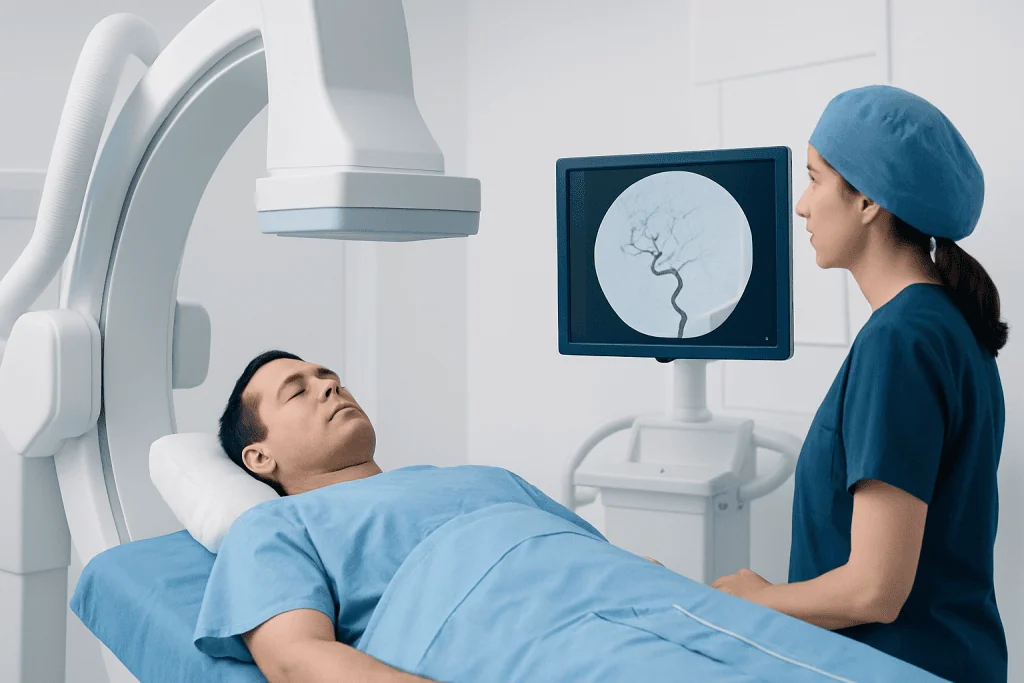
Cerebral angiography is often the most detailed test for treatment planning. During this procedure, contrast dye is injected through a catheter so specialists can see the blood vessels clearly in real time. This helps them assess the aneurysm’s neck, shape, branch vessels, and relationship to the parent artery, all of which affect whether coiling, a flow diverter, or a stent-assisted technique may work best.
Diagnosis also includes a full neurological assessment and review of medical history. The team may evaluate blood pressure, medications, kidney function, and bleeding or clotting risks before intervention. If there has been bleeding, doctors also focus on stabilizing the patient and watching for complications such as vessel spasm or hydrocephalus.
Coiling, Flow Diversion, and Stent-Assisted Treatment
In aneurysm coiling, the specialist places tiny soft platinum coils into the aneurysm through a microcatheter. The coils encourage clot formation inside the aneurysm, helping seal it off from circulation. This approach is commonly used for many saccular aneurysms, especially when the aneurysm shape allows the coils to stay securely in place. Patients looking for minimally invasive aneurysm treatment are often evaluated first to see whether coiling is technically suitable.
Some aneurysms have a wide neck or a shape that makes simple coiling less stable. In these cases, a stent may be placed in the parent artery to act as a scaffold while coils are inserted, a method called stent-assisted coiling. In other situations, a balloon may briefly support the artery during coil placement. Stents can also help reconstruct the vessel when anatomy is more complex, but they may require blood-thinning medication before and after treatment.
Flow diversion is a newer endovascular option used especially for certain large, wide-necked, or sidewall aneurysms. A flow diverter is a densely woven mesh tube placed across the aneurysm opening inside the artery. Rather than filling the aneurysm directly, it changes blood flow so the aneurysm gradually seals over time while the artery lining grows across the device. This technique is part of advanced interventional neuroradiology and may be useful for aneurysms that are difficult to treat with coils alone.
The best method depends on the aneurysm’s anatomy and whether it has ruptured. In some emergency cases, coiling may be preferred because it can secure the aneurysm quickly. In other cases, a flow diverter or intracranial stent offers a better long-term reconstruction of the vessel. Treatment planning is individualized and often discussed by a multidisciplinary team, sometimes including specialists in neurosurgery when more than one approach is possible.
What to Expect Before, During, and After the Procedure
Before treatment, patients usually have blood tests, medication review, and detailed imaging. The doctor explains the planned technique, possible benefits, and potential risks. Some people need antiplatelet medicines before stent or flow diverter placement to reduce the chance of clot formation on the device. The care team also gives instructions about eating, drinking, and regular medications before the procedure.
Most endovascular aneurysm procedures are done in a specialized angiography suite under general anesthesia or, in selected cases, sedation. A catheter is inserted through an artery and guided to the brain using real-time X-ray imaging. The specialist then deploys coils, a stent, a flow diverter, or a combination of these devices. The procedure length varies according to the aneurysm’s complexity.
Afterward, patients are monitored closely for blood pressure control, neurological changes, and access-site bleeding. Some go home within a short period, while others stay longer, especially after a ruptured aneurysm or more complex treatment. Follow-up imaging is essential because even a successfully treated aneurysm may need surveillance to make sure it remains closed and the artery stays open.
Benefits, Risks, and Recovery
The main benefit of neurointerventional treatment is that it can protect against aneurysm rupture or rebleeding without open brain surgery. For suitable patients, it may mean smaller incisions, less tissue disruption, and a faster return to normal activities. It also provides options for aneurysms located deep in the brain or in areas that may be challenging to reach surgically.
Like any medical procedure, endovascular treatment has risks. These may include bleeding, stroke, vessel injury, device movement, blood clots, contrast reactions, or recurrence of the aneurysm over time. Stents and flow diverters can also require ongoing blood-thinning medication for a period determined by the treating doctor. The exact risk profile varies widely depending on whether the aneurysm has ruptured and on the aneurysm’s anatomy.
Recovery differs from one person to another. After treatment for an unruptured aneurysm, many people gradually resume daily activities within days to weeks, depending on medical advice. Recovery is often longer after a ruptured aneurysm because the bleed itself can affect the brain and body. Good follow-up care, medication adherence, and imaging surveillance are important parts of long-term success.
Prevention, Follow-Up, and When to See a Doctor
It is not always possible to prevent a brain aneurysm, but reducing vascular risk factors can support overall blood vessel health. Important steps include controlling high blood pressure, avoiding smoking, limiting excessive alcohol use, and managing conditions such as high cholesterol. People with a strong family history of aneurysm should ask a doctor whether screening is appropriate.
Follow-up remains important whether an aneurysm is treated or monitored. Doctors may recommend periodic scans to check for aneurysm growth, recurrence, or healing after coiling or flow diversion. Patients should take prescribed medicines exactly as directed, especially antiplatelet drugs after stent-based treatment, and should not stop them without medical advice.
Immediate medical care is needed for a sudden severe headache, fainting, seizure, sudden confusion, or new weakness, numbness, speech difficulty, or vision changes. A scheduled specialist visit is appropriate for persistent headaches, known aneurysm follow-up, or questions about treatment options. At centers such as Acibadem International, multidisciplinary specialists in JCI-accredited hospitals evaluate and treat brain aneurysms for international patients using modern endovascular and surgical techniques.
Frequently asked questions
What is neurointerventional radiology for brain aneurysms?
It is a minimally invasive way to treat certain brain aneurysms from inside the blood vessels using catheters and imaging guidance. Common techniques include coiling, flow diversion, and stent-assisted treatment.
Is coiling better than open surgery for a brain aneurysm?
Neither option is automatically better for every patient. The best choice depends on the aneurysm’s size, shape, location, whether it has ruptured, and the patient’s overall health. A specialist team compares the risks and benefits of each approach.
How does a flow diverter work?
A flow diverter is a small mesh tube placed inside the parent artery across the aneurysm opening. It redirects blood flow away from the aneurysm, allowing the aneurysm to clot off gradually while the artery heals over the device.
Do stents for brain aneurysms require medication afterward?
Yes, many patients who receive an intracranial stent or flow diverter need antiplatelet medication for a period of time. This helps reduce the risk of clots forming on the device. The exact plan depends on the procedure and the doctor’s advice.
Can a treated brain aneurysm come back?
Some aneurysms can reopen or show residual filling over time, especially after coiling. That is why follow-up imaging is important even when treatment initially appears successful. If needed, additional treatment may be recommended.
How long is recovery after endovascular aneurysm treatment?
Recovery time varies according to the type of treatment and whether the aneurysm had ruptured. Many people treated for an unruptured aneurysm recover more quickly than those who had bleeding, but each case is different. The treating team gives individualized guidance on activity, medications, and follow-up.
References
- World Health Organization
- American Heart Association
- National Institute of Neurological Disorders and Stroke
- Society of NeuroInterventional Surgery
- Radiological Society of North America
This article is for general information only and is not a substitute for professional medical advice. Please consult a qualified doctor about your individual situation.
Heart care in Turkey — expert evaluation and treatment
JCI-accredited · board-certified surgeons · reply within 24h
Add us as a Preferred Source to see more of our trusted health content across Google Search, AI Overviews and Discover.
Check your numbers in seconds
BMI, calories, due date, blood pressure and 30+ more clinical calculators — free, instant, doctor-reviewed ranges.