Ocd Elbow Recovery: A Week-by-Week Timeline

Elbow osteochondritis dissecans (OCD) most often affects the capitellum, a part of the upper arm bone that meets the radius. Stable lesions may heal with activity modification, whereas loose or unstable fragments may need surgical treatment.
Key Takeaways
- Elbow osteochondritis dissecans (OCD) most often affects the capitellum, a part of the upper arm bone that meets the radius.
- Stable lesions may heal with activity modification, whereas loose or unstable fragments may need surgical treatment.
- Recovery is guided by symptoms, examination findings and follow-up imaging rather than the calendar alone.
- Throwing, gymnastics and heavy loading should resume only when strength, motion and healing criteria are met.
- Persistent locking, loss of motion, swelling or worsening pain warrants medical assessment.
OCD elbow recovery time varies with the size and stability of the cartilage-and-bone lesion, the person’s skeletal maturity, activity demands, and whether surgery is needed. Many stable lesions improve over several months with rest and rehabilitation, while recovery after a procedure commonly requires a gradual return to sport over approximately 4–9 months.
OCD Elbow Recovery Time: the Answer First
OCD elbow recovery time is commonly about 3–6 months for a stable lesion managed without surgery, although complete healing can take longer. After surgery, many people need roughly 4–9 months before returning to unrestricted throwing, gymnastics or other high-load activities; the exact timeline depends on the procedure, lesion healing and rehabilitation progress.
Osteochondritis dissecans (OCD) is a condition in which an area of bone beneath joint cartilage becomes damaged. In the elbow, it usually affects the capitellum, the rounded outer part of the humerus. The cartilage overlying the damaged bone may remain intact, crack, or separate with a piece of bone, producing an osteochondral fragment.
OCD is particularly seen in adolescents and young adults who repeatedly load the elbow, including baseball pitchers, gymnasts and racquet-sport athletes. Early assessment is important because stable lesions can sometimes heal with reduced loading, while unstable lesions may lead to persistent mechanical symptoms and require a procedure.
What Does OCD in the Elbow Feel Like?
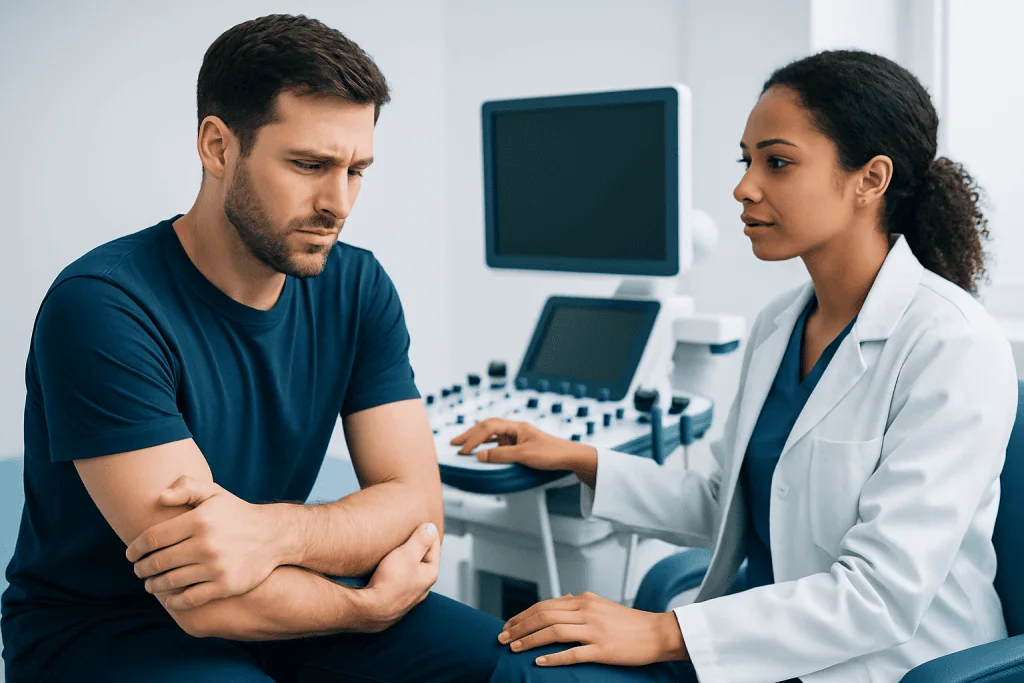
Elbow OCD often begins gradually. A person may notice aching pain on the outer side of the elbow during throwing, pushing off the hands, lifting, gymnastics or other repetitive activity. Pain may initially settle with rest but return when the activity resumes.
Other symptoms can include swelling after exercise, reduced ability to fully straighten or bend the elbow, clicking or catching, and a feeling that the elbow is not moving smoothly. If a cartilage-and-bone fragment becomes loose, the elbow can lock temporarily or feel as though something is moving inside the joint.
Symptoms vary. Some people have pain with sport but little discomfort in daily activities, while others develop more persistent pain and motion loss. Pain should not be pushed through, especially in a growing athlete, because continuing repetitive loading may worsen an unstable lesion.
How Long Does OCD in the Elbow Take to Heal?

There is no single healing time for elbow OCD. Stable lesions in younger people with open growth plates may respond best to rest from aggravating sport, activity modification and structured rehabilitation. Improvement in symptoms may occur over weeks, but bone and cartilage recovery is usually assessed over months with clinical reviews and, when appropriate, imaging.
A commonly used initial approach is to avoid throwing, gymnastics impact and other high-load elbow activity for a period directed by an orthopedic specialist. Range-of-motion and shoulder, forearm and trunk conditioning can often be introduced in a controlled way. Return to sport is considered only after pain has resolved, elbow movement and strength are restored, and the clinician is satisfied with healing progress.
Older adolescents, adults, and people with unstable or advanced lesions are less likely to heal with rest alone. Their recovery may require surgery followed by a longer, carefully progressed rehabilitation plan. A clinician can explain the expected outlook based on lesion location, size, stability and imaging findings.
Assessment, Staging and Stage 4 OCD
Diagnosis starts with a history of symptoms and sports participation, followed by an examination of elbow movement, tenderness, swelling and stability. X-rays may show changes in the capitellum. Magnetic resonance imaging (MRI) can provide more detail about cartilage, underlying bone and whether a fragment appears stable; computed tomography (CT) may be used to define bone structure in selected cases.
Staging systems differ between imaging and arthroscopic assessment, so the meaning of a numbered stage should always be confirmed with the treating specialist. In general, higher-stage disease describes a more advanced lesion with greater separation of the cartilage-bone fragment.
What happens during stage 4 of osteochondritis dissecans? Stage 4 commonly refers to a loose or displaced osteochondral fragment, sometimes called a loose body, within the joint. It can cause catching, locking, pain, swelling and loss of movement. Because the fragment is no longer securely attached, surgical assessment and treatment are often considered to remove, fix or restore the affected area, depending on the tissue quality and lesion characteristics.
Treatment Options and How Elbow OCD Procedures Work
Treatment aims to protect the joint surface, reduce symptoms, restore motion and help a person safely return to desired activity. For a stable lesion, non-surgical care may include avoiding painful loading, a supervised rehabilitation program, and correction of throwing mechanics or training factors where relevant. Follow-up is important because symptoms alone do not always show whether the lesion is healing.
Procedures are usually considered for unstable lesions, loose bodies, persistent symptoms despite appropriate conservative care, or lesions unlikely to heal on their own. Arthroscopy uses a small camera and specialized instruments through small incisions to inspect the joint. Depending on the findings, the surgeon may remove loose fragments, smooth damaged cartilage, stimulate healing of the underlying bone with drilling or microfracture, or fix a repairable fragment with small implants.
For larger or more complex defects, cartilage-restoration approaches may be discussed in selected patients. Candidacy depends on age, skeletal maturity, symptom severity, lesion size and location, cartilage condition, sporting goals and prior treatment. A specialist may recommend elbow arthroscopy when direct joint assessment and minimally invasive treatment are appropriate.
Potential benefits of surgery include removal of a mechanical block, reduction of pain and protection of remaining joint cartilage. Risks can include infection, bleeding, stiffness, nerve or blood-vessel injury, continued pain, incomplete healing, implant-related issues when fixation is used, and the possibility of later arthritis. The orthopedic team discusses individual risks before treatment.
Week-by-Week OCD Elbow Recovery Timeline
Weeks 0–2: After surgery, the early focus is pain and swelling control, wound care and protecting the treated area. A sling or brace may be used, depending on the procedure. Gentle hand, wrist, shoulder and prescribed elbow movement may begin, but weight-bearing, throwing and lifting are avoided. Non-surgical treatment also begins with stopping activities that reproduce symptoms.
Weeks 2–6: The rehabilitation plan commonly emphasizes gradually restoring comfortable elbow range of motion and maintaining strength in the shoulder blade, trunk and lower body. The permitted degree of elbow loading differs substantially after loose-body removal, drilling, fragment fixation or cartilage restoration. A fixation procedure generally requires more protection than a simple loose-body removal.
Weeks 6–12: If examination and healing progress are satisfactory, strengthening may become more active and sport-specific movement patterns may be introduced without impact or throwing. Follow-up imaging may be used for lesions treated with fixation or drilling. Any return of swelling, catching or increasing pain should be reported rather than trained through.
Months 3–6 and beyond: Progressive loading, a throwing progression or gymnastics-specific conditioning may start only when cleared by the treating team. Full return to competition often occurs later, particularly after fixation or restorative procedures, and may take 6–9 months or more. Return-to-sport decisions should be based on symptom-free function, near-normal motion, strength, sport demands and evidence of healing where relevant.
How Long Do Osteochondral Lesions Take to Heal?
Osteochondral lesions involve both cartilage and the bone beneath it, so healing is often slower than recovery from a simple muscle strain. Small, stable lesions in younger patients may improve over several months with unloading and rehabilitation. Larger, unstable or surgically treated lesions can require many months before they tolerate full sport-specific force.
Cartilage has a limited blood supply, while underlying bone healing depends on lesion stability and whether the blood supply can recover. This is why clinicians may use serial examinations and imaging rather than relying only on how the elbow feels. Feeling better is encouraging, but it does not always mean the joint is ready for unrestricted repetitive loading.
Adhering to activity restrictions, attending rehabilitation, following a gradual strengthening plan, and addressing training volume and technique can support recovery. Nicotine exposure may impair bone healing and should be discussed with a clinician. Nutrition, sleep and overall health also matter, although no supplement has been proven to replace appropriate protection and rehabilitation.
When to Seek Medical Care
Medical assessment is advisable for elbow pain that persists beyond a short period of rest, recurs with throwing or gymnastics, or limits normal sport participation. Young athletes with outer-elbow pain, loss of extension or swelling should be assessed by a qualified clinician, particularly if symptoms have been present for several weeks.
More urgent review is appropriate for a locked elbow, a sudden major loss of movement, marked swelling after an injury, numbness or weakness in the hand, or severe pain. These symptoms do not always mean OCD, but they need timely evaluation to identify the cause and protect joint function.
Acibadem International’s multidisciplinary orthopedic specialists and JCI-accredited hospitals diagnose and treat elbow conditions for international patients. Ongoing follow-up with an orthopedic surgeon and rehabilitation professional helps ensure that activity is advanced safely and at an individual pace.
Frequently asked questions
Can elbow OCD heal without surgery?
Yes. A stable lesion, particularly in a younger person who is still growing, may heal with reduced elbow loading, activity modification and supervised rehabilitation. Healing is monitored through symptoms, examination and sometimes repeat imaging; unstable lesions or persistent symptoms may need surgery.
When can an athlete throw again after elbow OCD?
Throwing should restart only after clearance from the treating clinician. The athlete should generally have no pain or swelling, adequate elbow movement and strength, and evidence of suitable healing when imaging is needed. Throwing is then progressed in stages rather than resumed at full intensity.
Is clicking in the elbow always a sign of a loose body?
No. Clicking can occur for several reasons, including tendon movement or normal joint mechanics. However, clicking associated with pain, catching, locking, swelling or reduced movement should be evaluated because it can occur with an unstable OCD lesion or loose body.
What is the difference between a stable and unstable OCD lesion?
In a stable lesion, the cartilage and underlying bone fragment remain attached to the surrounding bone. In an unstable lesion, the fragment is partly or fully separated, which can lead to mechanical symptoms such as catching or locking. Stability is assessed with imaging and, in some cases, arthroscopy.
Will elbow OCD cause arthritis?
An untreated or advanced lesion can damage the joint surface and may increase the long-term risk of degenerative changes. Early diagnosis, appropriate protection of the elbow and treatment when needed aim to preserve joint function. Individual long-term outlook depends on the lesion and response to treatment.
Can adults develop osteochondritis dissecans of the elbow?
Yes, although elbow OCD is often recognized in adolescents involved in repetitive overhead or impact sports. Adults may have persistent disease from earlier years, or symptoms may become apparent after accumulated elbow loading. Assessment is important because treatment options and healing potential may differ with skeletal maturity and lesion stability.
References
- American Academy of Orthopaedic Surgeons
- American Orthopaedic Society for Sports Medicine
- OrthoInfo
- National Institute of Arthritis and Musculoskeletal and Skin Diseases
This article is for general information only and is not a substitute for professional medical advice. Please consult a qualified doctor about your individual situation.
Explore treatments in Turkey — costs, top hospitals & a free quote
JCI-accredited · board-certified surgeons · reply within 24h
Add us as a Preferred Source to see more of our trusted health content across Google Search, AI Overviews and Discover.

More from the Health Library


Related Specialists
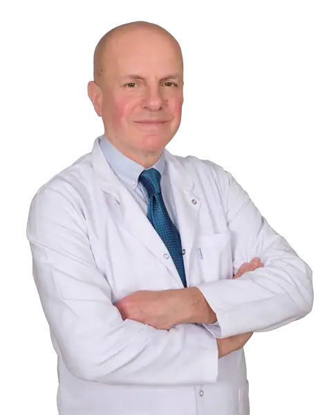
Dr. Güven Genç
Orthopedic Surgery & Traumatology
Prof. Dr. Güler Berkiten
Otorhinolaryngology
Dr. Canan Savaş İyigün
Dermatology
Assoc. Prof. Dr. Kamer Dere
Treatment of Pain Algology