Omphalocele Ultrasound: Preparation, Procedure and Results

Ultrasound can often identify omphalocele during routine prenatal screening, especially in the second trimester. The key finding is abdominal organs protruding into a membrane-covered sac at the umbilical cord insertion.
Key Takeaways
- Ultrasound can often identify omphalocele during routine prenatal screening, especially in the second trimester.
- The key finding is abdominal organs protruding into a membrane-covered sac at the umbilical cord insertion.
- Omphalocele may occur alone or alongside chromosomal, genetic, heart, or other structural differences.
- The scan itself is noninvasive and generally requires little preparation during pregnancy.
- Care commonly involves maternal-fetal medicine, genetics, neonatology, pediatric cardiology, and pediatric surgery teams.
- An omphalocele does not usually close before birth, but treatment after birth can repair the abdominal wall defect.
An omphalocele ultrasound is a prenatal or newborn imaging examination that helps clinicians see an abdominal wall opening at the base of the umbilical cord and assess which organs are involved. It is painless, does not use radiation, and helps guide genetic evaluation, delivery planning, and treatment after birth.
Omphalocele Ultrasound: What It Shows
An omphalocele ultrasound is an imaging examination used to look for an opening in a baby’s abdominal wall. In omphalocele, part of the bowel, liver, or other abdominal organs develops outside the abdomen within a protective, membrane-covered sac attached at the base of the umbilical cord. Ultrasound helps confirm this appearance and estimate the size and contents of the sac.
Most cases are found before birth during a routine pregnancy ultrasound. A detailed follow-up scan can help the care team look carefully at the baby’s anatomy, growth, placenta, and amniotic fluid. After birth, ultrasound may also be used as part of the newborn’s assessment, although the diagnosis is usually visible on physical examination.
The examination cannot predict every aspect of a baby’s outcome on its own. However, it provides important information for discussions about further testing, the safest place and timing for delivery, and the neonatal and surgical care that may be needed after birth.
How Omphalocele Ultrasound Works and Who May Need It

Ultrasound creates images using high-frequency sound waves. A trained sonographer moves a small handheld probe over the pregnant person’s abdomen. The probe sends sound waves into the body and receives returning echoes, which are converted into live images on a screen. It does not use ionizing radiation and is considered a standard part of prenatal care when performed for an appropriate medical reason.
A detailed omphalocele assessment may be recommended when a routine scan suggests an abdominal wall difference, when an early pregnancy scan shows a possible protrusion near the umbilical cord, or when other fetal findings raise concern for a genetic or structural condition. Specialist scanning is also useful to distinguish omphalocele from other abdominal wall defects, including gastroschisis, which has different imaging features and management considerations.
Clinicians may recommend additional imaging or evaluations after an omphalocele is identified. These can include a detailed fetal anatomy scan, fetal echocardiography to assess the heart, genetic counseling and testing options, and growth monitoring later in pregnancy. Recommendations are individualized according to the ultrasound findings, pregnancy stage, and family preferences.
Preparation and Step-by-Step Procedure
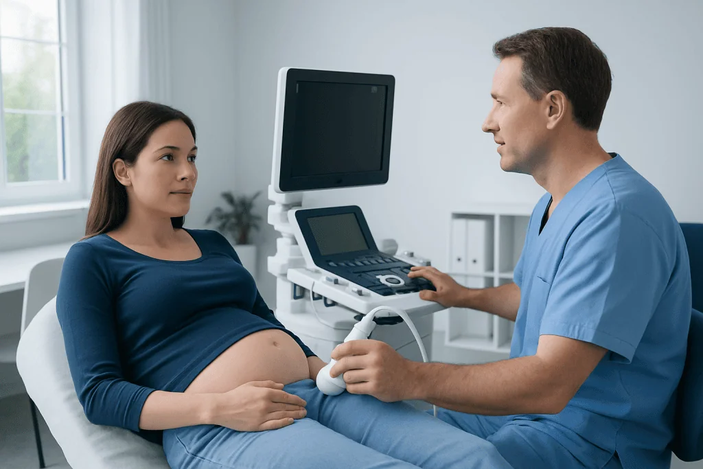
Preparation for a prenatal omphalocele ultrasound is usually simple. The imaging center may ask the patient to arrive with a moderately full bladder for an early pregnancy scan, because this can improve views of the uterus. Later in pregnancy, a full bladder is usually not necessary. Patients should follow the instructions given by their obstetric team or ultrasound facility.
For the examination, the patient lies comfortably on an examination bed. Gel is applied to the abdomen to help sound waves travel efficiently through the skin. The sonographer obtains images from several angles and may spend additional time examining the abdominal wall, umbilical cord insertion, organs within the sac, and other fetal structures. A detailed study may take longer than a routine scan because the goal is to assess the whole anatomy carefully.
The procedure is generally painless. Mild pressure from the probe may be felt, particularly if the sonographer needs to obtain a clearer view. In some cases, the baby’s position can limit imaging. The clinician may ask the patient to change position, walk briefly, return for another scan, or consider another imaging test such as fetal MRI when it could add useful information.
There is no physical recovery period after a prenatal ultrasound. Most people can return to normal activities immediately. The emotional impact of receiving unexpected findings can be significant, so prompt discussion with an obstetrician, maternal-fetal medicine specialist, genetic counselor, or pediatric surgical team can be helpful.
What Are the Sonographic Findings of Omphalocele?
The classic sonographic finding of omphalocele is a midline abdominal wall defect at the base of the umbilical cord. Abdominal contents protrude through the opening but remain covered by a smooth membrane or sac. The umbilical cord inserts into the top or center of this sac, which is an important feature that helps distinguish omphalocele from other abdominal wall defects.
The sac may contain bowel only, or it may contain bowel and part of the liver. A smaller defect with bowel outside the abdomen is sometimes described as a minor omphalocele, while a larger defect containing liver is often called a giant omphalocele. These descriptions help the care team anticipate potential surgical and respiratory needs, but they do not determine outcome by themselves.
During a detailed scan, clinicians also look for associated findings. These may include changes involving the heart, brain, kidneys, spine, limbs, growth, or other organs. They assess the amniotic fluid, fetal growth, and the relationship of the defect to the cord and abdominal wall. If the scan is performed early in pregnancy, repeat imaging may be needed because normal early developmental changes can sometimes resemble an abdominal wall protrusion.
Results, Benefits, Limits and Possible Risks
The ultrasound report typically describes the location and size of the abdominal wall defect, whether the membrane-covered sac is present, which organs appear to be involved, and whether other structural differences are seen. The findings are interpreted together with gestational age, medical history, and results of any genetic or cardiac evaluations. The referring obstetric clinician or maternal-fetal medicine specialist should explain what the results mean for the individual pregnancy.
The main benefit of omphalocele ultrasound is early, noninvasive information. It supports coordinated planning for delivery at a center with neonatal intensive care and pediatric surgery when appropriate. Prenatal diagnosis also gives families time to meet relevant specialists, discuss testing choices, and prepare for newborn treatment, including pediatric surgical care.
Ultrasound has limitations. Image quality may be affected by fetal position, pregnancy stage, maternal body tissues, or the amount of amniotic fluid. Not every associated condition can be seen before birth, and a normal-looking ultrasound elsewhere in the body cannot rule out all genetic differences. Diagnostic ultrasound is not known to harm the fetus when used appropriately by trained professionals, but it should be performed only when medically indicated and according to established safety principles.
Can Omphalocele Go Away?
An omphalocele usually does not go away during pregnancy because it is a structural opening in the abdominal wall. The sac and organs outside the abdomen may appear somewhat different as the baby grows, but the underlying defect generally remains present until it is treated after birth. Follow-up scans are used to monitor growth and refine care planning rather than to expect spontaneous closure.
After delivery, the care team protects the sac, keeps the baby warm, monitors breathing and circulation, and evaluates for associated medical needs. Repair may be completed in one operation if the abdominal cavity can safely accommodate the organs. For a larger omphalocele, treatment may be staged, allowing the organs to gradually move into the abdomen before final closure.
The timing and approach to repair depend on the defect’s size, the organs involved, the baby’s breathing and overall health, and associated conditions. Families benefit from discussing the expected plan with neonatology and pediatric surgery teams before delivery whenever possible.
What Syndromes Are Associated With Omphalocele?
Omphalocele can occur as an isolated finding, meaning no other condition is identified, but it can also be associated with chromosomal or genetic conditions. Examples include trisomy 18, trisomy 13, and trisomy 21. It may also be seen in Beckwith-Wiedemann syndrome and in some other, less common genetic syndromes.
Because the likelihood of an associated condition varies with the ultrasound pattern and individual circumstances, clinicians often offer genetic counseling after prenatal diagnosis. Counseling can explain screening and diagnostic testing options, what each test can and cannot tell the family, and how results may affect pregnancy and newborn planning. Testing is voluntary, and decisions should be made with appropriate medical support.
Heart differences are also more common in babies with omphalocele than in the general population. For this reason, fetal echocardiography is frequently recommended. A coordinated assessment helps identify needs early and ensures the delivery and newborn care plan is tailored to the baby.
When to Seek Medical Care
During pregnancy, patients should contact their obstetric clinician promptly if they have concerning symptoms such as vaginal bleeding, fluid leakage, persistent severe abdominal pain, fever, or a noticeable reduction in fetal movement after movements have become established. These symptoms do not necessarily indicate a problem related to omphalocele, but they need timely medical assessment.
When an ultrasound identifies a possible omphalocele, referral to maternal-fetal medicine should be arranged without unnecessary delay. A specialist team can confirm the finding, discuss further imaging and genetic counseling, and coordinate delivery planning. Seeking expert assessment is important, but families do not need to manage the diagnosis alone.
After birth, a visible membrane-covered sac at the umbilical cord area is an urgent neonatal concern requiring immediate medical care. The sac should be protected and the newborn should be assessed in a hospital setting. Acibadem International’s multidisciplinary specialists and JCI-accredited hospitals support diagnosis and treatment planning for international patients with complex fetal and newborn surgical conditions.
Frequently asked questions
Do I need to poop before an abdominal ultrasound?
No, a bowel movement is not usually required before an abdominal ultrasound. For a prenatal ultrasound, preparation depends mainly on the stage of pregnancy and the imaging center’s instructions; an early scan may sometimes require a comfortably full bladder. Patients should not use laxatives or change their usual routine unless their clinician specifically advises it.
What are the sonographic findings of omphalocele?
Ultrasound typically shows a midline opening in the abdominal wall at the umbilical cord insertion. Bowel, liver, or both may be seen within a membrane-covered sac, and the cord inserts into the sac. The scan also evaluates for other structural findings that may occur alongside omphalocele.
Can omphalocele go away?
Omphalocele does not usually close on its own during pregnancy. It is an abdominal wall defect that remains present until treatment after birth. Surgical care may be completed in one stage or in planned stages, depending on the size of the defect and the baby’s health.
What syndromes are associated with omphalocele?
Omphalocele may be associated with chromosomal conditions such as trisomy 18, trisomy 13, and trisomy 21, as well as genetic conditions including Beckwith-Wiedemann syndrome. Many babies do not have the same associated condition, and some have isolated omphalocele. Genetic counseling can help families understand appropriate testing options and what results may mean.
Is an omphalocele ultrasound painful or unsafe?
A prenatal ultrasound is generally painless, although light pressure from the probe may be uncomfortable for some people. It uses sound waves rather than radiation. When performed by qualified professionals for a medical reason, ultrasound is a standard and widely used prenatal imaging method.
What happens after omphalocele is found on ultrasound?
The care team commonly recommends a detailed anatomy assessment, fetal echocardiography, and discussion of genetic counseling or testing options. They may also arrange follow-up growth scans and plan delivery at a hospital with appropriate neonatal and pediatric surgical services. The exact plan depends on the ultrasound findings and the family’s individual needs.
References
- American College of Obstetricians and Gynecologists
- Society for Maternal-Fetal Medicine
- International Society of Ultrasound in Obstetrics and Gynecology
- Centers for Disease Control and Prevention
- MedlinePlus
This article is for general information only and is not a substitute for professional medical advice. Please consult a qualified doctor about your individual situation.
Ultrasound in Turkey — costs, top hospitals & a free quote
JCI-accredited · board-certified surgeons · reply within 24h
Add us as a Preferred Source to see more of our trusted health content across Google Search, AI Overviews and Discover.

Check-up & Preventive Medicine
Comprehensive health screenings designed for early detection and prevention.
1 specialists in this unit