Optic Chiasm: What Patients Need to Know
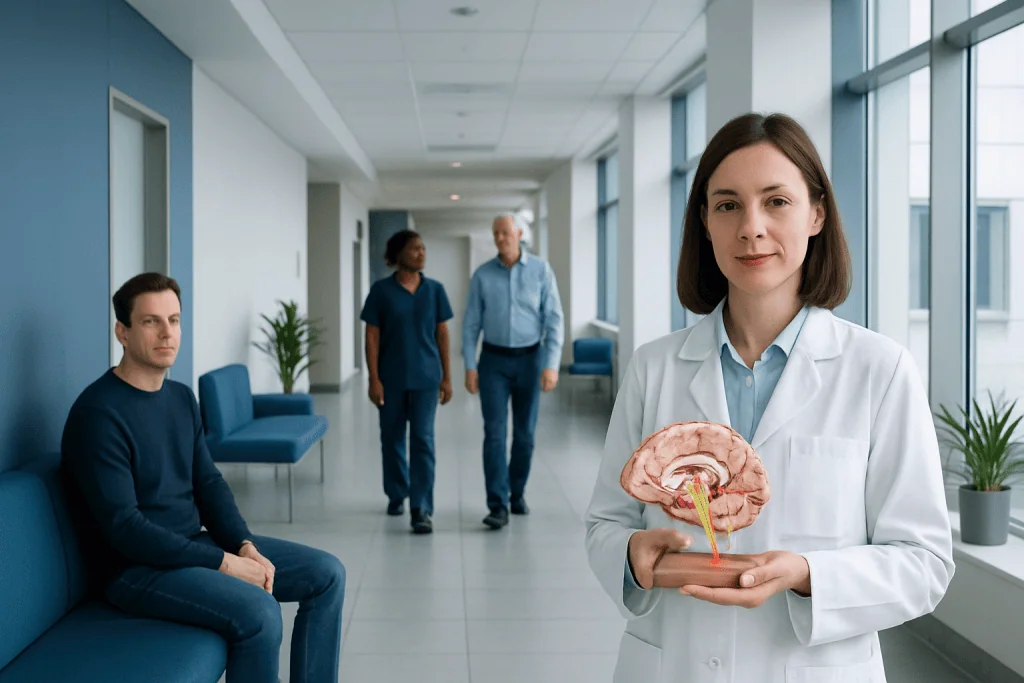
The optic chiasm helps the brain combine visual information from both eyes. Problems affecting the optic chiasm often cause loss of peripheral, or side, vision.
Key Takeaways
- The optic chiasm helps the brain combine visual information from both eyes.
- Problems affecting the optic chiasm often cause loss of peripheral, or side, vision.
- Common causes include pressure from nearby growths, inflammation, and less often injury.
- Diagnosis usually involves an eye exam, visual field testing, and brain imaging such as MRI.
- Treatment depends on the cause and may include monitoring, medication, surgery, or both.
- New or worsening vision changes should be evaluated promptly.
The optic chiasm is a small but important area at the base of the brain where some visual nerve fibers cross from one side to the other. When it is affected by pressure, inflammation, or injury, a person may notice changes in side vision, blurred sight, or other visual symptoms that should be assessed by a doctor.
What the optic chiasm is and why it matters
The optic chiasm is the place where some of the nerve fibers from each eye cross over on their way to the brain. This crossing allows visual information from the right and left sides of the visual world to be processed correctly. In simple terms, it helps both eyes work together so the brain can build one clear picture.
The optic chiasm sits near the base of the brain, close to important structures such as the pituitary gland and the hypothalamus. Because of this location, even a small problem nearby can sometimes affect vision. Patients often hear about the optic chiasm when a scan shows a lesion or when a doctor is investigating side-vision loss.
On its own, the optic chiasm is not a disease. It is a normal part of the visual pathway. However, disorders that press on, inflame, or damage this area can change how visual signals travel, leading to symptoms that may develop gradually or, less commonly, appear more suddenly.
How optic chiasm problems can affect vision
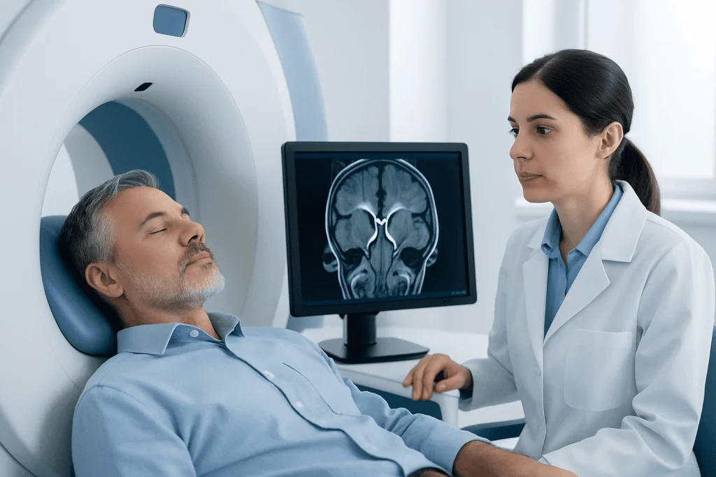
The pattern of symptoms often reflects the special job of the optic chiasm. Since crossing fibers carry information related to the outer, or temporal, visual fields, damage here commonly affects side vision in both eyes. A person may not notice this at first, especially if central vision remains fairly clear.
One classic pattern is called bitemporal hemianopia. This means loss of the outer half of the visual field in both eyes. People may describe bumping into objects, difficulty driving, trouble noticing movement off to the side, or feeling that their world has become narrower.
Other symptoms can occur depending on the cause and how much pressure or damage is present. These may include blurred vision, reduced visual sharpness, color vision changes, headaches, or eye strain. If the nearby pituitary gland is involved, there may also be hormonal symptoms such as changes in menstrual cycles, fatigue, sexual dysfunction, or unexplained weight changes.
- Reduced side vision
- Blurred or dim vision
- Difficulty reading or navigating spaces
- Headaches
- Hormonal symptoms if a nearby pituitary lesion is present
Common causes and risk factors

The most common reason the optic chiasm is discussed in clinical care is compression from a nearby structure. Because it sits just above the pituitary gland, a pituitary tumor is a well-known cause. Many of these growths are noncancerous, but they can still press on the optic chiasm and affect vision as they enlarge. This may be considered alongside conditions such as pituitary adenoma.
Other causes include meningiomas, craniopharyngiomas, aneurysms, inflammation, infections, and demyelinating disease. Less commonly, trauma or reduced blood supply can affect the visual pathway in this area. In children, congenital or developmental conditions may also be considered depending on symptoms and imaging findings.
Risk factors depend on the underlying condition rather than the optic chiasm itself. For example, a person with a known pituitary lesion, a history of neurologic disease, or symptoms suggesting hormonal imbalance may have a higher likelihood of a nearby process affecting the chiasm. Still, some people have no obvious risk factors before symptoms appear.
Not every optic chiasm abnormality causes permanent injury. In some cases, vision changes improve when the underlying cause is treated early. That is why prompt evaluation of unexplained visual field loss is important.
How doctors diagnose optic chiasm disorders
Diagnosis begins with a careful history and examination. A doctor may ask when symptoms started, whether one or both eyes are affected, whether headaches or hormonal changes are present, and whether symptoms are stable or progressing. A basic eye exam can check visual acuity, pupils, eye movements, and the appearance of the optic nerves.
Visual field testing is especially useful because it can show the pattern of sight loss. This helps doctors identify whether the problem may involve the retina, optic nerve, optic chiasm, or brain. A characteristic side-vision defect in both eyes often points toward the optic chiasm.
Imaging is usually needed to find the cause. An MRI of the brain and the sellar region is commonly preferred because it gives detailed pictures of the optic pathways and nearby structures. In some situations, CT scanning or blood tests may also be used, especially if there is concern about bone changes, bleeding, inflammation, or hormonal disorders.
Depending on the findings, patients may be assessed by ophthalmology, neuro-ophthalmology, endocrinology, neurology, or neurosurgery. When imaging or symptoms suggest a lesion near the pituitary region, further work-up may include hormone testing and consultation about brain tumor treatment or neurosurgical care.
Treatment options and what recovery may look like
Treatment for an optic chiasm problem depends entirely on the cause. If a lesion is pressing on the area, the main goal is to relieve pressure and protect vision. This may involve observation with repeat scans, medication, surgery, radiation therapy, or a combination of these approaches. Doctors tailor treatment to the size, location, behavior, and likely diagnosis of the underlying lesion.
For pituitary tumors, treatment may include hormone-related medication or surgery to remove or shrink the mass when appropriate. When inflammation is responsible, anti-inflammatory treatment may be considered. If an aneurysm or another vascular problem is identified, specialized care is needed to reduce the risk of further damage.
Visual recovery is variable. Some patients improve significantly after treatment, particularly when pressure is relieved early. Others may continue to have some degree of visual field loss if nerve fibers have been damaged for a longer time. Follow-up testing helps show whether vision is stable, improving, or needs further attention.
Rehabilitation may also be helpful. This can include low-vision support, safety strategies for reduced side vision, and regular monitoring. In selected cases, care may involve endoscopic pituitary surgery or broader neurology evaluation to guide treatment planning.
Living with symptoms: self-care and daily precautions
Self-care cannot treat the optic chiasm itself, but it can help a person cope safely while awaiting diagnosis or during recovery. Patients with side-vision loss may benefit from turning the head more deliberately when walking, using extra caution on stairs, and avoiding driving until a doctor confirms it is safe. Good lighting at home can also make navigation easier.
Keeping follow-up appointments is important because subtle visual changes are not always easy to notice. A person may adapt to gradual vision loss without realizing how much the visual field has narrowed. Repeat visual field tests and imaging can show whether the condition is stable or changing.
If the underlying cause involves the pituitary gland or another endocrine issue, following advice about hormone testing and treatment is also part of self-care. Patients should tell their doctor about headaches, fatigue, menstrual changes, sexual symptoms, unusual thirst, or changes in weight, because these details may help identify a related hormonal condition.
Support from eye specialists, neurologists, and endocrine teams can make daily life easier and improve treatment decisions. Near the end of the care pathway, some international patients choose multidisciplinary assessment at centers such as Acibadem International, where JCI-accredited hospitals diagnose and treat conditions affecting the optic pathways.
When to seek medical care
Any new, unexplained change in vision should be discussed with a qualified doctor. This is particularly important if a person notices reduced side vision, blurry vision in both eyes, repeated collisions with objects, or visual symptoms that are steadily getting worse. Even gradual changes deserve evaluation because some optic chiasm conditions develop slowly.
Urgent medical attention is appropriate if vision loss appears suddenly, if there is severe headache, double vision, confusion, weakness, vomiting, or symptoms of a neurologic emergency. These features do not always mean the optic chiasm is the cause, but they require prompt assessment.
People with a known pituitary tumor or another lesion near the optic pathways should report any visual change without delay. Early diagnosis can help preserve vision and guide the most suitable treatment plan.
Frequently asked questions
What does the optic chiasm do?
The optic chiasm is where some nerve fibers from each eye cross to the opposite side. This helps the brain combine visual information properly so a person can interpret the right and left sides of the visual world.
What are the first signs of an optic chiasm problem?
Early signs often involve side-vision loss rather than complete blindness. Some people notice bumping into objects, trouble driving, headaches, or a sense that their field of view is becoming narrower.
Can an optic chiasm problem be caused by a pituitary tumor?
Yes. A pituitary tumor is one of the best-known causes because the pituitary gland lies very close to the optic chiasm. Even a noncancerous tumor can affect vision if it grows large enough to press on nearby nerve fibers.
Is optic chiasm damage reversible?
Sometimes vision improves after the cause is treated, especially if pressure is relieved early. Recovery depends on how long the optic pathway has been affected and whether the nerve fibers have sustained lasting damage.
How is an optic chiasm disorder diagnosed?
Doctors usually combine a medical history, eye examination, and visual field testing with imaging such as MRI. If a lesion near the pituitary gland is suspected, hormone tests and specialist evaluation may also be needed.
Does every optic chiasm finding need surgery?
No. Treatment depends on the underlying cause, the severity of symptoms, and whether the condition is stable or progressing. Some cases are monitored, while others need medication, surgery, radiation, or multidisciplinary care.
This article is for general information only and is not a substitute for professional medical advice. Please consult a qualified doctor about your individual situation.
Explore treatments in Turkey — costs, top hospitals & a free quote
JCI-accredited · board-certified surgeons · reply within 24h
Add us as a Preferred Source to see more of our trusted health content across Google Search, AI Overviews and Discover.

More from the Health Library

Vitamin d blood test: A Complete Medical Guide for Patients
Way Brotin: An Evidence-Based Guide for Patients

Strawberry Birthmark — Explained by Medical Evidence, Not Myths

Peripheral Edema: A Complete Medical Overview

Turbid Urine — Explained by Medical Evidence, Not Myths