

Optic Nerve Atrophy: Causes, Imaging, and Neuro-Ophthalmology Evaluation

Optic nerve atrophy is a sign of previous or ongoing optic nerve damage, not a single disease. Symptoms may include blurred vision, reduced color brightness, blind spots, or loss of side vision.
Key Takeaways
- Optic nerve atrophy is a sign of previous or ongoing optic nerve damage, not a single disease.
- Symptoms may include blurred vision, reduced color brightness, blind spots, or loss of side vision.
- Common evaluations include a dilated eye examination, OCT scans, visual field testing, and sometimes MRI of the brain and orbits.
- Treatment focuses on the underlying cause, such as glaucoma, inflammation, compression, nutritional deficiency, or inherited disease.
- Prompt medical attention is important for sudden vision loss, eye pain, new neurologic symptoms, or rapidly changing vision.
Optic nerve atrophy describes damage and loss of nerve fibers that carry visual information from the eye to the brain. A careful neuro-ophthalmology evaluation can help identify the cause, estimate the outlook, and guide treatment to protect remaining vision whenever possible.
Overview
Optic nerve atrophy is a term doctors use when the optic nerve has lost some of its nerve fibers. The optic nerve is the cable that carries visual signals from the retina at the back of the eye to the brain. When those fibers are damaged, the optic disc may look pale during an eye examination, and vision may be affected in different ways.
It is important to understand that optic nerve atrophy is usually a finding or result, not a diagnosis by itself. Many different conditions can injure the optic nerve, including glaucoma, inflammation, poor blood supply, compression from a mass, inherited optic neuropathies, trauma, nutritional deficiencies, and toxic exposures. Some causes are stable, while others may be treatable if identified early.
A neuro-ophthalmology evaluation brings together eye examination, neurologic assessment, imaging, and targeted laboratory testing when needed. The goal is to determine where the damage occurred, whether it is still active, and what can be done to protect the patient’s remaining vision and overall health.
Symptoms and Vision Changes
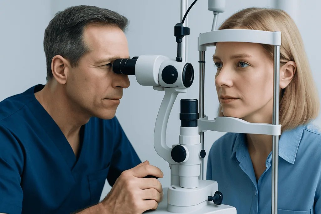
Symptoms of optic nerve atrophy vary depending on the number of nerve fibers affected and the underlying cause. Some people notice gradual blurring, dimming, or a sense that one eye does not see as clearly as the other. Others may first become aware of difficulty reading, reduced contrast, or trouble seeing in low light.
Color vision changes are common in optic nerve disease. Red objects may look faded or less bright through the affected eye. Visual field loss can also occur, meaning there may be blind spots, missing areas in central vision, or reduced side vision. In some cases, the person may not notice these changes until formal visual field testing is performed.
Possible symptoms include:
- Blurred or reduced vision in one or both eyes
- Dim vision or reduced brightness
- Faded color perception, especially red color desaturation
- Blind spots or missing areas in the visual field
- Difficulty reading or recognizing faces
- Poor night vision or reduced contrast sensitivity
- Eye pain with movement in some inflammatory conditions, such as optic neuritis
The pattern of symptoms can provide important clues. Sudden painless vision loss may suggest a blood supply problem, while gradual loss of side vision may be seen in glaucoma. Slowly progressive vision loss can sometimes point to compression, inherited disease, or toxic and nutritional causes.
Causes and Risk Factors

Optic nerve atrophy can develop after many types of optic nerve injury. One of the most common causes worldwide is glaucoma, in which damage to the optic nerve is often related to eye pressure and individual susceptibility. Glaucoma-related optic nerve damage typically affects peripheral vision first and may progress slowly if not detected and treated.
Inflammatory optic nerve disease, such as optic neuritis, can also lead to optic atrophy after an episode of swelling or inflammation. Optic neuritis may be associated with autoimmune conditions, demyelinating diseases, infections, or other inflammatory disorders. Reduced blood supply to the optic nerve, known as ischemic optic neuropathy, is another important cause, especially in adults with vascular risk factors.
Compression of the optic nerve or visual pathway may occur due to tumors, aneurysms, enlarged blood vessels, thyroid eye disease, or other structural problems near the orbit or brain. This is one reason neuro-imaging can be essential when the examination pattern suggests pressure on the optic nerve. Traumatic injury to the eye socket or head can also damage optic nerve fibers directly or indirectly.
Other causes include hereditary optic neuropathies, such as Leber hereditary optic neuropathy or dominant optic atrophy, and toxic or nutritional optic neuropathies related to certain medications, alcohol misuse, vitamin deficiencies, or exposure to toxins. Risk factors that may guide testing include family history, previous optic neuritis, diabetes, high blood pressure, high cholesterol, smoking, autoimmune disease, cancer history, and recent medication or toxin exposure.
Diagnosis and Neuro-Ophthalmology Evaluation
A detailed evaluation usually begins with a careful medical and vision history. The doctor asks when symptoms started, whether they are stable or changing, whether one or both eyes are affected, and whether there is pain, headache, neurologic symptoms, family history, medication use, or toxin exposure. This history helps separate longstanding optic atrophy from active or progressive disease.
The eye examination includes measurement of visual acuity, pupil reactions, color vision, eye pressure, eye movements, and a dilated examination of the retina and optic disc. A relative afferent pupillary defect may be present when one optic nerve is more affected than the other. The optic disc may appear pale, cupped, swollen, or otherwise abnormal depending on the cause and timing of injury.
Formal visual field testing is often central to the diagnosis. It maps areas of vision that may be missing, such as central scotomas, arcuate defects, altitudinal defects, or bitemporal field loss. These patterns can help localize disease to the optic nerve, optic chiasm, or further back in the visual pathway.
Neuro-ophthalmologists combine these findings to decide whether additional tests are needed. Blood tests may be considered for inflammatory, infectious, nutritional, autoimmune, or metabolic causes. Genetic counseling and genetic testing may be appropriate when the history and examination suggest an inherited optic neuropathy.
Imaging and Eye Tests
Optical coherence tomography, often called OCT, is a noninvasive scan that measures the thickness of the retinal nerve fiber layer and the ganglion cell complex. These layers contain the nerve cells and fibers that form the optic nerve. OCT can help document optic nerve atrophy, compare both eyes, and monitor change over time.
Fundus photography is useful for recording the appearance of the optic disc and retina. In some patients, additional retinal imaging or fluorescein angiography may be considered to evaluate blood flow or retinal disease that could mimic optic nerve problems. Visual evoked potential testing may be used in selected cases to assess how well signals travel from the eye to the visual cortex, particularly when demyelinating disease is suspected.
MRI of the brain and orbits with contrast is commonly recommended when there are signs of optic nerve inflammation, compression, unexplained optic atrophy, asymmetry between the eyes, or visual field patterns suggesting a lesion near the optic chiasm. MRI can show the optic nerves, optic chiasm, brain, and surrounding structures. In some situations, CT imaging may be preferred, especially when bone injury, calcification, or certain orbital conditions are being assessed.
Imaging decisions are individualized. Not every patient with optic atrophy needs every test, but unexplained, progressive, asymmetric, or neurologically associated findings usually require a broader evaluation. The purpose is to avoid missing treatable conditions while keeping testing focused and appropriate.
Treatment Options
Treatment for optic nerve atrophy depends on the cause. Once nerve fibers have been permanently lost, they usually cannot be restored with current medical treatments. However, identifying and treating the underlying condition can often help stabilize vision, prevent further damage, or address related health risks.
For glaucoma, treatment may include prescription eye drops, laser procedures, or surgery to lower eye pressure and reduce ongoing optic nerve damage. In inflammatory optic neuropathies, treatment may involve corticosteroids or other immune therapies under specialist guidance, depending on the diagnosis. Ischemic optic neuropathy care focuses on identifying vascular risk factors and, when appropriate, coordinating care with internal medicine, cardiology, neurology, or rheumatology specialists.
If imaging shows compression of the optic nerve or visual pathway, management may involve neurosurgery, endocrinology, oncology, or radiation oncology depending on the cause. Nutritional deficiencies are treated by correcting the deficiency and addressing dietary or absorption problems. Toxic optic neuropathy requires stopping or replacing the harmful exposure whenever medically appropriate, under supervision.
Low vision rehabilitation can be very helpful when vision loss affects reading, mobility, work, or daily activities. This may include magnification devices, contrast enhancement, lighting strategies, digital accessibility tools, and orientation support. The aim is to help the person use remaining vision as effectively and safely as possible.
Prevention, Monitoring, and Self-Care
Not all causes of optic nerve atrophy are preventable, but regular eye care can reduce the risk of avoidable vision loss. People with glaucoma risk factors, diabetes, high blood pressure, strong family history of eye disease, or previous optic nerve problems should follow the examination schedule recommended by their eye specialist. Monitoring is especially important because some optic nerve diseases progress silently.
Self-care focuses on supporting general vascular and nerve health. Patients are encouraged to avoid smoking, manage blood pressure and blood sugar, maintain a balanced diet, limit alcohol according to medical guidance, and review medications with their doctor if new visual symptoms occur. No supplement should be used as a substitute for diagnosis and treatment, and high-dose vitamins should be taken only when recommended by a healthcare professional.
At follow-up visits, doctors may repeat OCT, visual field testing, optic disc photographs, and vision measurements. Comparing results over time helps determine whether the condition is stable or progressing. Patients can also help by noting changes in reading ability, color perception, contrast, or side vision and reporting them promptly.
For international patients who need coordinated assessment, Acibadem International offers multidisciplinary evaluation through ophthalmology, neurology, radiology, and related specialties in JCI-accredited hospitals. This can be useful when optic nerve atrophy requires both eye-focused testing and broader neurologic or systemic assessment.
When to See a Doctor
A person should see an eye doctor if vision becomes blurred, dim, distorted, or noticeably different between the two eyes. Evaluation is also important for reduced color brightness, unexplained blind spots, trouble seeing to the side, or a known history of optic neuritis, glaucoma, neurologic disease, or optic nerve injury.
Prompt medical attention is needed for sudden vision loss, rapidly worsening vision, new double vision, severe headache, weakness, numbness, difficulty speaking, or eye pain with vision changes. These symptoms do not always mean a serious condition is present, but they deserve timely assessment because some causes are more treatable when addressed early.
Patients who have already been told they have optic nerve atrophy should keep scheduled follow-up visits, even if vision seems stable. Ongoing monitoring helps detect progression, adjust treatment, and support daily visual function. A qualified ophthalmologist or neuro-ophthalmologist can explain what the test results mean for the individual patient’s situation.
Frequently asked questions
Is optic nerve atrophy the same as blindness?
No. Optic nerve atrophy means there has been loss of some optic nerve fibers, but the amount of vision loss varies widely. Some people have mild changes found only on testing, while others have more significant visual impairment. The cause, severity, and whether the condition is still progressing are key factors.
Can optic nerve atrophy be reversed?
In most cases, optic nerve fibers that have been permanently lost cannot be regenerated with current treatments. Treatment focuses on the underlying cause and on preventing additional damage when possible. Low vision rehabilitation can also improve day-to-day function even when vision cannot be fully restored.
Why is MRI sometimes needed for optic nerve atrophy?
MRI can show the optic nerves, optic chiasm, brain, and surrounding tissues in detail. It is often used when optic atrophy is unexplained, progressive, asymmetric, or associated with symptoms suggesting inflammation or compression. The doctor decides whether MRI is needed based on the examination and test results.
What does OCT show in optic nerve atrophy?
OCT measures the thickness of retinal layers that contain optic nerve fibers and related nerve cells. Thinning on OCT can support the diagnosis of optic nerve atrophy and help monitor stability or progression over time. OCT results are interpreted together with vision tests, visual fields, and the clinical examination.
Can glaucoma cause optic nerve atrophy?
Yes. Glaucoma is a common cause of optic nerve damage and can lead to optic nerve atrophy over time. Because glaucoma often affects side vision first and may not cause early symptoms, regular eye examinations are important for people at risk.
Should family members be tested if optic nerve atrophy is found?
Family testing depends on the suspected cause. If the pattern suggests an inherited optic neuropathy, the doctor may recommend genetic counseling and, in some cases, genetic testing for the patient and selected relatives. Many cases are not inherited, so the decision is individualized.
Which specialist evaluates optic nerve atrophy?
An ophthalmologist can evaluate many optic nerve problems, especially glaucoma and retinal-related conditions. A neuro-ophthalmologist is often involved when the cause is unclear or when neurologic, inflammatory, compressive, or complex visual pathway disease is suspected. Care may also include neurology, radiology, endocrinology, rheumatology, or other specialists depending on the findings.
References
- American Academy of Ophthalmology
- National Eye Institute
- North American Neuro-Ophthalmology Society
- European Glaucoma Society
- Mayo Clinic
This article is for general information only and is not a substitute for professional medical advice. Please consult a qualified doctor about your individual situation.
Explore treatments in Turkey — costs, top hospitals & a free quote
JCI-accredited · board-certified surgeons · reply within 24h
Add us as a Preferred Source to see more of our trusted health content across Google Search, AI Overviews and Discover.
Check your numbers in seconds
BMI, calories, due date, blood pressure and 30+ more clinical calculators — free, instant, doctor-reviewed ranges.

More from the Health Library



Related Specialists

Dr. Sema Eryılmaz
Pediatric Endocrinology
Prof. Dr. Ayşe Korkmaz Toygar
Neonatal Intensive Care Unit
Dr. Arzu Ertem Cengiz
Pulmonology
Dt. Avi Karako
Oral & Dental Health