Optic Neuritis: Diagnosis, Outlook, and Modern Treatment Approaches

Optic neuritis commonly causes eye pain, especially with eye movement, and blurred or dim vision in one eye. It may occur on its own or be linked to conditions such as multiple sclerosis, neuromyelitis optica spectrum disorder, or MOG antibody-associated disease.
Key Takeaways
- Optic neuritis commonly causes eye pain, especially with eye movement, and blurred or dim vision in one eye.
- It may occur on its own or be linked to conditions such as multiple sclerosis, neuromyelitis optica spectrum disorder, or MOG antibody-associated disease.
- Diagnosis usually combines an eye examination with brain and orbit MRI, and sometimes blood tests and other neurologic studies.
- Treatment depends on severity and cause; some cases improve on their own, while others need corticosteroids or targeted immune therapy.
- New or sudden vision loss should be assessed promptly to rule out other urgent eye or neurologic problems.
Optic neuritis is inflammation of the optic nerve, often causing eye pain and reduced vision in one eye over hours to days. Outlook is frequently good, but timely evaluation is important because treatment and long-term follow-up depend on the underlying cause.
Overview: what optic neuritis means
Optic neuritis is inflammation of the optic nerve, the structure that carries visual information from the eye to the brain. It often causes eye pain and a noticeable drop in vision, usually affecting one eye and developing over a short period of time. For many people, vision improves over weeks to months, but the best approach depends on why the inflammation happened.
Doctors use the term optic neuritis for a specific pattern of optic nerve inflammation, but it is not a single disease with one cause. In some people, it appears after a viral illness or as an isolated event. In others, it can be associated with immune-related conditions that affect the brain, spinal cord, or optic nerves, including multiple sclerosis.
Because the optic nerve is part of the central nervous system, optic neuritis is often evaluated by both eye specialists and neurologists. This combined approach helps confirm the diagnosis, identify related conditions, and guide treatment decisions. A careful workup is especially important when symptoms are severe, involve both eyes, or do not follow the usual pattern.
Symptoms and how vision may change
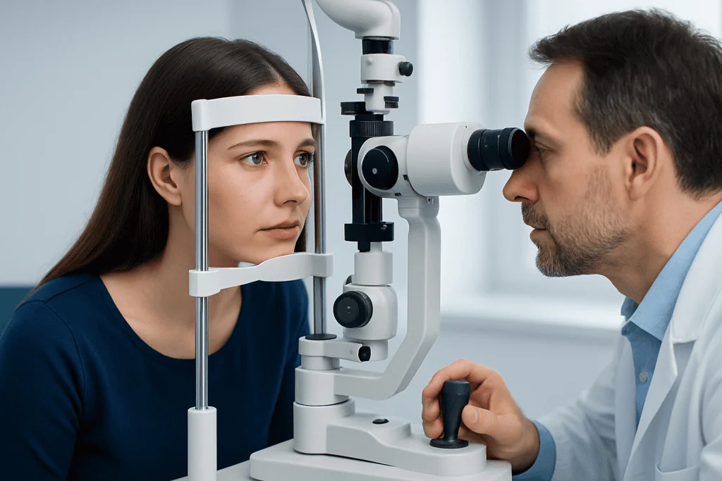
The most common symptoms of optic neuritis are pain around the eye and reduced vision. Pain often becomes more noticeable when the person moves the affected eye. Vision changes can include blurring, dimming, reduced sharpness, or a dark spot in the center of vision. Colors, especially red, may look less vivid than usual.
Symptoms often worsen over several days before stabilizing. Some people notice flashing lights or increased visual discomfort with exercise or heat, a temporary effect sometimes linked to prior optic nerve inflammation. In classic cases, one eye is affected, but both eyes can be involved, especially in some immune-mediated disorders.
Doctors also look for signs that help distinguish optic neuritis from other eye problems. These may include:
- Reduced color vision
- A relative afferent pupillary defect in one eye
- Loss of part of the visual field
- Optic disc swelling in some cases, though the optic nerve can also look normal early on
Not every episode of blurred vision is optic neuritis. Other causes of sudden vision loss, such as retinal disease, glaucoma, ischemic optic neuropathy, or compression of the optic nerve, may need a different and sometimes more urgent treatment plan.
Causes, triggers, and related conditions

Optic neuritis can happen for several reasons. In many younger adults, it is linked to inflammatory demyelination, meaning the protective covering around nerve fibers is damaged by an immune response. This is why optic neuritis can be associated with conditions such as multiple sclerosis, neuromyelitis optica, and MOG antibody-associated disease.
In other cases, optic neuritis may follow an infection or occur with broader autoimmune diseases. Less commonly, inflammation of the optic nerve may be related to sarcoidosis, lupus, or certain infections. Some people have an atypical presentation, which prompts doctors to broaden the evaluation and look carefully for causes beyond classic demyelinating optic neuritis.
Features that may suggest a non-classic cause include very severe vision loss, no pain, prominent swelling of the optic nerve, recurrent attacks, both eyes being affected, or poor recovery after the first episode. Age also matters: optic neuritis can occur in children and older adults, but the list of possible causes may be different in these groups.
Identifying the underlying condition is important because the outlook and treatment may differ significantly. A person with isolated optic neuritis may only need monitoring after recovery, while someone with recurrent inflammation may need longer-term neuroimmunology care.
How optic neuritis is diagnosed
Diagnosis begins with a detailed history and eye examination. Doctors ask when the symptoms began, whether pain occurs with eye movement, whether one or both eyes are affected, and if there have been previous neurologic symptoms such as numbness, weakness, balance changes, or bladder problems. Vision testing includes visual acuity, color vision, pupil responses, and a dilated examination of the optic nerve and retina.
MRI of the brain and orbits with contrast is one of the most useful tests. It can show inflammation of the optic nerve and may also reveal brain lesions that raise the likelihood of an associated demyelinating condition. Depending on the pattern, clinicians may arrange MRI scanning as part of the early evaluation.
Additional tests may include optical coherence tomography, which measures the retinal nerve fiber layer; visual field testing; and blood tests for antibodies such as aquaporin-4 and MOG when indicated. Some patients also need lumbar puncture or other neurologic investigations, especially if the diagnosis is uncertain or there are signs of disease elsewhere in the nervous system. In selected cases, a comprehensive eye examination and coordinated neuro-ophthalmology assessment help clarify the diagnosis.
The goal of testing is not only to confirm optic neuritis, but also to separate typical cases from atypical ones. That distinction helps predict outlook and supports decisions about whether a person may benefit from observation, corticosteroids, or longer-term immune treatment.
Modern treatment approaches and recovery
Treatment depends on symptom severity and the suspected cause. Some mild, typical cases of optic neuritis improve without specific treatment, and doctors may recommend close follow-up rather than immediate medication. When vision loss is more significant, intravenous corticosteroids are often used to speed recovery, although they may not change the final level of vision in every case.
If optic neuritis is linked to neuromyelitis optica spectrum disorder, MOG antibody-associated disease, or another relapsing inflammatory condition, treatment can be different from standard management of a one-time episode. In severe or steroid-resistant cases, plasma exchange may be considered. Long-term immunotherapy may also be recommended to reduce the risk of future attacks when a relapsing disorder is confirmed.
Recovery usually begins within a few weeks, but improvement can continue for months. Some people regain near-normal sight, while others may continue to notice reduced contrast sensitivity, color desaturation, or subtle visual field loss even after the main episode resolves. If a broader neurologic condition is identified, care may include neurology evaluation and follow-up to discuss ongoing monitoring or preventive therapy.
For patients who need coordinated care, Acibadem International’s multidisciplinary specialists in neuro-ophthalmology, neurology, and imaging work in JCI-accredited hospitals to diagnose and treat optic neuritis and related disorders for international patients.
Outlook, recurrence, and long-term follow-up
The outlook for optic neuritis varies with the cause. In classic demyelinating optic neuritis, vision often improves substantially over time. However, a good visual acuity result does not always mean vision feels completely normal, since contrast, depth perception, and color quality may remain slightly changed.
One of the most important long-term questions is whether optic neuritis is an isolated event or part of a recurring neurologic disease. MRI findings, antibody testing, and the clinical pattern all help estimate that risk. Patients with recurrent episodes, bilateral involvement, or severe attacks usually need more structured follow-up than those with a single typical event.
Follow-up visits may include repeat visual field testing, optical coherence tomography, and neurologic review. These appointments help track recovery, identify residual nerve damage, and detect signs of relapse early. If there is concern for a related disorder such as multiple sclerosis, a doctor may discuss additional imaging or preventive treatment options over time.
Emotional reassurance is also important. Sudden vision symptoms can be frightening, but many people recover well, especially when the diagnosis is made promptly and follow-up is tailored to the underlying cause.
Self-care and when to seek medical care
There is no home remedy that can treat optic neuritis itself, but practical self-care can support recovery. Rest, taking prescribed medicines exactly as directed, and attending follow-up appointments are all important. People may also find it helpful to reduce activities that strain vision while symptoms are most noticeable and to avoid overheating if heat temporarily worsens visual symptoms.
Because optic neuritis can overlap with other eye and neurologic conditions, new vision loss should not be self-diagnosed. Prompt assessment is the safest way to confirm the cause and protect vision. Patients should seek medical care urgently if they develop sudden loss of vision, severe eye pain, symptoms in both eyes, new weakness or numbness, trouble speaking, or a severe headache with visual changes.
It is also important to contact a doctor if symptoms return after recovery, if vision continues to worsen after the first several days, or if there is little improvement over the expected recovery period. Early re-evaluation can help detect recurrence, reconsider the diagnosis, or identify a condition that needs a different treatment plan.
Frequently asked questions
Is optic neuritis the same as multiple sclerosis?
No. Optic neuritis is inflammation of the optic nerve, while multiple sclerosis is a broader disease that affects the central nervous system. Optic neuritis can occur on its own, but in some people it is the first sign of multiple sclerosis or another neuroinflammatory condition.
Can optic neuritis go away on its own?
Some cases improve without specific treatment, especially typical mild episodes. Even so, medical evaluation is important because not all vision loss is optic neuritis, and some underlying causes need prompt therapy or long-term follow-up.
How long does vision recovery take after optic neuritis?
Many people begin to improve within a few weeks, and recovery may continue for several months. The degree of recovery varies, and some people notice lingering changes in color vision, contrast, or visual clarity even if standard eye chart vision improves.
Does optic neuritis always affect one eye?
No. It often affects one eye in classic cases, but both eyes can be involved, particularly in some immune-related conditions such as MOG antibody-associated disease or neuromyelitis optica spectrum disorder. Bilateral symptoms usually require careful evaluation.
What tests are used to confirm optic neuritis?
Doctors commonly use an eye examination, vision testing, and MRI of the brain and orbits. Depending on the clinical picture, they may also order visual field testing, optical coherence tomography, blood tests for specific antibodies, or other neurologic studies.
Can optic neuritis come back?
Yes, it can recur, especially when it is linked to an underlying inflammatory or autoimmune disorder. Recurrence risk depends on the cause, MRI findings, and antibody results, which is why follow-up care is an important part of management.
References
- American Academy of Ophthalmology
- National Eye Institute
- National Institute of Neurological Disorders and Stroke
- Mayo Clinic
- MS International Federation
This article is for general information only and is not a substitute for professional medical advice. Please consult a qualified doctor about your individual situation.
Explore treatments in Turkey — costs, top hospitals & a free quote
JCI-accredited · board-certified surgeons · reply within 24h
Add us as a Preferred Source to see more of our trusted health content across Google Search, AI Overviews and Discover.