Orbital Bone — Explained by Medical Evidence, Not Myths

The orbital bone usually refers to the bones that form the eye socket rather than one separate bone. The orbit protects the eye and contains muscles, nerves, blood vessels, and fatty tissue that help the eye move and function.
Key Takeaways
- The orbital bone usually refers to the bones that form the eye socket rather than one separate bone.
- The orbit protects the eye and contains muscles, nerves, blood vessels, and fatty tissue that help the eye move and function.
- Orbital injuries can cause pain, swelling, double vision, numbness, or changes in eye position.
- Diagnosis often involves an eye examination and imaging such as CT scans.
- Treatment depends on the cause and may range from observation to surgery.
- Urgent medical assessment is important after facial trauma or sudden vision changes.
The orbital bone is not a single bone but part of the bony eye socket, called the orbit, which protects the eye and supports muscles, nerves, and blood vessels. Understanding its structure helps explain how eye injuries, fractures, and some sinus or facial conditions can affect vision, eye movement, and comfort.
Overview: what the orbital bone is
The term orbital bone is commonly used to describe the bones that make up the eye socket, also called the orbit. In medical terms, the orbit is a bony cavity in the skull that holds and protects the eyeball along with the muscles, nerves, blood vessels, connective tissue, and fat that support vision and eye movement.
Rather than being one single bone, the orbit is formed by several facial and skull bones that fit together like a protective frame. This matters because different parts of the orbit can be affected by trauma, infection, inflammation, tumors, or congenital differences, and each area may cause slightly different symptoms.
The orbit does more than simply surround the eye. Its shape helps position the eye correctly, protects it from external force, and provides passageways for important structures such as the optic nerve and the nerves that control sensation and eye movement. Because of this close anatomy, conditions involving the orbital bones may affect not only appearance but also vision and comfort.
How the orbit is built and why it matters
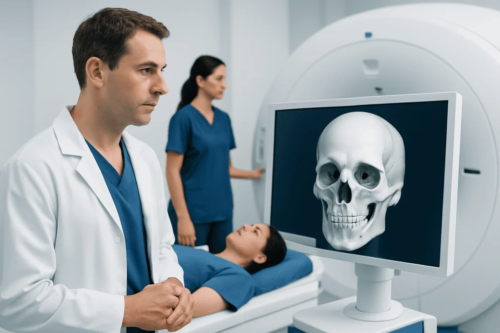
The bony orbit is formed by seven bones: the frontal, sphenoid, zygomatic, maxillary, palatine, lacrimal, and ethmoid bones. These bones create the roof, floor, medial wall, and lateral wall of the eye socket. Some portions are thick and strong, while others are very thin and more vulnerable to fracture.
The orbital floor lies above the maxillary sinus, and the medial wall lies next to the ethmoid sinuses. This close relationship explains why infections, inflammation, or trauma can sometimes involve both the orbit and the sinuses. The orbit also contains small openings and canals that allow nerves and blood vessels to pass between the eye and the brain.
Inside the orbit are structures essential for sight and movement. These include the optic nerve, the muscles that move the eye, the lacrimal system, and cushioning fat. When the orbit is injured or inflamed, pressure can build in this tight space, which may affect eye movement, cause pain, or in some cases threaten vision.
Understanding anatomy helps separate facts from myths. For example, many people think the eye itself is the only structure injured in facial trauma, but orbital bone injuries can significantly affect the eye even when the eyeball looks intact at first.
Symptoms linked to orbital bone problems

Symptoms depend on the underlying problem. A fracture after trauma may cause pain around the eye, swelling, bruising, tenderness, numbness in the cheek or upper lip, double vision, or a feeling that the eye is sunken or out of place. In more serious injuries, there may be reduced vision or difficulty moving the eye.
Not all orbital conditions are caused by injury. Infections and inflammatory conditions can lead to redness, eyelid swelling, eye pain, fever, bulging of the eye, or pain when moving the eye. Certain thyroid-related eye conditions can also change the position of the eye and affect movement. In some cases, symptoms overlap with glaucoma or other eye problems, which is why proper evaluation is important.
Children and adults may describe symptoms differently. A child with an orbital injury may mainly seem nauseated, refuse to move the eye, or complain of blurry or double vision. Some fractures, especially so-called trapdoor injuries in children, may look less dramatic on the outside than they are internally.
- Pain or pressure around the eye
- Swelling or bruising of the eyelids
- Double vision
- Numbness of the cheek, nose, or upper lip
- Sunken or bulging appearance of the eye
- Reduced vision or difficulty moving the eye
Causes and risk factors
The most common cause of orbital bone injury is blunt facial trauma. Falls, sports injuries, traffic accidents, workplace injuries, and physical assaults can all lead to orbital fractures. A direct blow from a ball, elbow, fist, or other object may suddenly increase pressure within the orbit and cause one of the thinner walls to break.
Different fracture patterns can occur. A blowout fracture usually affects the orbital floor or medial wall, allowing tissue to herniate into a nearby sinus. Other injuries may involve the rim of the orbit, multiple facial bones, or nearby structures such as the nose and cheekbone. Some patients may also have associated eye injuries, which can be more urgent than the fracture itself.
Orbital problems are not limited to fractures. Infection spreading from the sinuses may cause orbital cellulitis, an important condition related to sinusitis. Inflammatory disease, thyroid eye disease, tumors, and congenital abnormalities can also involve the orbital bones or nearby tissues. Risk increases when a person has significant facial trauma, untreated sinus infection, contact sports exposure, or previous facial surgery.
How doctors diagnose orbital bone conditions
Diagnosis starts with a careful history and physical examination. Doctors ask how the symptoms began, whether there was trauma, and whether there is double vision, numbness, pain with eye movement, or changes in vision. They also examine the eyelids, the position of the eye, pupil reactions, eye movements, and the surface and inside of the eye.
Imaging is often needed when an orbital fracture or deeper orbital disease is suspected. A CT scan is commonly used because it shows the bony walls of the orbit and can reveal trapped muscles, displaced fractures, bleeding, or sinus involvement. In selected situations, MRI may be used to evaluate soft tissues, inflammation, or tumors more clearly.
Doctors may also check visual acuity, color vision, eye pressure, and the back of the eye. If the problem seems related to the tear system, muscles, or optic nerve, referral to ophthalmology or related specialists may be appropriate. Depending on the findings, treatment may be coordinated with maxillofacial surgery, ENT, neurology, or radiology teams, and imaging may be part of a broader MRI scan or CT scan work-up.
Treatment options and recovery
Treatment depends on the cause, severity, and whether vision is threatened. Minor orbital fractures without significant double vision, muscle entrapment, or cosmetic change may be managed conservatively. This often includes rest, cold compresses, pain control as advised by a doctor, and avoiding nose blowing because air can be forced from the sinus into the orbit.
If there is severe fracture displacement, persistent double vision, trapped muscle, sunken eye position, or functional problems, surgery may be recommended. The goal is to restore the orbital walls and relieve entrapment while protecting the eye and nearby nerves. Some patients may need orbital surgery or a broader facial reconstruction approach depending on the injury pattern.
When infection is present, treatment may include antibiotics and close monitoring, sometimes in hospital if symptoms are severe. Inflammatory or thyroid-related orbital disease is managed differently and may involve medications or specialist-led care. If a person also has an internal eye problem, treatment may overlap with retina treatment or other eye-specific care.
Recovery time varies. Swelling and bruising often improve over days to weeks, while healing of bone and restoration of eye movement may take longer. Follow-up matters because some issues, such as persistent double vision or delayed changes in eye position, may become clearer after the initial swelling settles.
Prevention, self-care, and common myths
Many orbital injuries can be reduced with practical prevention. Protective eyewear during sports, helmets when appropriate, seat belt use, and workplace safety measures all help lower the risk of facial trauma. Prompt treatment of sinus infections may also reduce the chance of serious orbital spread in susceptible cases.
At home after minor trauma, people often use ice packs and rest, but home care should not replace medical evaluation when symptoms involve vision changes, worsening swelling, or trouble moving the eye. Self-care is supportive, not diagnostic. It is especially important not to apply pressure to the eye or ignore numbness and double vision.
Several myths can be misleading. One myth is that if the eye looks normal, the orbit must be fine; in reality, a fracture can exist with limited external signs. Another myth is that every orbital fracture needs surgery; many do not, and careful monitoring may be enough. A third myth is that bruising alone proves the problem is minor, when in fact internal injuries can coexist.
For international patients who need coordinated assessment, Acibadem International’s multidisciplinary specialists in JCI-accredited hospitals diagnose and treat orbital and related eye conditions using imaging and specialist eye care.
When to seek medical care
Medical care should be sought promptly after any significant blow to the eye or face, especially if there is double vision, reduced vision, severe pain, numbness, increasing swelling, nausea with eye movement, or the sense that the eye has moved backward or forward. These symptoms can suggest an orbital fracture, trapped muscle, bleeding, or another urgent eye problem.
Urgent care is also needed if there are signs of infection around the eye, such as fever, eyelid redness, bulging, or pain with eye movement. In children, reluctance to open the eye, vomiting after facial trauma, or unusual sleepiness should also be assessed quickly.
Even when symptoms seem mild, persistent discomfort, bruising that does not improve, or ongoing changes in vision deserve professional evaluation. Early diagnosis can help protect both eye function and appearance.
Frequently asked questions
Is the orbital bone a single bone?
Not usually. In everyday language, orbital bone refers to the bones that form the eye socket, called the orbit. The orbit is made of several bones that work together to protect and support the eye.
Can an orbital bone fracture heal on its own?
Some small orbital fractures can heal without surgery if the eye is moving well, vision is stable, and there is no major change in eye position. Doctors monitor these injuries closely because symptoms such as double vision or sinking of the eye may appear or persist as swelling decreases.
What does an orbital fracture feel like?
It may cause pain, swelling, bruising, numbness of the cheek or upper lip, and double vision. Some people also feel pressure around the eye or notice that the eye looks sunken or difficult to move.
Is every black eye an orbital bone injury?
No. A black eye can happen from soft tissue bruising alone without a fracture. However, a black eye can also occur with an orbital fracture or other eye injury, so associated symptoms such as blurred vision, double vision, or severe pain should be checked.
Why does an orbital fracture sometimes affect the sinuses?
Part of the eye socket shares thin walls with the nearby sinuses, especially the maxillary and ethmoid sinuses. If one of these walls breaks, air or tissue may move between the orbit and the sinus, which can cause swelling, pressure, or trapped muscles.
When is orbital bone surgery needed?
Surgery may be considered when there is trapped eye muscle, persistent double vision, significant fracture displacement, vision-threatening complications, or a noticeable change in eye position. The decision is individualized and based on examination findings and imaging results.
References
- American Academy of Ophthalmology
- National Eye Institute
- Merck Manual Professional Edition
- American Association for Pediatric Ophthalmology and Strabismus
- Mayo Clinic
This article is for general information only and is not a substitute for professional medical advice. Please consult a qualified doctor about your individual situation.
Joint and spine care in Turkey — expert assessment & treatment
JCI-accredited · board-certified surgeons · reply within 24h
Add us as a Preferred Source to see more of our trusted health content across Google Search, AI Overviews and Discover.