Osteochondritis Knee Surgery: Procedure, Recovery and Results

Osteochondritis dissecans affects cartilage and the bone directly beneath it, most often on the inner part of the knee joint. Surgery is usually considered for unstable, detached, or persistent lesions, especially after non-surgical treatment has not led to healing.
Key Takeaways
- Osteochondritis dissecans affects cartilage and the bone directly beneath it, most often on the inner part of the knee joint.
- Surgery is usually considered for unstable, detached, or persistent lesions, especially after non-surgical treatment has not led to healing.
- The procedure may involve drilling, fixation of the original fragment, cartilage restoration, or removal of loose tissue depending on the lesion.
- Many patients need crutches and limited weight-bearing for several weeks, while return to impact sports often takes months.
- Long-term results depend on lesion stability, size, location, cartilage condition, skeletal maturity, and commitment to rehabilitation.
Osteochondritis knee surgery may be recommended when an osteochondritis dissecans (OCD) lesion does not heal with non-surgical care, is unstable, or has detached from the joint surface. The procedure aims to protect the knee joint by preserving, repairing, or replacing the affected cartilage and underlying bone, followed by a carefully guided rehabilitation program.
Overview: What osteochondritis knee surgery treats
Osteochondritis knee surgery treats osteochondritis dissecans (OCD), a condition in which a small area of bone beneath knee cartilage has reduced blood supply or becomes damaged. The overlying cartilage may remain intact at first, but the involved bone-and-cartilage segment can become unstable and, in some cases, separate from the joint surface.
The goal of surgery is to preserve as much of the person’s natural joint surface as possible. Depending on the lesion, an orthopedic surgeon may stimulate healing in the bone, secure the original fragment with small implants, or use a cartilage-restoration technique when the cartilage surface cannot be preserved.
Not every OCD lesion requires an operation. Stable lesions, particularly in children and adolescents whose growth plates are still open, can sometimes heal with activity modification, monitoring, and rehabilitation. Surgery is more often discussed when symptoms persist, imaging suggests instability, or a fragment has become loose within the knee.
How surgeons decide whether surgery is appropriate
Candidacy for osteochondritis knee surgery is based on more than pain alone. The orthopedic team considers the person’s age, activity level, duration of symptoms, physical examination findings, and imaging results. MRI is particularly useful because it can show the condition of the cartilage, bone, and signs that a lesion may be unstable.
Surgery may be advised when a lesion is unstable, detached, large, or associated with catching, locking, recurrent swelling, or reduced knee motion. It may also be considered when a stable lesion has not shown healing after an appropriate period of non-surgical care. A loose body in the knee generally needs assessment because it can damage other joint surfaces.
Children and teenagers with open growth plates may have a stronger capacity for healing than adults. However, treatment decisions remain individual. The surgeon will explain whether the aim is to encourage the existing lesion to heal, fix the fragment in place, or restore an area where cartilage and bone have been lost.
- Stable lesion: cartilage surface appears intact and the fragment remains attached.
- Unstable lesion: the fragment shows movement or separation but may still be repairable.
- Detached lesion: a fragment has separated, creating a loose body or a defect in the joint surface.
How osteochondritis knee surgery works: procedures and steps
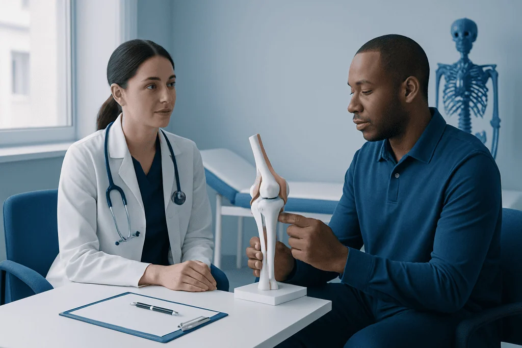
Most procedures are performed using arthroscopy, in which the surgeon places a camera and fine instruments through small incisions around the knee. Arthroscopy allows direct inspection of the cartilage and OCD lesion. In some cases, a larger incision is needed to access and reconstruct a difficult-to-reach area safely.
For a stable lesion, the surgeon may perform drilling. Small channels are made into the affected bone to encourage blood flow and healing. When the fragment is unstable but still healthy enough to preserve, it may be repositioned and fixed with pins, screws, or bioabsorbable implants. The surgeon may also prepare the underlying bone to support healing before fixation.
If the fragment is too damaged to repair or has been detached for some time, cartilage restoration may be considered. Options can include marrow-stimulation techniques for selected small defects, osteochondral grafting that transfers cartilage and bone, or cell-based cartilage procedures in carefully selected cases. The recommended approach depends on the defect’s size, depth, location, and the condition of the surrounding cartilage.
Before surgery, patients commonly have a medical review, imaging assessment, and discussion about anesthesia, rehabilitation, time away from sport or work, and realistic goals. Knee arthroscopy can be used both to assess the lesion and to carry out many OCD repair procedures.
Recovery timeline after OCD knee surgery
Recovery after osteochondritis knee surgery varies considerably because the operation may range from drilling of a stable lesion to fixation or a more extensive cartilage-restoration procedure. Early recovery focuses on controlling swelling, protecting the repair, restoring knee extension, and activating the thigh muscles safely.
Many patients use crutches initially. Weight-bearing may be restricted for several weeks after fixation or cartilage-restoration surgery, while some simpler procedures allow earlier protected walking. A brace may be used to limit motion or provide support, particularly if the repaired area needs protection during the first phase of healing.
Physiotherapy is central to recovery. Exercises are progressed from movement and muscle activation to balance, strength, and functional training. Running, jumping, pivoting, and contact sports are delayed until the surgeon and rehabilitation team confirm that symptoms, strength, motion, and healing are appropriate for a gradual return.
A typical recovery may involve daily activities becoming easier over the first several weeks, progressive strengthening over the following months, and return to high-impact sport often taking six months or longer. Follow-up appointments and, when needed, repeat imaging help the team assess bone healing and the integrity of the repaired joint surface.
How long does it take to recover from OCD knee surgery?
Recovery from OCD knee surgery commonly takes several months, and full return to running or pivoting sports may take six to twelve months depending on the procedure. Drilling for a stable juvenile lesion may have a shorter recovery than fixation of an unstable fragment or a cartilage-and-bone restoration procedure.
During the first few weeks, the knee is protected from excessive load while swelling, pain, and movement are managed. The individual then progresses through supervised rehabilitation to restore range of motion, leg strength, balance, and confidence with movement. The timeline should be guided by the operating surgeon and physiotherapist rather than by the calendar alone.
Healing of the underlying bone can take time even when the knee starts to feel better. Returning to sport too early can place stress on the repair, so clinicians usually look for a pain-free knee, minimal swelling, near-normal strength and motion, and evidence of healing when imaging is appropriate.
How long after knee cartilage surgery can you walk?
Walking after knee cartilage surgery depends on exactly what was repaired and where the lesion is located. Some patients can begin partial or protected weight-bearing soon after surgery, while others need to avoid or significantly limit weight-bearing for approximately four to eight weeks. Crutches are commonly used during this protected period.
After drilling or fixation of an OCD lesion, the surgeon may allow a gradual increase in weight-bearing as healing progresses. Procedures that reconstruct cartilage and bone may require more cautious protection. A person should follow the individualized instructions about crutches, bracing, stairs, driving, and work activities rather than testing the knee independently.
Walking comfortably does not always mean that the repair has fully healed. New or increasing swelling, pain, catching, or a feeling that the knee is giving way should be reported to the orthopedic team. Structured physical therapy and rehabilitation can help restore a safe and efficient walking pattern as restrictions are lifted.
What happens during stage 4 of osteochondritis dissecans?
Stage 4 osteochondritis dissecans generally describes an advanced lesion in which the osteochondral fragment has become detached from the underlying bone. The fragment may remain near its original location or become a loose body within the knee joint. Classification systems differ somewhat, so the treating clinician will interpret the stage together with MRI and arthroscopic findings.
A detached fragment can cause mechanical symptoms such as catching, locking, sudden pain, swelling, or reduced movement. It can also leave an area of exposed or damaged joint surface. Prompt orthopedic assessment is important because treatment may be needed to remove, repair, or restore the affected area.
Whether a stage 4 lesion can be fixed depends on the quality and size of the fragment, the condition of the cartilage and bone, and how long it has been detached. When preservation is not possible, the surgical plan may focus on restoring the defect and reducing the risk of ongoing joint damage.
Benefits, risks and expected results
The potential benefit of osteochondritis knee surgery is improved lesion healing or joint-surface restoration, with the aim of reducing pain, swelling, mechanical symptoms, and future cartilage damage. When an original fragment can be preserved and heals successfully, this may offer the best opportunity to maintain the person’s own cartilage surface.
As with any operation, there are risks. These include infection, bleeding, blood clots, anesthesia-related complications, stiffness, persistent pain or swelling, failure of the lesion to heal, implant-related problems, and need for further surgery. Cartilage damage can also increase the long-term risk of degenerative knee changes, even after appropriate treatment.
Results cannot be summarized by one reliable success rate because studies include different ages, lesion stages, surgical techniques, definitions of success, and follow-up periods. In general, outcomes tend to be more favorable when lesions are treated before extensive cartilage loss occurs, when the fragment is repairable, and when rehabilitation is completed as advised.
Acibadem International’s multidisciplinary orthopedic specialists at JCI-accredited hospitals can assess OCD lesions and coordinate surgical care and rehabilitation for international patients.
When to seek medical care
A person should arrange medical assessment for persistent knee pain, swelling after activity, limping, repeated clicking or catching, reduced knee motion, or pain that does not improve with rest. These symptoms can have several causes, and an examination helps identify whether OCD or another knee condition may be involved.
More urgent evaluation is appropriate if the knee locks, cannot fully straighten, suddenly becomes very swollen, or cannot bear weight after an injury. These symptoms may indicate a loose body, significant injury, or another problem requiring timely care.
After surgery, the patient should contact the surgical team for worsening redness, drainage, fever, calf pain or swelling, uncontrolled pain, new numbness, or a sudden decline in knee function. Attending follow-up visits is important because healing may need to be assessed even if symptoms are improving.
Frequently asked questions
What is osteochondritis knee surgery?
Osteochondritis knee surgery is an operation used to treat osteochondritis dissecans in the knee when a damaged area of bone and cartilage is unstable, detached, or unlikely to heal without surgery. The procedure may involve drilling, fixation of a fragment, removal of loose tissue, or cartilage restoration.
How long does it take to recover from OCD knee surgery?
Recovery often takes several months, while return to impact sports may take six to twelve months. The exact timeline depends on the lesion, the surgical technique, bone healing, and progress in rehabilitation.
What happens during stage 4 of osteochondritis dissecans?
Stage 4 usually means that the bone-and-cartilage fragment has detached from the underlying area. It may act as a loose body and cause locking, catching, swelling, or loss of movement, and it often requires orthopedic evaluation for possible surgical treatment.
How long after knee cartilage surgery can you walk?
Some patients begin protected walking soon after surgery, but many need crutches and limited weight-bearing for several weeks. The surgeon’s instructions vary according to the location and type of cartilage or OCD repair performed.
What is the success rate of OCD knee surgery?
There is no single success rate that applies to every OCD knee operation because outcomes differ by age, lesion stability, lesion size, cartilage damage, procedure type, and length of follow-up. Surgery can provide good results in appropriately selected patients, especially when the joint surface can be preserved and rehabilitation is completed carefully.
Can OCD return after knee surgery?
A repaired lesion may fail to heal completely or symptoms may recur, particularly if the lesion was advanced or the knee is overloaded before recovery is complete. Regular follow-up and a gradual, clinician-guided return to activity can help identify concerns early.
References
- American Academy of Orthopaedic Surgeons
- OrthoInfo – American Academy of Orthopaedic Surgeons
- American Orthopaedic Society for Sports Medicine
- National Institute of Arthritis and Musculoskeletal and Skin Diseases
- International Cartilage Regeneration & Joint Preservation Society
This article is for general information only and is not a substitute for professional medical advice. Please consult a qualified doctor about your individual situation.
Joint and spine care in Turkey — expert assessment & treatment
JCI-accredited · board-certified surgeons · reply within 24h
Add us as a Preferred Source to see more of our trusted health content across Google Search, AI Overviews and Discover.