Palinopsia: A Complete Medical Overview

Palinopsia means seeing persistent or recurring images after the original visual stimulus is gone. It can occur with migraine, certain medications, seizures, head injury, or structural changes affecting visual pathways.
Key Takeaways
- Palinopsia means seeing persistent or recurring images after the original visual stimulus is gone.
- It can occur with migraine, certain medications, seizures, head injury, or structural changes affecting visual pathways.
- A careful history, eye exam, and sometimes brain imaging help identify the underlying cause.
- Treatment focuses on the reason for the symptom rather than the afterimage alone.
- New, sudden, or worsening palinopsia should be assessed by a qualified doctor, especially if it occurs with headache or neurological symptoms.
Palinopsia is a visual symptom in which an image continues, repeats, or reappears after the original object is no longer in view. It is not a disease itself, but a sign that may be related to migraine, medication effects, seizures, eye or brain conditions, so proper evaluation is important.
Overview: what palinopsia means
Palinopsia is a visual symptom in which a person continues to see an image after the object has disappeared, or sees that image return later without the object being present. People may describe this as a lingering afterimage, a repeated visual scene, or a “trail” following movement. The experience can affect one or both eyes and may happen occasionally or more often, depending on the cause.
Importantly, palinopsia is not a diagnosis by itself. It is a clue that the visual system is being affected somewhere along the pathway from the eyes to the brain. In some people it is linked to migraine or medication effects. In others, it may be related to seizures, head trauma, metabolic problems, or structural changes in the parts of the brain that process vision.
Doctors often separate palinopsia into two broad patterns. One pattern involves long-lasting, clearer recurrences of a previous image. Another involves less distinct effects such as prolonged afterimages, light streaking, visual trailing, or repeated outlines. This distinction can help guide the medical evaluation, although patients do not need to identify the subtype themselves before seeking care.
How palinopsia can look and feel
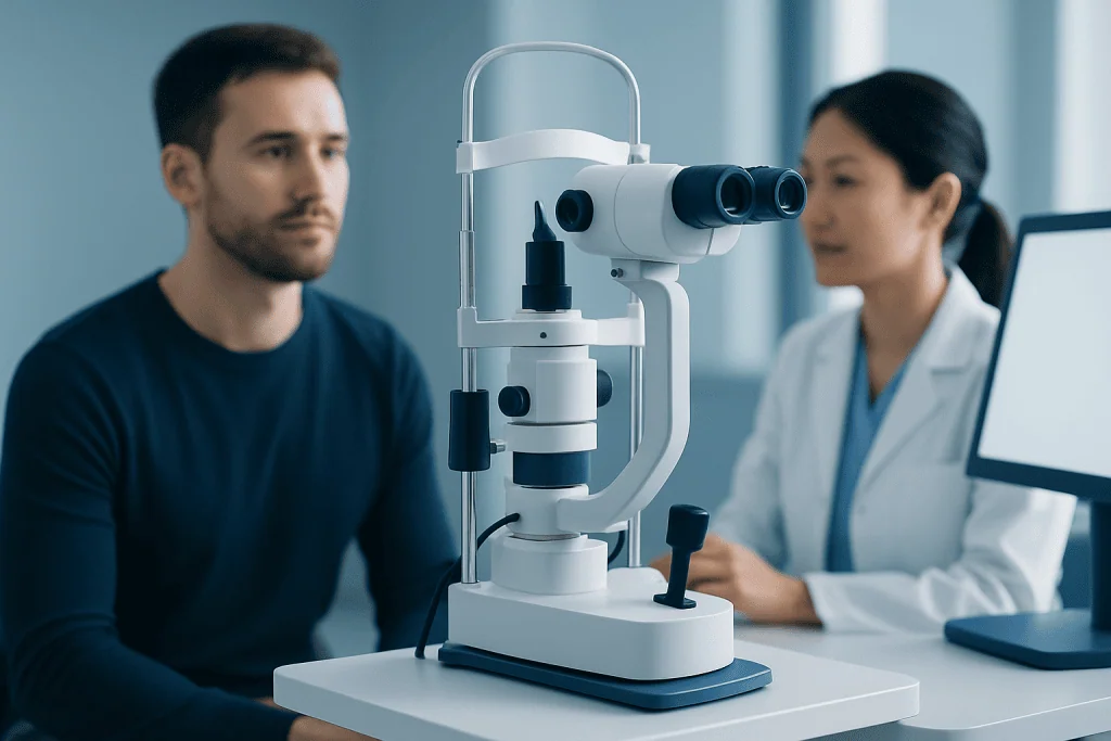
Palinopsia can appear in different ways. Some people notice that an object leaves a persistent visual impression for longer than expected after they look away. Others see copies of a moving object, repeated shapes, or a recently viewed image appearing again in a different part of the visual field. Symptoms may be more noticeable in bright light, with high-contrast patterns, or when a person is tired.
The exact experience can vary from mild and brief to disruptive. A person may have trouble reading, driving, using screens, or following moving objects. Palinopsia can also be unsettling because it changes how the environment appears, even when the eyes themselves seem otherwise normal.
Symptoms sometimes occur together with other visual changes or neurological symptoms, such as:
- Blurred vision or difficulty focusing
- Sensitivity to light
- Flashing lights or visual aura
- Headache, especially migraine-type headache
- Dizziness or imbalance
- Numbness, weakness, or changes in speech
When symptoms are accompanied by other neurological features, doctors may consider conditions affecting the brain’s visual processing centers. For example, palinopsia may sometimes appear alongside symptoms seen in migraine or other neurological disorders that need a structured assessment.
Common causes and risk factors

Palinopsia can have several possible causes. One of the more common associations is migraine, particularly migraine with visual aura. In these cases, the symptom may happen before, during, after, or even between headache episodes. Some people develop persistent visual disturbances related to migraine even when pain is not the main issue.
Medications can also play a role. Certain drugs that affect the brain or visual processing have been linked to afterimages and visual trailing in some patients. Illicit substances and withdrawal states may produce similar effects. Because of this, a complete medication and substance history is an important part of evaluation, including recently started, stopped, or dose-adjusted treatments.
Other causes include seizures arising from the visual cortex, head injury, stroke, brain tumors, inflammation, and metabolic disturbances. Sometimes the issue comes from the eye or retina, while in other cases the eye exam is normal and the problem lies in the nervous system. Rarely, palinopsia may occur in the setting of psychological distress, but organic causes are usually considered first.
Risk factors depend on the underlying condition rather than palinopsia itself. They may include a personal history of migraine, epilepsy, recent concussion, use of certain psychoactive or neurological medications, prior stroke, or known eye disease. Because the range of causes is broad, new symptoms should not be self-diagnosed.
How doctors evaluate palinopsia
Diagnosis begins with a detailed history. The doctor will ask what the images look like, how long they last, whether they occur after movement or bright light, and whether they affect one eye or both. It is also helpful to describe when the problem started, how often it happens, and whether there are triggers such as headache, sleep loss, medication changes, or recent illness or injury.
A physical examination usually includes a full eye assessment and a neurological examination. Vision, pupils, eye movements, visual fields, and the health of the retina and optic nerve may be checked. If symptoms suggest a neurological rather than purely eye-related cause, referral to neurology, neuro-ophthalmology, or both may be appropriate.
Further tests depend on the clinical picture. These may include visual field testing, blood tests, electroencephalography if seizures are suspected, or brain imaging such as MRI scanning to look at the visual pathways and surrounding brain structures. Imaging is especially important when symptoms are sudden, focal, progressive, or associated with other neurological changes.
Because some serious conditions can affect vision, doctors aim to identify whether palinopsia is benign and self-limited or a sign of a treatable underlying disorder. The goal is not only symptom relief, but also protection of neurological and visual health.
Treatment options and symptom management
There is no single treatment that fits all cases of palinopsia. Care is directed at the underlying cause. If migraine is responsible, migraine management may reduce symptoms over time. If a medication is contributing, the prescribing doctor may consider adjusting the treatment plan. If seizures, inflammation, or a structural brain condition are found, treatment focuses on those disorders.
Some patients benefit from environmental adjustments while the evaluation is underway. Reducing glare, limiting prolonged screen exposure, improving sleep, and avoiding known migraine triggers may help lessen the impact of symptoms. Tinted lenses or carefully selected visual aids are occasionally suggested by specialists when light sensitivity or contrast effects are prominent.
When an underlying neurological cause is suspected or confirmed, a patient may be referred for more targeted care, such as neurology evaluation or additional tests to clarify the diagnosis. If seizures are part of the picture, management may involve a structured review by epilepsy specialists. When migraine or other brain-related visual symptoms are persistent, coordinated care between ophthalmology and neurology is often helpful.
In selected cases, further assessment of the brain and nervous system may include advanced imaging or consultation in services such as brain MRI pathways, depending on the person’s symptoms and examination findings. Near the end of the care pathway, Acibadem International’s multidisciplinary specialists and JCI-accredited hospitals evaluate and treat visual and neurological conditions in international patients when this is clinically appropriate.
Self-care, daily adjustments, and prevention
Prevention depends on the reason palinopsia is happening. For people with migraine, maintaining regular sleep, hydration, meals, and stress management may reduce attacks and visual symptoms. Avoiding known personal triggers, such as bright flickering light, alcohol, or skipped meals, can also be useful when these factors are relevant.
If symptoms began after starting a new medicine, it is important not to stop treatment abruptly without medical advice. Instead, the person should speak with the prescribing clinician promptly. The doctor can review whether the timing suggests a medication effect and whether safer alternatives or changes are possible.
At home, practical steps may make symptoms easier to cope with:
- Use softer indoor lighting and reduce glare from windows or screens
- Take regular breaks during reading or computer work
- Keep a symptom diary noting triggers, duration, and associated features
- Avoid driving or hazardous tasks if vision feels unreliable
- Follow treatment plans for migraine, epilepsy, or other known conditions
While these steps may improve comfort, they do not replace medical assessment. Persistent or unexplained visual symptoms should still be discussed with a qualified doctor.
When to seek medical care
Palinopsia should be medically assessed when it is new, recurrent, or interfering with daily life. A person should arrange timely care if symptoms are becoming more frequent, lasting longer, or appearing together with headache, dizziness, confusion, or visual field loss. Evaluation is also advisable after a head injury or after starting or changing a medication.
Urgent medical attention is important if palinopsia begins suddenly and is accompanied by weakness, numbness, trouble speaking, severe headache, seizure-like activity, loss of consciousness, or a major change in vision. These features can signal a more serious neurological problem that should not be delayed.
Patients may start with an eye doctor, neurologist, or primary care physician depending on local access and associated symptoms. In some situations, coordinated work-up for visual pathway disorders may overlap with evaluation for conditions such as brain tumor or other causes of neurological visual symptoms, although many cases turn out to be related to less dangerous explanations such as migraine or medication effects.
Frequently asked questions
Is palinopsia an eye disease?
Palinopsia is a symptom, not a disease by itself. It can result from problems involving the eyes, the brain’s visual pathways, migraine, medication effects, or other neurological conditions.
Can migraine cause palinopsia?
Yes. Migraine, especially migraine with aura, is one of the better-known causes of palinopsia and other visual disturbances. In some people, visual symptoms may happen even when headache is mild or absent.
What is the difference between normal afterimages and palinopsia?
Brief afterimages after looking at a bright light can be normal. Palinopsia usually refers to afterimages or recurring images that are unusually persistent, repetitive, or disruptive, and may occur without a typical visual trigger.
Can medications trigger palinopsia?
Yes, some medications can contribute to visual trailing or persistent afterimages in certain people. A doctor will review current and recent medicines carefully before deciding whether they may be involved.
Will palinopsia go away on its own?
Sometimes it improves when the trigger is temporary, such as a migraine episode or a reversible medication effect. However, because it can also signal a condition needing treatment, persistent or new palinopsia should be evaluated rather than watched indefinitely.
What tests are usually needed for palinopsia?
The work-up often starts with a medical history, eye examination, and neurological examination. Depending on the symptoms, doctors may recommend visual field testing, brain imaging, or tests for seizures or other underlying causes.
References
- American Academy of Ophthalmology
- National Eye Institute
- National Institute of Neurological Disorders and Stroke
- Merck Manual Professional Edition
- Mayo Clinic
This article is for general information only and is not a substitute for professional medical advice. Please consult a qualified doctor about your individual situation.
Explore treatments in Turkey — costs, top hospitals & a free quote
JCI-accredited · board-certified surgeons · reply within 24h
Add us as a Preferred Source to see more of our trusted health content across Google Search, AI Overviews and Discover.