Panorex: An Evidence-Based Guide for Patients
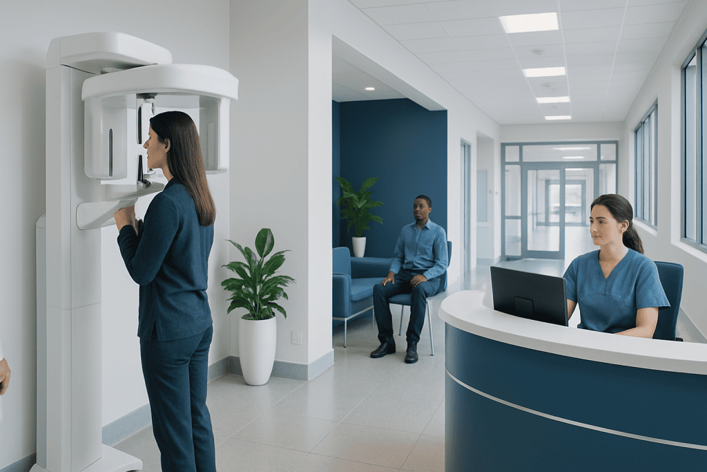
A Panorex provides a wide, two-dimensional overview of the teeth, jaws and surrounding facial structures. Dentists may use it to assess impacted teeth, tooth development, jawbone changes, infections, cysts and treatment needs.
Key Takeaways
- A Panorex provides a wide, two-dimensional overview of the teeth, jaws and surrounding facial structures.
- Dentists may use it to assess impacted teeth, tooth development, jawbone changes, infections, cysts and treatment needs.
- The examination does not require needles or contrast material and usually takes only a few minutes.
- Panoramic X-rays use ionizing radiation, so they are requested only when the expected diagnostic benefit is useful.
- A Panorex complements, rather than replaces, a clinical dental examination and other targeted dental images when needed.
A Panorex, also called a panoramic dental X-ray or panoramic radiograph, is an imaging test that captures the teeth, upper and lower jaws, jaw joints and nearby facial structures in one broad image. It is quick, painless and commonly used by dentists and oral health specialists to identify concerns that may not be visible during a routine mouth examination.
What Is a Panorex?
A Panorex is a common name for a panoramic dental X-ray, also known as a panoramic radiograph. It produces one wide image of the mouth and face, including the upper and lower teeth, the jawbones, tooth roots, areas beneath the gums, the temporomandibular joints (TMJs), and parts of the nasal and sinus regions. The image gives a dentist a broad view that cannot be obtained from looking inside the mouth alone.
Unlike small dental X-rays that focus on a few teeth at a time, a panoramic image is created while the X-ray unit moves in an arc around the patient’s head. The result is a two-dimensional overview rather than a highly detailed image of every individual tooth. For this reason, it is often used as part of an assessment, while close-up X-rays or other imaging may be added if a specific area needs closer evaluation.
“Panorex” is often used informally to describe the examination, although panoramic radiograph is the general clinical term. The test is performed in dental clinics, oral and maxillofacial radiology services, and some hospital imaging departments.
What Can a Panoramic Dental X-Ray Show?
A Panorex can help a clinician evaluate the number, position and general condition of the teeth. It is particularly useful for seeing teeth that have not erupted through the gums, including wisdom teeth, and for checking whether developing teeth are present and positioned as expected. It may also show retained baby teeth, missing teeth, extra teeth, or roots with unusual shapes.
The image can reveal changes in the jawbone around tooth roots, larger areas of bone loss, signs that may be associated with infection, and some cysts or other jaw lesions. It can also help assess the jaw before procedures such as tooth removal, orthodontic treatment, dental implants or oral surgery. When a suspicious finding is seen, the dentist may recommend a targeted X-ray, cone-beam computed tomography (CBCT), clinical examination, or referral to an appropriate specialist.
- Impacted, unerupted or displaced teeth
- Wisdom tooth position and relationship to nearby structures
- General jawbone shape, symmetry and larger bone changes
- Some dental infections, cysts and abnormalities around tooth roots
- Tooth development in children and adolescents
- Planning information for orthodontic, implant or surgical care
A panoramic image is valuable for overview and planning, but it does not diagnose every dental problem. Very early tooth decay, subtle gum disease changes and fine fractures may be better assessed with other types of dental imaging and a direct examination.
Why a Dentist May Recommend a Panorex
A dentist may recommend a Panorex when a comprehensive view of the mouth and jaws is likely to help answer a clinical question. It may be taken during an initial assessment when a person has complex dental needs, before certain procedures, or when symptoms suggest a concern beyond a single tooth. The decision should be based on the person’s age, symptoms, oral health history and previous imaging.
For example, panoramic X-rays are frequently used to assess wisdom teeth before removal, especially if their roots appear close to important structures in the lower jaw. They may also support orthodontic planning by showing the development and position of permanent teeth. In people considering dental implants, a panoramic image can offer an initial view of available jawbone and important anatomical landmarks, although more detailed imaging may sometimes be needed.
Jaw pain, facial swelling, difficulty opening the mouth, unexplained tooth movement or a suspected jaw lesion can also lead a clinician to request panoramic imaging. A Panorex does not determine the cause of every symptom by itself. Its findings are interpreted together with the patient’s history, examination and, where appropriate, additional tests.
What Happens During the Examination?
A panoramic dental X-ray is usually simple and comfortable. Before the scan, the patient may be asked to remove glasses, earrings, necklaces, removable dentures, hearing devices or other metal objects around the head and neck. These items can create shadows or obscure parts of the image.
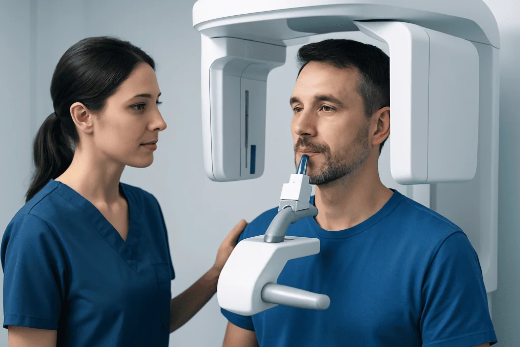
The patient stands or sits in the machine and gently bites on a small positioning guide. A radiographer or trained dental professional helps place the head correctly, often using light guides. It is important to remain still and keep the tongue positioned as instructed while the X-ray arm rotates around the head. The equipment does not normally touch the face during its movement.
The actual scan typically lasts less than a minute, while preparation and positioning may take a few minutes. It is painless and does not involve injections, sedation or recovery time. Normal activities, eating and drinking can continue immediately afterward unless the person is having another procedure at the same appointment.
Patients should tell the dental team about pregnancy, a possible pregnancy, recent dental imaging, or any difficulty standing still or following positioning instructions. These details help the team determine whether the examination is needed and how it can be performed safely.
Radiation Safety and the Limits of the Image
Like other X-ray examinations, a Panorex uses ionizing radiation. Dental professionals follow the principle of using the lowest radiation exposure reasonably achievable while obtaining the information needed for care. Modern digital panoramic systems are designed to use lower exposures than older film-based systems, but an X-ray should still be taken only when it is clinically justified.
Clinicians review whether useful recent images already exist before requesting new ones. The right type and frequency of imaging vary from person to person; there is no single schedule that suits everyone. Children may need imaging for development or treatment planning, but their assessment should account for their individual needs and sensitivity to radiation.
Pregnancy does not automatically mean that all dental X-rays must be avoided. If imaging is necessary for diagnosis or urgent treatment, the dental team can assess the situation and use appropriate protective practices. A patient who is pregnant or thinks they may be pregnant should mention this before the scan.
A Panorex has technical limitations. Because it is a broad, two-dimensional image, structures can appear enlarged, overlapped or slightly distorted. It may not show fine details as clearly as intraoral X-rays, and it may not provide the three-dimensional information needed for certain surgical or implant decisions. Further imaging is recommended only when it is expected to change or improve care.
Understanding the Results and Next Steps
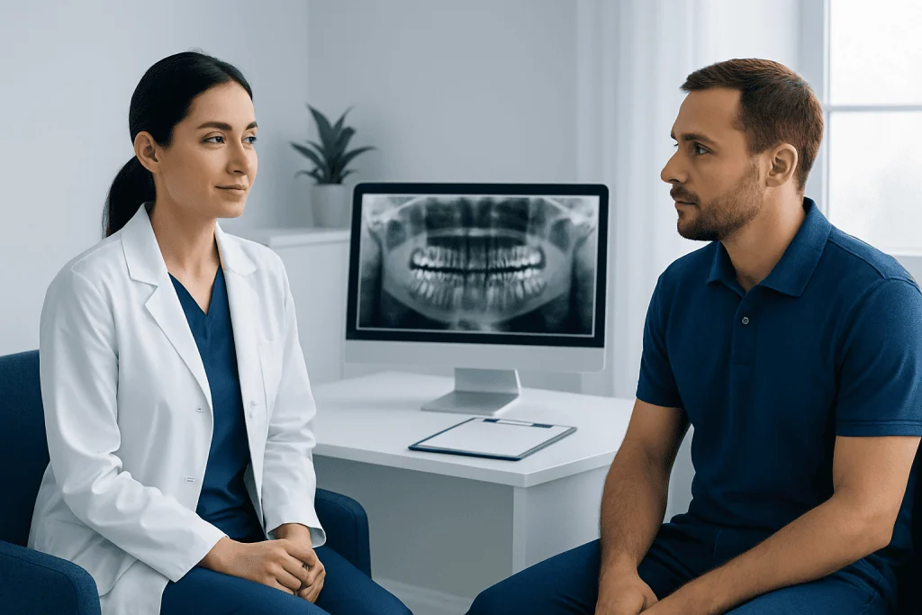
A dentist, oral and maxillofacial radiologist, oral surgeon or another qualified clinician reviews the image. They consider both the image quality and what is visible within the context of symptoms, dental history and findings from the mouth examination. In many clinics, preliminary results may be discussed at the same visit, although formal review and care planning can take longer.
A normal or reassuring Panorex does not necessarily rule out all causes of dental discomfort. Small cavities, early gum changes, cracks in teeth, and some soft-tissue conditions may not be apparent on a panoramic image. Conversely, an area that looks unusual on a Panorex does not always indicate a serious problem; additional views may be needed to clarify what it represents.
If the image identifies an impacted tooth, infection, bone change or another concern, the next step depends on the finding. Options may include monitoring, preventive dental care, focused X-rays, referral to an endodontist, orthodontist or oral surgeon, or treatment of the underlying condition. Patients can ask what the image shows, whether more imaging is necessary, and how the result affects the proposed treatment plan.
For international patients who need coordinated dental and medical assessment, Acibadem International’s multidisciplinary specialists and JCI-accredited hospitals can support diagnosis and treatment planning where appropriate.
When to Seek Medical Care
A Panorex is not usually an emergency test, but prompt dental assessment is important when symptoms suggest an infection or significant dental problem. A person should contact a dentist soon for persistent toothache, swelling of the gums or face, a tooth that has become loose without explanation, pain around a wisdom tooth, difficulty chewing, or a jaw that feels increasingly stiff or painful.
Urgent medical or dental care is needed for facial swelling accompanied by fever, trouble swallowing, difficulty breathing, confusion, severe weakness, or rapidly worsening pain. These symptoms can indicate a potentially serious infection that requires timely assessment. Emergency services should be used if breathing is affected or the person cannot swallow saliva.
People should also arrange a dental review after a facial or jaw injury, especially if the bite suddenly feels different, teeth are displaced, there is numbness of the lip or chin, or opening and closing the mouth is difficult. Imaging may be part of the assessment, but the appropriate test depends on the nature of the injury and the clinician’s examination.
Frequently asked questions
Is a Panorex the same as a regular dental X-ray?
A Panorex is a type of dental X-ray, but it differs from small intraoral X-rays. It captures a broad view of both jaws and surrounding structures in one image, while intraoral X-rays usually provide more detailed views of individual teeth and the bone around them. A dentist may use one or both types depending on the clinical question.
Does a Panorex hurt?
No. A panoramic dental X-ray is painless and does not involve needles, contrast dye or instruments placed deep inside the mouth. The patient only needs to stay still and bite gently on a positioning guide while the machine moves around the head.
How long does a Panorex take?
The scan itself generally takes less than a minute. Allowing time for removing metal items, positioning and checking the image, the appointment portion is usually only a few minutes. There is no recovery period afterward.
Can a Panorex show cavities?
A Panorex may show some larger or advanced areas of tooth decay, but it is not the most sensitive test for detecting early cavities between teeth. Dentists often use bitewing or other close-up dental X-rays when evaluating possible decay. Clinical examination remains important as well.
Is a panoramic dental X-ray safe during pregnancy?
Dental imaging is considered only when it is needed for diagnosis or treatment planning, including during pregnancy. A patient should always tell the dental team if they are pregnant or may be pregnant so the need for imaging can be reviewed and appropriate protective measures can be used. Delaying necessary care may not always be the safest option.
Why might I need another scan after a Panorex?
A Panorex provides a useful overview, but its broad two-dimensional format can limit fine detail and depth assessment. If an area needs closer evaluation, a dentist may recommend a targeted dental X-ray or, in selected situations, three-dimensional imaging. Additional imaging should have a clear clinical purpose.
References
- American Dental Association
- U.S. Food and Drug Administration
- International Atomic Energy Agency
- Radiological Society of North America
- American Academy of Oral and Maxillofacial Radiology
This article is for general information only and is not a substitute for professional medical advice. Please consult a qualified doctor about your individual situation.
Joint and spine care in Turkey — expert assessment & treatment
JCI-accredited · board-certified surgeons · reply within 24h
Add us as a Preferred Source to see more of our trusted health content across Google Search, AI Overviews and Discover.