Papilledema: Swollen Optic Nerves, Raised Brain Pressure, and Urgent Warning Signs

Papilledema means the optic nerve is swollen because pressure inside the skull is too high. It can cause headaches, brief vision changes, blurred vision, nausea, or double vision, but some people have few symptoms at first.
Key Takeaways
- Papilledema means the optic nerve is swollen because pressure inside the skull is too high.
- It can cause headaches, brief vision changes, blurred vision, nausea, or double vision, but some people have few symptoms at first.
- Diagnosis often involves an eye examination, brain imaging, and sometimes a lumbar puncture to measure pressure.
- Treatment focuses on lowering intracranial pressure and treating the underlying cause.
- Sudden vision loss, severe headache, vomiting, confusion, or neurological symptoms need urgent medical care.
Papilledema is swelling of the optic nerve caused by increased pressure inside the skull. It is not a disease by itself, but an important warning sign that needs prompt medical evaluation to protect vision and identify the underlying cause.
Overview
Papilledema is swelling of the optic disc, the visible front part of the optic nerve at the back of the eye. This swelling happens when pressure inside the skull, also called intracranial pressure, becomes elevated and is transmitted to the optic nerve. Because the optic nerve connects the eye to the brain, papilledema is considered a neurological and ophthalmic warning sign rather than a separate disease.
It can affect one or both eyes, although it is usually seen in both. In some people, vision remains normal early on, while in others there may be blurred vision, brief episodes of visual dimming, or other symptoms related to raised pressure in the brain. Without timely treatment, ongoing pressure may damage the optic nerve and lead to lasting visual loss.
Papilledema should always be assessed promptly by a qualified doctor. The main goal is to find out why the pressure is high. Causes may range from conditions such as idiopathic intracranial hypertension to more urgent problems involving bleeding, infection, inflammation, or a mass inside the skull. Care often involves specialists in eye health, neurology, and brain imaging.
Symptoms of Papilledema
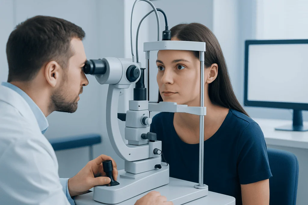
The symptoms of papilledema can vary depending on how high the intracranial pressure is, how long it has been present, and what is causing it. Some people notice symptoms related to vision, while others mainly experience headache or other general symptoms. In a number of cases, papilledema is first discovered during an eye examination before major visual symptoms appear.
Common symptoms may include:
- Headache, often worse in the morning or when coughing, straining, or bending
- Brief episodes of blurred or dim vision, sometimes lasting only seconds
- Persistent blurred vision
- Double vision
- Nausea or vomiting
- A whooshing sound in the ears that follows the heartbeat
- Difficulty with side vision if optic nerve damage develops
Visual symptoms may come and go at first. A person may describe momentary graying out of vision when standing up quickly or changing posture. Over time, if pressure remains high, visual field loss can become more noticeable and may not fully recover if treatment is delayed.
The underlying cause of the raised pressure may also produce additional symptoms. For example, fever and neck stiffness may suggest infection, while weakness, confusion, seizures, or speech difficulty may point to a more serious neurological problem that needs immediate assessment.
Causes and Risk Factors

Papilledema happens because intracranial pressure rises and affects the optic nerves. Many different conditions can lead to this pressure increase. Identifying the cause is essential, because treatment depends on the underlying problem.
Possible causes include:
- Idiopathic intracranial hypertension, a condition of raised pressure without an obvious structural cause
- Brain tumors or other masses
- Bleeding inside the skull
- Brain swelling after injury
- Meningitis or other infections of the brain and surrounding tissues
- Inflammatory disorders
- Hydrocephalus, where cerebrospinal fluid builds up
- Blood clots in the veins that drain the brain
- Certain medicines in some cases
One of the better-known associations is idiopathic intracranial hypertension, which often affects younger adults and is more common in people with obesity. However, papilledema should never be assumed to come from this cause alone without proper evaluation, because more urgent conditions can look similar.
Risk factors depend on the cause. Recent head injury, a history of clotting disorders, active infection, some medications, and conditions that alter cerebrospinal fluid flow may increase the likelihood of raised intracranial pressure. Even so, papilledema can occur in people without obvious risk factors, which is why a thorough medical assessment is so important.
How Papilledema Is Diagnosed
Diagnosis begins with a careful history and examination. A doctor will ask about headaches, visual symptoms, recent weight changes, medications, infections, neurological symptoms, and past medical conditions. The eye examination is especially important. Looking at the back of the eye may reveal a swollen optic disc with blurred margins, and additional tests can help assess how the swelling is affecting vision.
Common eye-related tests may include visual acuity testing, visual field testing, and imaging of the optic nerve and retina. Some people are referred to a specialist in neuro-ophthalmology evaluation when the diagnosis is uncertain or when both eye and neurological findings need to be interpreted together.
Because papilledema suggests raised pressure in the skull, brain imaging is often needed urgently. This may include MRI or, in some situations, other imaging to look for a mass, swelling, hydrocephalus, or venous sinus thrombosis. After imaging has excluded conditions that make it unsafe, a lumbar puncture may be performed to measure cerebrospinal fluid pressure and analyze the fluid for clues such as infection or inflammation.
Doctors also consider other causes of optic disc swelling that are not true papilledema, such as optic neuritis, ischemic optic neuropathy, or congenital disc appearance differences. This distinction matters because the treatment and urgency can be very different.
Treatment Options
Treatment for papilledema focuses on two goals: lowering intracranial pressure and treating the cause. The right approach depends on what is found during the evaluation. Because untreated papilledema can threaten vision, follow-up is often close and may involve repeat eye examinations and visual field testing.
If papilledema is due to idiopathic intracranial hypertension, treatment may include weight management when appropriate, medicines that reduce cerebrospinal fluid production, and regular monitoring of vision. If a medication is contributing, the doctor may review whether it should be adjusted or stopped. When the cause is infection, a blood clot, bleeding, or inflammation, treatment is directed specifically at that condition.
In some situations, procedures or surgery are needed to protect vision or reduce pressure. Depending on the cause, this may involve neurosurgical treatment to remove a mass, relieve hydrocephalus, or address another structural problem. When vision is at immediate risk, doctors may discuss additional interventions to relieve pressure around the optic nerve or divert cerebrospinal fluid.
People with significant headaches or nausea may also receive supportive treatment, but symptom relief alone is not enough. The main priority is identifying and correcting the reason for the raised pressure. At centers with multidisciplinary care, specialists in neurology, ophthalmology, radiology, and neurosurgery work together to plan treatment.
Prevention and Self-care
There is no single way to prevent papilledema because it is a sign of many different medical conditions. The most helpful step is early recognition of symptoms and prompt assessment. Ongoing headaches with visual changes should not be ignored, especially if symptoms are worsening or recurring.
For people with idiopathic intracranial hypertension, following the care plan may help reduce pressure and protect vision. This may include attending regular follow-up visits, monitoring changes in headaches or eyesight, and discussing healthy weight management strategies with a healthcare professional if advised.
General self-care can support overall neurological and eye health:
- Take prescribed medicines exactly as directed
- Report new or worsening visual symptoms promptly
- Keep scheduled eye and neurology appointments
- Avoid stopping important medicines without medical advice
- Seek medical review if severe headache, vomiting, or neurological symptoms develop
It is important not to self-diagnose papilledema based on symptoms alone. Headache and blurred vision can occur with many conditions, including migraine and glaucoma, but papilledema specifically requires examination of the optic nerve and often brain imaging to confirm the cause.
When to See a Doctor
Papilledema is a reason for urgent medical evaluation because it can signal a serious increase in pressure inside the skull. A person should seek prompt care if there is a new severe headache, repeated vomiting, blurred or dim vision, double vision, or a whooshing sound in the ears together with headache or visual symptoms.
Emergency care is especially important if symptoms come with confusion, fainting, seizures, weakness, numbness, trouble speaking, fever, or a recent head injury. Sudden or progressive vision loss should never be delayed, even if it improves briefly between episodes.
After diagnosis, regular follow-up is essential because papilledema can change over time. Vision may worsen even if headaches improve, so repeat eye examinations and visual field testing help guide treatment decisions. Near the end of the care pathway, some international patients may choose evaluation at centers such as Acibadem International, where multidisciplinary specialists and JCI-accredited hospitals diagnose and treat papilledema and its underlying causes.
Prompt attention offers the best chance of preserving vision and addressing the reason pressure is elevated. Anyone who suspects papilledema or has concerning neurological or visual symptoms should consult a qualified doctor without delay.
Frequently asked questions
Is papilledema an emergency?
Papilledema needs urgent medical evaluation because it can be a sign of raised pressure inside the skull. Not every cause is life-threatening, but some are serious and require prompt treatment to protect vision and brain health.
Can papilledema cause permanent vision loss?
Yes, it can if the pressure remains high long enough to damage the optic nerve. Early diagnosis and treatment greatly improve the chances of preserving vision.
What does papilledema feel like?
Papilledema itself may not cause pain in the eye, but many people have headaches, nausea, or brief episodes of blurred or dim vision. Some people also notice double vision or a pulsing sound in the ears.
How is papilledema different from other optic nerve swelling?
Papilledema specifically means optic disc swelling caused by increased intracranial pressure. Other conditions can also swell the optic nerve, such as inflammation or poor blood flow, but they have different causes and treatments.
Does papilledema always mean a brain tumor?
No. A brain tumor is only one possible cause of raised intracranial pressure. Papilledema can also occur with idiopathic intracranial hypertension, infection, bleeding, hydrocephalus, or blood clots in the brain's veins.
What tests are usually done for papilledema?
Doctors usually perform a detailed eye examination and assess vision, including visual fields. Brain imaging is commonly needed, and a lumbar puncture may be done after imaging to measure cerebrospinal fluid pressure and look for the cause.
References
- National Eye Institute
- American Academy of Ophthalmology
- National Institute of Neurological Disorders and Stroke
- Mayo Clinic
- Merck Manual Professional Edition
This article is for general information only and is not a substitute for professional medical advice. Please consult a qualified doctor about your individual situation.
Explore treatments in Turkey — costs, top hospitals & a free quote
JCI-accredited · board-certified surgeons · reply within 24h
Add us as a Preferred Source to see more of our trusted health content across Google Search, AI Overviews and Discover.