Papilledema: Symptoms, Causes, and Why Urgent Evaluation Matters

Papilledema is not a disease itself but a sign of raised intracranial pressure. Symptoms may include headache, brief vision changes, nausea, double vision, or ringing in the ears.
Key Takeaways
- Papilledema is not a disease itself but a sign of raised intracranial pressure.
- Symptoms may include headache, brief vision changes, nausea, double vision, or ringing in the ears.
- Urgent evaluation is needed to identify the cause and prevent vision loss or complications.
- Diagnosis usually involves an eye examination, brain imaging, and sometimes a lumbar puncture.
- Treatment focuses on lowering pressure and addressing the underlying condition.
Papilledema is swelling of the optic disc caused by increased pressure inside the skull. Because it can signal a serious underlying problem and may threaten vision, prompt medical assessment is important.
Overview
Papilledema is swelling of the optic disc, the point where the optic nerve enters the back of the eye. This swelling happens when pressure inside the skull rises and is transmitted along the optic nerve. Papilledema usually affects both eyes, although symptoms may not always feel the same on each side.
It is important to understand that papilledema is a clinical sign rather than a diagnosis by itself. In other words, it tells doctors that something is increasing intracranial pressure, and the reason for that pressure must be found. Causes can range from conditions that are treatable to problems that require urgent specialist care.
Some people with papilledema notice headaches or temporary visual dimming, while others have few symptoms and are diagnosed during an eye examination. Even when symptoms seem mild, papilledema should not be ignored because ongoing pressure can damage the optic nerve and affect vision over time.
Symptoms of Papilledema
Symptoms of papilledema often reflect both the swelling of the optic nerve and the raised pressure inside the skull. A common complaint is headache, which may be worse in the morning, with coughing, or when bending over. Some people also feel nausea or may vomit, especially if intracranial pressure is significantly elevated.
Vision symptoms can be subtle at first. A person may experience brief episodes of blurred or dim vision lasting seconds, often when standing up, changing position, or straining. Double vision can also occur, especially if increased pressure affects the nerves that control eye movement.
Other possible symptoms include pulsatile tinnitus, a whooshing sound in the ears that follows the heartbeat, and difficulty with peripheral vision as pressure persists. In some cases, visual acuity remains normal early on, which is why a normal reading chart result does not rule out papilledema.
- Headache
- Brief episodes of blurred or darkened vision
- Double vision
- Nausea or vomiting
- Pulsatile tinnitus
- Progressive loss of side vision in more advanced cases
Causes and Risk Factors

Papilledema develops when intracranial pressure rises for any reason. Important causes include brain tumors, bleeding in or around the brain, head injury, hydrocephalus, infections such as meningitis, inflammation, or impaired drainage of cerebrospinal fluid. Blood clots in the veins that drain the brain can also lead to increased pressure and papilledema.
One well-known cause is idiopathic intracranial hypertension, a condition in which pressure around the brain rises without an obvious structural cause on imaging. This condition is more common in women of childbearing age and is often associated with obesity, although it can affect other groups as well. In that setting, papilledema may be found alongside headaches and transient visual symptoms.
Certain medicines are also linked to raised intracranial pressure in some people. Examples may include vitamin A derivatives, tetracycline-class antibiotics, growth hormone, or steroid withdrawal. Doctors will also consider conditions that can mimic papilledema, such as optic neuritis or pseudopapilledema caused by optic disc drusen, because these require different management.
How Papilledema Is Diagnosed
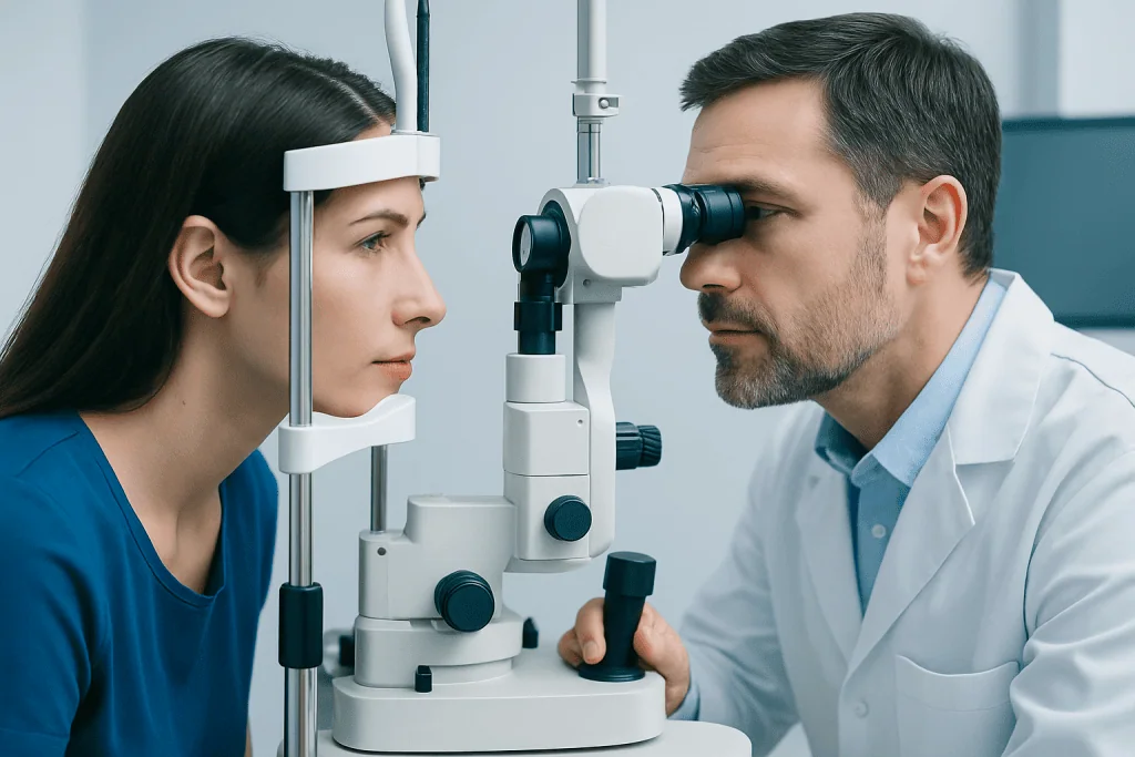
Diagnosis begins with a careful history and examination. An eye doctor or neuro-ophthalmology specialist examines the optic discs after dilating the pupils and looks for signs of swelling. They may also assess visual acuity, color vision, pupil responses, eye movements, and the visual field to understand whether the optic nerve is being affected.
If papilledema is suspected, urgent brain imaging is usually the next step to look for a cause of raised pressure and to make sure it is safe to proceed with further testing. Depending on the situation, this may involve MRI or CT imaging. Imaging can help identify masses, bleeding, hydrocephalus, or venous sinus problems.
After imaging, some patients need a lumbar puncture to measure cerebrospinal fluid pressure and check the fluid for infection, inflammation, or other abnormalities. Additional tests may be used to document the eyes in detail, such as optical coherence tomography, fundus photography, or formal visual field testing. Because the causes vary, diagnosis often involves coordination between ophthalmology, neurology, neurosurgery, and radiology.
Treatment Options
Treatment for papilledema is aimed at the underlying cause and at reducing pressure quickly enough to protect vision and brain function. The approach depends entirely on why pressure is elevated. For example, infections may require urgent antimicrobial treatment, bleeding or a mass may need neurosurgical care, and a clot in the brain’s venous drainage system may call for specific medical treatment.
When papilledema is related to idiopathic intracranial hypertension, treatment may include weight management, medicines that reduce cerebrospinal fluid production, and close monitoring of vision. In selected cases where pressure remains high or vision is worsening, procedures may be considered, such as cerebrospinal fluid shunting or optic nerve sheath fenestration. If a structural problem is found, a patient may be referred for neurosurgical treatment when appropriate.
Regular follow-up is important because symptoms alone do not always show whether the optic nerves are improving or deteriorating. Repeat eye examinations and visual field tests help guide treatment decisions. If papilledema is linked to another neurological condition, the care team may also evaluate for related disorders such as brain tumors when suggested by symptoms and imaging findings.
Prevention and Self-Care
Papilledema itself cannot always be prevented, because it is a sign of many different conditions. However, early recognition can make a major difference. Anyone with persistent headache, unexplained visual changes, or double vision should seek medical advice rather than trying to manage symptoms alone.
For people diagnosed with idiopathic intracranial hypertension, following the treatment plan closely is an important part of self-care. This may include attending regular eye and neurology appointments, taking medicines as prescribed, and discussing a safe weight-management strategy with a doctor if recommended. Medications should not be started or stopped without professional guidance, especially if they may affect intracranial pressure.
General habits that support neurological and eye health include staying engaged with routine medical care, reporting new symptoms promptly, and reviewing medication lists with a clinician. It may also help to keep a simple record of headaches, vision episodes, and triggers, which can support more accurate follow-up discussions.
When to See a Doctor
Papilledema requires urgent evaluation because the underlying cause may be serious and some conditions need immediate treatment. A person should seek prompt medical care for a new severe headache, repeated vomiting, sudden or recurring vision loss, double vision, confusion, or symptoms after head injury. These features can point to dangerous increases in intracranial pressure.
Even less dramatic symptoms deserve attention when they are persistent or unexplained. Brief episodes of dim vision, a whooshing noise in the ears, or headaches that are becoming more frequent should be assessed by a qualified doctor, especially if they occur together.
Near the end of the diagnostic pathway, some patients benefit from multidisciplinary care involving neuro-ophthalmology, neurology, radiology, and neurosurgery. Acibadem International’s multidisciplinary specialists and JCI-accredited hospitals evaluate and treat papilledema and its causes for international patients when advanced assessment is needed.
Frequently asked questions
Is papilledema an emergency?
Papilledema should be treated as an urgent medical finding because it can signal increased pressure inside the skull. The cause may range from manageable to serious, so prompt assessment helps protect vision and identify whether emergency treatment is needed.
Can papilledema cause permanent vision loss?
Yes, if raised pressure continues and damages the optic nerve, vision loss can become permanent. Early diagnosis and treatment improve the chance of preserving vision.
What does papilledema feel like?
Papilledema itself may not cause pain in the eye, but many people notice headaches, brief dimming of vision, double vision, or a whooshing sound in the ears. Some people have very mild symptoms, which is why an eye examination is important when concerns arise.
How is papilledema different from optic neuritis?
Papilledema is optic disc swelling caused by increased intracranial pressure, while optic neuritis is inflammation of the optic nerve. They can both affect vision, but their causes, associated symptoms, and treatments are different.
Will papilledema go away on its own?
Papilledema usually does not resolve safely without addressing the reason for the increased pressure. Symptoms may fluctuate, but the underlying problem still needs medical evaluation and monitoring.
What tests are usually needed for papilledema?
Doctors typically perform a dilated eye examination and visual testing first. Brain imaging is usually needed, and some patients also need a lumbar puncture to measure cerebrospinal fluid pressure and look for infection or inflammation.
References
- American Academy of Ophthalmology
- National Eye Institute
- National Institute of Neurological Disorders and Stroke
- Merck Manual Professional Edition
- Mayo Clinic
This article is for general information only and is not a substitute for professional medical advice. Please consult a qualified doctor about your individual situation.
Explore treatments in Turkey — costs, top hospitals & a free quote
JCI-accredited · board-certified surgeons · reply within 24h
Add us as a Preferred Source to see more of our trusted health content across Google Search, AI Overviews and Discover.