Patellar Tendon Rupture: What Patients Need to Know
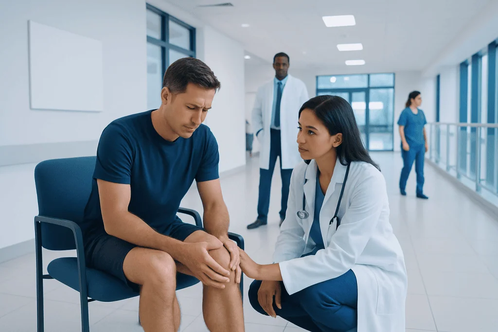
A patellar tendon rupture disrupts the knee’s ability to straighten and support walking. Common signs include sudden pain, swelling, a popping sensation, and trouble lifting the leg.
Key Takeaways
- A patellar tendon rupture disrupts the knee’s ability to straighten and support walking.
- Common signs include sudden pain, swelling, a popping sensation, and trouble lifting the leg.
- Complete ruptures usually require surgery, while some partial tears may be treated without surgery.
- Early diagnosis and rehabilitation can improve strength, function, and recovery.
- People with tendon degeneration, chronic disease, or repeated jumping sports may have higher risk.
Patellar tendon rupture is a serious knee injury in which the tendon connecting the kneecap to the shinbone partially or completely tears. It usually causes sudden pain, swelling, weakness, and difficulty straightening the knee, and prompt medical assessment is important because complete tears often need surgical repair.
Overview: what a patellar tendon rupture means
A patellar tendon rupture is a tear in the strong band of tissue that connects the kneecap (patella) to the shinbone (tibia). This tendon works together with the quadriceps muscle and kneecap to straighten the knee, stand up from a chair, climb stairs, and support movement during walking, running, and jumping. When the tendon tears, the knee’s extensor mechanism is disrupted, which can make active knee straightening difficult or impossible.
Patients often ask whether this injury is the same as ordinary tendon pain. It is not. Tendinitis or tendinopathy may cause ongoing pain and stiffness, but a rupture means the tendon fibers have torn. The tear may be partial, where some fibers remain intact, or complete, where the tendon is fully disrupted and the kneecap may shift out of its normal position.
This injury is less common than many other knee problems, but it is clinically important because delayed diagnosis can affect treatment and recovery. A complete tear usually needs timely surgical repair, while selected partial tears may heal with immobilization and rehabilitation under specialist guidance. Because other knee injuries can cause similar symptoms, an orthopedic assessment is the safest way to confirm the diagnosis.
Symptoms and how the injury usually feels
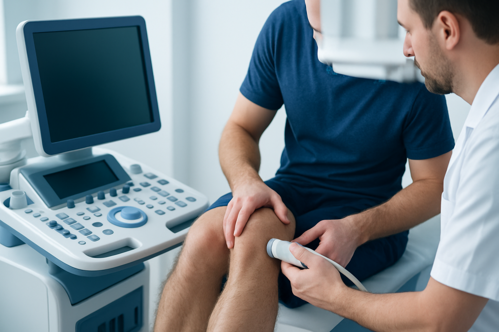
Patellar tendon rupture often happens suddenly. Many patients describe a sharp pain below the kneecap during a jump, landing, change in direction, or forceful contraction of the thigh muscle. Some notice a popping or tearing sensation at the time of injury. Swelling usually develops quickly, and the front of the knee may feel unstable or weak.
A key symptom is difficulty straightening the knee or raising the leg while keeping it straight. Walking may become hard, especially on stairs or uneven ground. In a complete rupture, the person may be unable to actively extend the knee at all. There may also be bruising, tenderness, and a visible or palpable gap below the kneecap.
Another common clue is a change in the position of the kneecap. Because the torn tendon can no longer hold the patella in its usual place, the kneecap may sit higher than normal. However, not every patient can recognize this, and swelling can hide the abnormality. Symptoms can overlap with knee pain from fractures, ligament injuries, or severe tendon inflammation, which is why a structured examination matters.
- Sudden pain at the front of the knee
- Swelling and tenderness below the kneecap
- A popping sensation during injury
- Trouble straightening the knee
- Difficulty walking or bearing weight
- Weakness when standing, climbing, or rising from a chair
Causes and risk factors
Most patellar tendon ruptures occur when the tendon is placed under a sudden, heavy load. This often happens during sports that involve jumping, sprinting, or abrupt stopping and turning. A forceful quadriceps contraction while the knee is bent, such as landing from a jump, is a typical mechanism. Direct trauma to the front of the knee can also cause a tear, though this is less common.
In many adults, the tendon does not rupture solely because of one movement. The tissue may already be weakened by repeated strain or degeneration. Longstanding patellar tendinopathy, sometimes called jumper’s knee, can increase vulnerability. Repeated microtrauma over time can reduce tendon quality, making a rupture more likely during a high-stress movement.
Certain medical conditions and treatments may also raise risk. These can include chronic kidney disease, diabetes, inflammatory conditions, previous knee surgery, and long-term use of medications that can affect tendon health, such as corticosteroids in some settings. Age-related tendon changes, obesity, and reduced conditioning may also contribute. In a patient with risk factors and front-of-knee symptoms, doctors may also consider related orthopedic conditions such as meniscus tear or ligament injury before confirming the diagnosis.
How doctors diagnose patellar tendon rupture
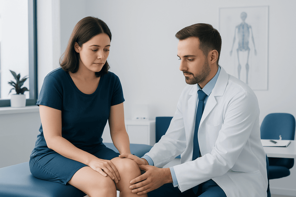
Diagnosis begins with the story of the injury and a physical examination. A doctor will ask how the symptoms started, whether there was a pop, and whether the patient can straighten the knee. During the exam, they may assess swelling, tenderness, kneecap position, and the ability to perform an active straight-leg raise. The inability to actively extend the knee is an important finding in complete ruptures.
Imaging helps confirm the diagnosis and assess the extent of injury. Plain X-rays do not show the tendon itself well, but they can reveal the position of the kneecap and help rule out fractures or other bony injuries. Ultrasound can sometimes identify a tendon tear, especially when performed by an experienced clinician. Magnetic resonance imaging is often the most detailed test when the diagnosis is uncertain or when partial tearing, associated injuries, or surgical planning need to be evaluated.
Because management depends on whether the tear is partial or complete, accurate assessment is important. Early evaluation can also help reduce the chance that a complete rupture is mistaken for a severe sprain or simple strain. In patients with significant knee trauma, broader evaluation may include MRI imaging to define the injury clearly and support a tailored treatment plan.
Treatment options and what recovery may involve
Treatment depends mainly on the severity of the tear, the patient’s activity level, and overall health. Complete patellar tendon ruptures are usually treated with surgery because the tendon must be reattached to restore the knee’s extension mechanism. Surgeons typically repair the torn tendon and then protect the knee in a brace while healing begins. Partial tears may sometimes be managed without surgery if the knee can still extend and imaging shows enough tendon continuity.
Non-surgical care generally includes a period of immobilization, reduced weight-bearing as advised, pain control, and carefully supervised rehabilitation. Surgical care is followed by a structured recovery program designed to protect the repair while gradually restoring motion, strength, and function. The exact pace varies from one patient to another and should follow the treating surgeon’s guidance.
Physical therapy is a central part of both surgical and non-surgical treatment. Early goals often include controlling swelling, protecting the tendon, and maintaining safe mobility. Later stages focus on improving range of motion, rebuilding quadriceps strength, balance, and return to daily activity or sport. Depending on the overall knee injury pattern, some patients may be assessed by teams experienced in knee surgery and physical therapy and rehabilitation to support recovery.
Recovery time varies. Some patients return to basic daily activities within weeks, but full recovery of strength and confidence can take several months. A careful rehabilitation plan matters because returning to demanding activity too soon may place stress on a healing tendon.
Self-care, rehabilitation, and protecting the knee after injury
After diagnosis, self-care should support medical treatment rather than replace it. In the early phase, patients are commonly advised to rest the injured knee, limit activities that increase pain, and follow instructions on bracing, crutches, and weight-bearing. Ice and elevation may help reduce swelling in the first days after injury, if the treating clinician recommends them.
Rehabilitation is not only about exercise intensity; it is also about timing and technique. Progressing too fast may risk pain or re-injury, while progressing too slowly may lead to stiffness and muscle weakness. For this reason, home exercises should match the stage of healing and the plan provided by the orthopedic team or physiotherapist.
As strength returns, movement training becomes important. Patients may work on balance, walking mechanics, stair use, and gradual return to sport-specific tasks. The quadriceps, hamstrings, hip muscles, and core all contribute to knee control. A guided, stepwise program is usually safer than self-directed high-load exercise in the early months after a rupture.
Can patellar tendon rupture be prevented?
Not every patellar tendon rupture can be prevented, especially those caused by high-force accidents. Still, some practical steps may reduce risk. Good conditioning, gradual increases in training load, proper technique in jumping and landing, and adequate recovery between intense sessions can all help limit repeated tendon strain. Addressing persistent tendon pain early may be especially valuable in athletes and active adults.
People with chronic patellar tendinopathy should avoid ignoring symptoms that continue for weeks or keep returning. Ongoing pain below the kneecap can signal tendon overload. Evaluation and treatment of overuse problems may improve tendon health and lower the chance of a more serious tear. Supportive strategies can include load modification, strengthening programs, and correction of movement patterns when appropriate.
General health also matters. Managing long-term conditions such as diabetes or kidney disease, discussing medication risks with a doctor, and maintaining a healthy body weight may support overall tendon health. Warm-up routines, flexibility work, and strength training do not guarantee prevention, but they can improve knee resilience and control during activity.
When to seek medical care
Medical care should be sought promptly after a knee injury if there is sudden front-of-knee pain, a popping sensation, rapid swelling, or inability to straighten the leg. These features may suggest a patellar tendon rupture or another significant knee injury that should not be managed as a minor strain at home. Urgent assessment is especially important if walking is difficult or the knee feels unstable.
Patients should also see a doctor if they have persistent pain below the kneecap that does not improve with rest, especially if they play sports involving jumping or have known tendon problems. Early review can help identify tendon degeneration before a major tear occurs. Immediate emergency care may be needed if there is severe deformity, intense pain after trauma, numbness, coldness in the leg, or concern for fracture or dislocation.
For international patients who need specialist evaluation, Acibadem International’s multidisciplinary teams in JCI-accredited hospitals diagnose and treat knee tendon injuries with orthopedic assessment, imaging, surgery when needed, and rehabilitation planning.
Frequently asked questions
Can a patellar tendon rupture heal on its own?
A small partial tear may sometimes heal without surgery if the knee can still straighten properly and the tendon remains sufficiently intact. A complete patellar tendon rupture usually does not heal well on its own and often requires surgical repair. A doctor should decide the safest approach after examination and imaging.
How is patellar tendon rupture different from patellar tendinitis?
Patellar tendinitis or tendinopathy usually develops gradually and causes pain from overuse, often below the kneecap. A patellar tendon rupture is an actual tear of the tendon fibers and often causes sudden pain, weakness, and loss of knee extension. Tendinopathy can sometimes increase the risk of rupture if the tendon becomes weakened over time.
Will a person always hear or feel a pop?
Not always, but many patients report a popping or tearing sensation at the time of injury. Even without a pop, a rupture may still be present if there is sudden pain, swelling, and difficulty straightening the knee. Because symptoms vary, medical evaluation is important.
How long does recovery usually take?
Recovery time varies depending on whether the tear is partial or complete, whether surgery is needed, and how the rehabilitation progresses. Many patients need several months to regain strength, movement, and confidence in the knee. Returning to sports or heavy activity usually takes longer than returning to basic daily tasks.
Can someone walk with a patellar tendon rupture?
Some people can still take a few steps, especially with a partial tear, but walking is often painful and difficult. In a complete rupture, the knee may buckle and active straightening is usually severely limited. The ability to walk does not rule out a serious injury.
What tests are used to confirm the diagnosis?
Doctors often begin with a physical examination and knee X-rays to assess kneecap position and rule out fractures. Ultrasound or MRI may be used to visualize the tendon directly and determine whether the tear is partial or complete. MRI is especially useful when the diagnosis is uncertain or surgery is being planned.
This article is for general information only and is not a substitute for professional medical advice. Please consult a qualified doctor about your individual situation.
Joint and spine care in Turkey — expert assessment & treatment
JCI-accredited · board-certified surgeons · reply within 24h
Add us as a Preferred Source to see more of our trusted health content across Google Search, AI Overviews and Discover.

More from the Health Library
Bariumenema: What Patients Need to Know

Blackstrap Molasses: A Complete Medical Overview

Fodmap: An Evidence-Based Guide for Patients
Corn on Foot: An Evidence-Based Guide for Patients

Idioventricular Rhythm: A Complete Medical Overview

Amiloride: A Complete Medical Overview
Orthopedic Surgery & Traumatology Specialists at Acibadem

Assoc. Prof. Dr. Altuğ Yücekul
Orthopedic Surgery & Traumatology
Prof. Dr. Arel Gereli
Orthopedic Surgery & Traumatology
Fzt. Asım Sakallı
Physical Medicine & Rehabilitation
Fzt. Aslı Hacıoğlu
Physical Medicine & Rehabilitation