PET-MRI vs PET-CT: What Is the Difference for Patients?
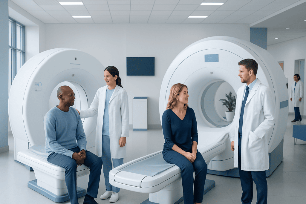
Both PET-MRI and PET-CT combine PET with another imaging method to provide more complete information. PET-MRI generally uses less radiation than PET-CT because MRI does not use X-rays.
Key Takeaways
- Both PET-MRI and PET-CT combine PET with another imaging method to provide more complete information.
- PET-MRI generally uses less radiation than PET-CT because MRI does not use X-rays.
- PET-CT is usually faster, more widely available, and commonly used in cancer imaging.
- PET-MRI can be especially helpful for soft tissues such as the brain, liver, pelvis, and some children’s imaging.
- The best choice depends on the medical reason for the scan, the body area being examined, and patient-specific factors.
PET-MRI and PET-CT are advanced imaging tests that combine positron emission tomography with another scan to show both body structure and activity. For patients, the main differences usually involve radiation exposure, scan time, comfort, availability, and how well each test answers a specific medical question.
Overview: What PET-MRI and PET-CT Are
PET-MRI and PET-CT are both hybrid imaging tests. This means they combine positron emission tomography (PET), which shows how tissues and organs are functioning, with another imaging technology that shows anatomy in detail. PET highlights metabolic activity by detecting a small amount of injected radioactive tracer, while CT or MRI provides a detailed picture of the body’s structures.
In a PET-CT scan, PET is combined with computed tomography (CT). CT uses X-rays to create cross-sectional images of the body. In a PET-MRI scan, PET is combined with magnetic resonance imaging (MRI), which uses a strong magnetic field and radio waves instead of X-rays.
For patients, these tests may look similar because both usually involve an injection, a waiting period while the tracer circulates, and then lying still on a scan table. However, the experience and the kind of information the scan provides can differ. Doctors choose between them based on the clinical question, the part of the body being examined, and practical issues such as speed and availability.
How the Two Scans Differ
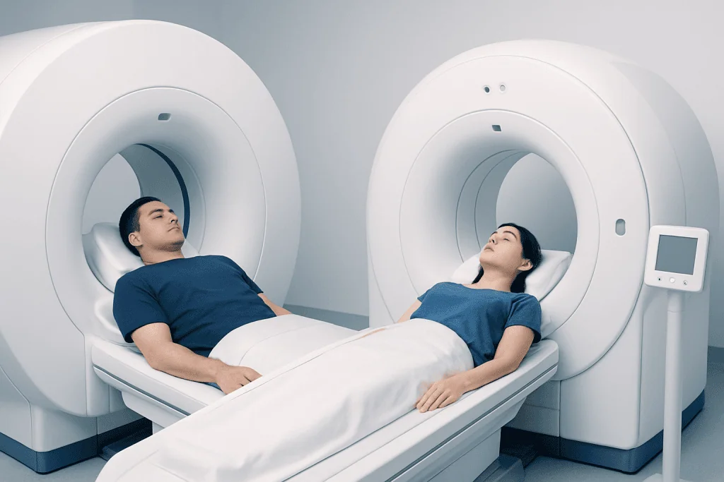
The biggest technical difference is the second imaging method paired with PET. PET-CT combines PET with CT, which is excellent for showing bones, lungs, and general body anatomy quickly. PET-MRI combines PET with MRI, which is especially good for soft tissues such as the brain, muscles, liver, pelvis, and some cancers affecting these areas.
Radiation exposure is another important difference. Both tests involve radiation from the PET tracer itself. PET-CT also adds radiation from the CT portion, while MRI does not use ionizing radiation. As a result, PET-MRI usually exposes patients to less total radiation than PET-CT, which can be particularly relevant for children, younger adults, or people who need repeated imaging over time.
Scan length also matters. PET-CT is generally faster and often easier to schedule because it is more widely available in many hospitals. PET-MRI typically takes longer, and MRI can feel more enclosed and noisy. Some people find MRI more difficult if they feel claustrophobic or have trouble lying still for a longer period.
Image quality differs by body region and medical purpose. In some situations, PET-CT remains the standard approach because it balances speed, accuracy, and access very well. In others, PET-MRI may offer an advantage by providing more detailed soft-tissue information without extra radiation.
When Doctors May Prefer PET-CT or PET-MRI

PET-CT is widely used in oncology because it is efficient and effective for many types of cancer. It may be used to help diagnose disease, stage cancer, evaluate response to treatment, or check for recurrence. It is often especially useful when doctors need a whole-body overview in a relatively short time, and it commonly works alongside other tests such as PET-CT imaging and standard CT.
PET-MRI may be preferred when soft-tissue detail is especially important. This can include selected brain disorders, some head and neck conditions, liver lesions, pelvic tumors, and certain pediatric cases. It can also be helpful when reducing cumulative radiation is a priority, such as in younger patients or those who require multiple follow-up scans.
Neither test is automatically “better” in all circumstances. The right choice depends on what the doctor needs to learn. For example, if lung detail is a key concern, CT may be more practical and informative. If the question involves the brain, spinal cord, or pelvic soft tissues, MRI may add valuable detail.
These scans may also be considered as part of a broader imaging pathway. Depending on symptoms and diagnosis, a patient might also need targeted tests such as MRI, computed tomography, or ultrasound. The goal is always to select the method that answers the clinical question most clearly and safely.
What Patients Can Expect Before, During, and After the Scan
Preparation for PET-MRI and PET-CT is often similar. Patients are commonly asked not to eat for several hours before the scan, although water may be allowed. Blood sugar may need attention, especially for people with diabetes, because high glucose levels can affect how some PET tracers work. The imaging team will explain any specific instructions in advance.
At the appointment, a small amount of radioactive tracer is injected into a vein. After that, there is usually a waiting period to allow the tracer to circulate through the body. During the scan itself, the patient lies on a table that moves through the scanner. It is important to stay as still as possible so the images are clear.
The experience can feel different depending on the machine. PET-CT is usually shorter and quieter. PET-MRI often takes longer, and MRI scanners can produce loud tapping or knocking sounds, so ear protection is typically provided. Some MRI scanners also feel more enclosed, which can be challenging for patients with claustrophobia.
After either test, most people can return to normal activities unless they were told otherwise. Drinking fluids may help clear the tracer from the body more quickly. The radioactive tracer used in PET imaging generally leaves the body over time, and the medical team will provide any needed safety advice.
Benefits, Limitations, and Safety Considerations
PET-CT offers several practical advantages. It is fast, widely available, and well established in everyday clinical care. It is often an excellent option when doctors need timely answers, especially in cancer care, emergency pathways, or when chest and bone detail are important. It is also commonly used in the assessment of diseases such as lung cancer and other cancers where whole-body staging is needed.
PET-MRI offers the important benefit of lower overall radiation exposure compared with PET-CT. It can also provide superior soft-tissue contrast, which may help in selected cancers, neurological conditions, and pediatric imaging. In some cases, PET-MRI can bring metabolic and highly detailed anatomical information together in a way that reduces the need for multiple separate tests.
There are limitations to both. PET-CT involves more radiation than PET-MRI because of the CT component. PET-MRI can be more expensive, less available, and longer to perform. MRI also has its own restrictions: people with certain implanted devices, metal fragments, or severe claustrophobia may not be suitable candidates, although modern MRI safety screening can clarify this.
Contrast agents are sometimes used with CT or MRI, depending on the study. These are different from the PET tracer and may carry separate considerations, such as allergy history or kidney function. The imaging team reviews these factors carefully so the scan can be performed as safely as possible.
How Doctors Decide Which Scan Is Right
Choosing between PET-MRI and PET-CT is a medical decision based on the patient’s symptoms, diagnosis, age, treatment history, and the body area being examined. Doctors also consider how urgently the scan is needed, whether prior images are available for comparison, and which test is most likely to provide the clearest answer.
For example, PET-CT is often favored when speed, broad availability, and evaluation of the lungs or skeleton are priorities. PET-MRI may be more attractive when repeated imaging is expected, when radiation reduction matters, or when the question involves soft tissues in the brain, abdomen, or pelvis. In cancer care, the exact tumor type and treatment stage often guide the choice.
It is also important to remember that imaging is only one part of diagnosis. Doctors interpret scan results together with the patient’s history, physical examination, blood tests, and other investigations. If needed, imaging findings may lead to further steps such as biopsy, surgery, or follow-up scanning.
In international centers, patients may be assessed by a multidisciplinary team that includes radiologists, nuclear medicine physicians, oncologists, neurologists, and surgeons. Near the end of the care pathway, some patients also choose institutions such as Acibadem International, where multidisciplinary specialists in JCI-accredited hospitals evaluate and treat international patients using advanced imaging and condition-specific care pathways.
Questions to Ask and When to Speak With a Doctor
Patients do not need to choose between PET-MRI and PET-CT on their own. The best next step is to ask why the scan is being recommended and what information it is expected to provide. Understanding the reason for the test can make the process feel more manageable and less stressful.
Helpful questions may include:
- Why is this scan recommended for this condition?
- Would PET-MRI or PET-CT be more useful in this case?
- How should the patient prepare before the appointment?
- Will contrast be used, and are there any kidney or allergy concerns?
- How long will the scan take, and when will the results be ready?
Patients should tell their doctor before the scan if they are pregnant, breastfeeding, have diabetes, feel severely claustrophobic, or have an implanted medical device. They should also mention any previous allergic reactions to contrast agents and any difficulty lying flat or still.
Medical advice is especially important if the scan has been ordered to investigate serious symptoms, monitor a known condition, or guide cancer treatment. If there is uncertainty, a qualified doctor can explain the risks, benefits, and alternatives in the context of the individual patient.
Frequently asked questions
Is PET-MRI better than PET-CT?
Neither scan is always better in every situation. PET-MRI may be more helpful for soft tissues and lower radiation exposure, while PET-CT is often faster, more available, and very useful for many cancers. The best choice depends on the medical question being asked.
Does PET-MRI have less radiation than PET-CT?
Yes, PET-MRI usually involves less total radiation because MRI does not use X-rays. Both tests still include a PET tracer, which is radioactive, but PET-CT adds radiation from the CT portion. This difference may matter more for children or people needing repeated scans.
Which scan takes longer, PET-MRI or PET-CT?
PET-MRI usually takes longer than PET-CT. MRI images generally require more time to acquire, and the machine can feel noisier and more enclosed. PET-CT is often the quicker option, which can be helpful for people who have difficulty remaining still.
Are PET-MRI and PET-CT used for cancer?
Yes, both scans can be used in cancer care. Doctors may use them to help diagnose cancer, determine how far it has spread, assess response to treatment, or look for recurrence. The preferred scan depends on the cancer type and the part of the body being evaluated.
Can everyone have a PET-MRI?
Not always. Some patients cannot safely undergo MRI because of certain implants, metal fragments, or severe claustrophobia. The imaging team reviews a person’s medical history carefully to decide whether PET-MRI is appropriate.
How should a patient prepare for one of these scans?
Preparation often includes fasting for several hours, following instructions about medications, and arriving on time for the tracer injection and waiting period. People with diabetes may receive special guidance because blood sugar can affect PET imaging. The hospital will provide exact instructions based on the reason for the scan.
References
- World Health Organization
- Radiological Society of North America
- Society of Nuclear Medicine and Molecular Imaging
- American College of Radiology
- National Cancer Institute
This article is for general information only and is not a substitute for professional medical advice. Please consult a qualified doctor about your individual situation.
Explore treatments in Turkey — costs, top hospitals & a free quote
JCI-accredited · board-certified surgeons · reply within 24h
Add us as a Preferred Source to see more of our trusted health content across Google Search, AI Overviews and Discover.