Preparing for a Brain or Spine MRI: Contrast, Claustrophobia, and Safety Questions
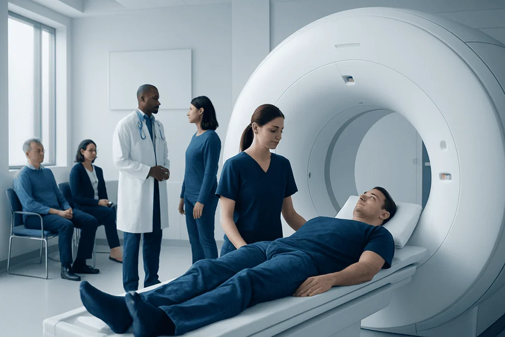
Most brain and spine MRI scans are safe and painless, but careful screening for metal and implants is essential. Contrast dye is not needed for every MRI; when used, it helps highlight certain tissues, blood vessels, inflammation, or tumors.
Key Takeaways
- Most brain and spine MRI scans are safe and painless, but careful screening for metal and implants is essential.
- Contrast dye is not needed for every MRI; when used, it helps highlight certain tissues, blood vessels, inflammation, or tumors.
- Claustrophobia is common and can often be managed with planning, reassurance, relaxation techniques, or sedation when appropriate.
- Patients should tell the imaging team about kidney problems, pregnancy, implanted devices, allergies, or prior reactions to contrast.
- Following instructions about clothing, jewelry, eating, and arrival time helps the scan go more smoothly.
A brain or spine MRI is a common imaging test that helps doctors see detailed structures without using ionizing radiation. Preparing well, understanding contrast, and knowing what to expect with claustrophobia or implanted devices can make the experience smoother and more comfortable.
Overview: What a Brain or Spine MRI Involves
A brain or spine MRI is a type of imaging test that uses a strong magnetic field and radio waves to create detailed pictures of the brain, spinal cord, nerves, discs, and surrounding tissues. It does not use X-rays or other ionizing radiation. Doctors often request it to evaluate symptoms such as headaches, weakness, numbness, dizziness, seizures, back pain, neck pain, or suspected nerve compression.
MRI is especially helpful because it shows soft tissues more clearly than many other imaging tests. In the brain, it can help assess inflammation, stroke, tumors, infection, bleeding, or changes linked to conditions such as multiple sclerosis. In the spine, it can show disc problems, spinal stenosis, nerve root compression, spinal cord changes, or signs of infection and tumor.
During the scan, the patient lies still on a table that moves into the MRI machine. The machine makes loud tapping or thumping sounds while it captures images. The test is painless, but staying still is important because movement can blur the images and reduce their quality.
How to Prepare Before the Appointment
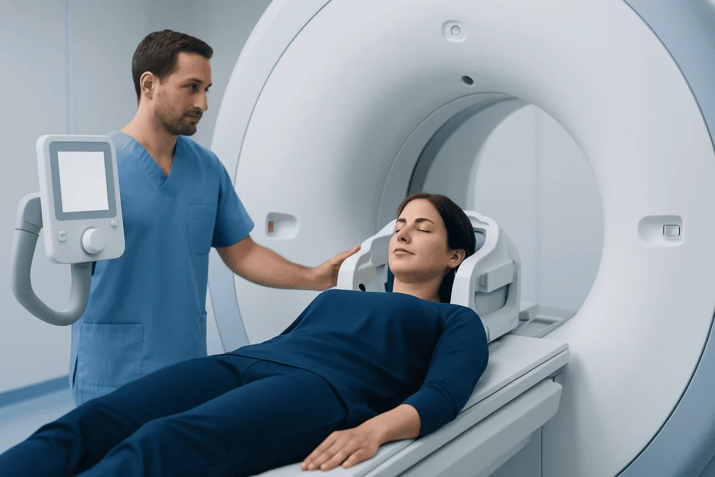
Preparation for a brain or spine MRI usually begins before arrival. Patients are commonly asked to complete a safety questionnaire about pacemakers, aneurysm clips, cochlear implants, neurostimulators, metal fragments, prior surgeries, and any history of working with metal. This information helps the imaging team confirm that the MRI can be performed safely.
On the day of the scan, comfortable clothing without metal fasteners is often best, although some centers provide a gown. Jewelry, watches, hairpins, hearing aids, removable dental work, and credit cards should be removed before entering the MRI room. Phones and other electronic items must also stay outside because the magnet can damage them or pull on metal components.
If contrast may be used, the team may ask about kidney disease, prior contrast reactions, or recent blood test results. Some patients can eat and drink normally, while others may be told not to eat for a few hours, especially if sedation is planned. It is helpful to arrive early to review forms, ask questions, and discuss any concerns about anxiety, pain, or difficulty lying flat.
- Bring a list of medications and past surgeries.
- Tell the team about pregnancy or possible pregnancy.
- Mention tattoos, permanent makeup, or medication patches.
- Ask in advance if a companion may come, especially if sedation is planned.
MRI Contrast: Why It Is Used and Common Safety Questions

Not every brain or spine MRI needs contrast. When used, MRI contrast is typically a gadolinium-based agent given through a vein. It can help doctors see areas of inflammation, abnormal blood vessels, infection, breakdown of the blood-brain barrier, or certain tumors more clearly. This may be useful in detailed studies such as MRI scan protocols for the brain or spine.
For most people, gadolinium contrast is well tolerated. Mild side effects such as a cool feeling at the injection site, a metallic taste, nausea, or headache may occur but are usually brief. Serious allergic reactions are uncommon, but patients should always report any previous reaction to MRI contrast, as well as asthma, multiple allergies, or prior severe reactions to other injectable medicines.
Kidney function matters when contrast is being considered. People with advanced kidney disease may need additional evaluation before receiving gadolinium because certain contrast agents can carry added risk in severe renal impairment. If there is any question, the imaging team may request recent kidney blood tests or choose a non-contrast study if that can answer the clinical question.
Many patients also ask about contrast retention in the body. Small amounts of gadolinium can remain in tissues after some examinations, but the clinical significance in people with normal kidney function remains uncertain. The decision to use contrast is based on whether the expected diagnostic benefit outweighs the potential risk, and patients should feel comfortable asking why contrast is recommended in their case.
Claustrophobia and Anxiety During MRI
Feeling nervous in an MRI scanner is common, especially when the head is inside the machine for a brain MRI or when a longer spine study is planned. Claustrophobia does not mean a person cannot complete the scan. In many cases, a few practical steps make the experience much easier.
Patients should tell the care team in advance if they have panic symptoms, severe anxiety, chronic pain, or trouble lying still. The team may be able to schedule extra time, offer a scanner with a wider bore, provide ear protection and music, or explain exactly how long each part of the exam will last. Knowing there is two-way communication with the technologist and that the scan can be paused if necessary often helps people feel more in control.
Relaxation techniques can also be useful. Slow breathing, keeping the eyes closed before entering the scanner, guided imagery, and focusing on the rhythm of the sounds may reduce distress. For some people, a mild sedative prescribed in advance is appropriate, but this requires planning because the patient should not drive afterward.
Children and adults who cannot remain still despite reassurance may sometimes need monitored sedation. This decision depends on age, medical history, and the length of the exam. If sedation is being considered, patients should ask about fasting instructions, medication review, and post-procedure supervision.
Metal, Implants, Pregnancy, and Other MRI Safety Issues
The most important MRI safety rule is to report any implanted or metal-containing device before the scan. Some devices are MRI-safe or MRI-conditional, meaning they can be scanned only under specific conditions. Others may be unsafe. This includes certain pacemakers, older aneurysm clips, cochlear implants, infusion pumps, spinal cord stimulators, and retained metal fragments in the eye or body.
Past surgery does not automatically prevent MRI, but details matter. Joint replacements, dental implants, surgical clips, stents, and hardware used in spine surgery may be compatible with MRI, yet the team usually needs the device name, model, or surgical records to confirm. Patients should also report any bullet fragments, shrapnel, or history of metalwork injuries, especially if metal entered the eye.
Pregnancy is another common concern. MRI may be performed during pregnancy when medically necessary, particularly if the information is important for the health of the mother or baby. However, contrast is usually avoided unless there is a clear reason to use it. Patients who are pregnant or trying to conceive should let the referring doctor and MRI staff know before the appointment.
People with chronic medical conditions should also mention kidney disease, severe liver disease, seizure disorders, or difficulty lying flat because of breathing problems or pain. In some cases, scan planning can be adapted. Patients being assessed for neurological symptoms may also undergo related testing depending on the clinical picture, such as CT scan in emergency situations or other targeted imaging studies.
What Happens During and After the Scan
Once the patient is positioned on the table, the technologist may place a special device called a coil around the head or spine area to improve image quality. Cushions or straps may be used to help the patient stay comfortable and still. The table then moves into the scanner, and the technologist operates the exam from the next room while maintaining communication throughout the test.
MRI sequences are performed in stages, and each one lasts several minutes. The main task for the patient is to remain as still as possible. If contrast is needed, it may be injected partway through the exam through an IV line. Some spine examinations and advanced neurological studies may take longer than a routine scan.
After the MRI, most people can return to normal activities right away. If contrast was used, drinking fluids as usual is generally sufficient unless a doctor gives different advice. If sedation was given, the patient will need observation until fully alert and should arrange for someone else to drive home.
The images are reviewed by a radiologist, often a neuroradiologist for brain and spine studies. Results are then sent to the referring doctor, who explains what they mean and whether further tests are needed. Depending on the findings, the next step may involve follow-up imaging, neurological evaluation, or treatment planning such as brain tumor surgery when appropriate for a confirmed diagnosis.
When to Contact a Doctor or the Imaging Center
Patients should contact the imaging center before the appointment if they discover new information about an implant, develop an illness that affects their ability to lie flat, become pregnant, or realize they may need help with claustrophobia. It is much easier to adjust plans before the scan than to cancel at the last minute.
After the exam, most people feel completely fine. However, medical advice should be sought promptly if there are symptoms that could suggest a contrast reaction, such as difficulty breathing, widespread rash, facial swelling, or severe persistent vomiting. New or worsening neurological symptoms, including sudden weakness, confusion, seizure, or loss of bladder or bowel control, also require urgent medical attention regardless of when the MRI was performed.
Questions about test results should be directed to the referring clinician, who can explain the findings in context. A scan may show age-related changes, an incidental finding, or evidence of a condition that needs follow-up. Clear communication helps patients understand what is important and what the next step should be.
For international patients who need coordinated neurological imaging and follow-up care, Acibadem International’s multidisciplinary specialists and JCI-accredited hospitals diagnose and treat brain and spine conditions with patient-centered planning.
Frequently asked questions
Do all brain or spine MRI scans use contrast?
No. Many MRI exams are done without contrast, and in some cases non-contrast images provide enough information. Contrast is used only when it is likely to improve the accuracy of the study or answer a specific clinical question.
Is MRI contrast safe?
For most people, gadolinium-based contrast is considered safe and well tolerated. The imaging team still needs to know about kidney disease, prior contrast reactions, pregnancy, and other relevant medical history before deciding whether contrast should be used.
What if the patient is claustrophobic?
Claustrophobia is common and can often be managed successfully. Helpful options include discussing concerns in advance, using relaxation techniques, listening to music if available, choosing a wider scanner when possible, or arranging sedation if medically appropriate.
Can a person with metal implants have an MRI?
Sometimes yes, but the implant must be checked carefully first. Many modern implants are MRI-compatible or MRI-conditional, while some older or specialized devices may require special precautions or may not be safe in the MRI environment.
Should patients fast before a brain or spine MRI?
Often no, but instructions vary by center and by whether contrast or sedation is planned. It is best to follow the specific preparation instructions provided by the imaging department.
How long does a brain or spine MRI take?
The timing depends on the body area being scanned, the number of sequences needed, and whether contrast is used. Many exams take between 30 and 60 minutes, though some specialized studies can take longer.
References
- Radiological Society of North America
- American College of Radiology
- National Institute of Neurological Disorders and Stroke
- National Institute of Biomedical Imaging and Bioengineering
- U.S. Food and Drug Administration
This article is for general information only and is not a substitute for professional medical advice. Please consult a qualified doctor about your individual situation.
Joint and spine care in Turkey — expert assessment & treatment
JCI-accredited · board-certified surgeons · reply within 24h
Add us as a Preferred Source to see more of our trusted health content across Google Search, AI Overviews and Discover.