Pseudotumor Cerebri: Headache, Visual Symptoms, and Neuro-Ophthalmology Assessment

Pseudotumor cerebri means increased pressure around the brain without an actual tumor. Common symptoms include headache, temporary visual dimming, double vision, and a whooshing sound in the ears.
Key Takeaways
- Pseudotumor cerebri means increased pressure around the brain without an actual tumor.
- Common symptoms include headache, temporary visual dimming, double vision, and a whooshing sound in the ears.
- Eye examination is essential because swelling of the optic nerve can threaten vision if untreated.
- Diagnosis usually involves brain imaging, a detailed eye exam, and lumbar puncture to measure pressure.
- Treatment may include weight management, medicines to reduce cerebrospinal fluid pressure, and procedures when vision is at risk.
Pseudotumor cerebri, also called idiopathic intracranial hypertension, is a condition in which pressure inside the skull becomes too high without a brain tumor or other space-occupying lesion. It commonly causes headache and visual symptoms, so careful neurological and eye assessment is important to protect vision.
Overview
Pseudotumor cerebri is a condition in which the pressure of cerebrospinal fluid around the brain is higher than normal, even though there is no brain tumor. The term literally means “false brain tumor” because the symptoms can resemble those caused by a mass inside the skull. Today, many doctors use the name idiopathic intracranial hypertension, or IIH, especially when no clear cause is found.
This condition can affect adults and children, but it is seen most often in women of childbearing age. Headache is a common symptom, but visual complaints are especially important because high pressure can cause swelling of the optic nerves, called papilledema. Without timely treatment, some people may develop permanent vision loss.
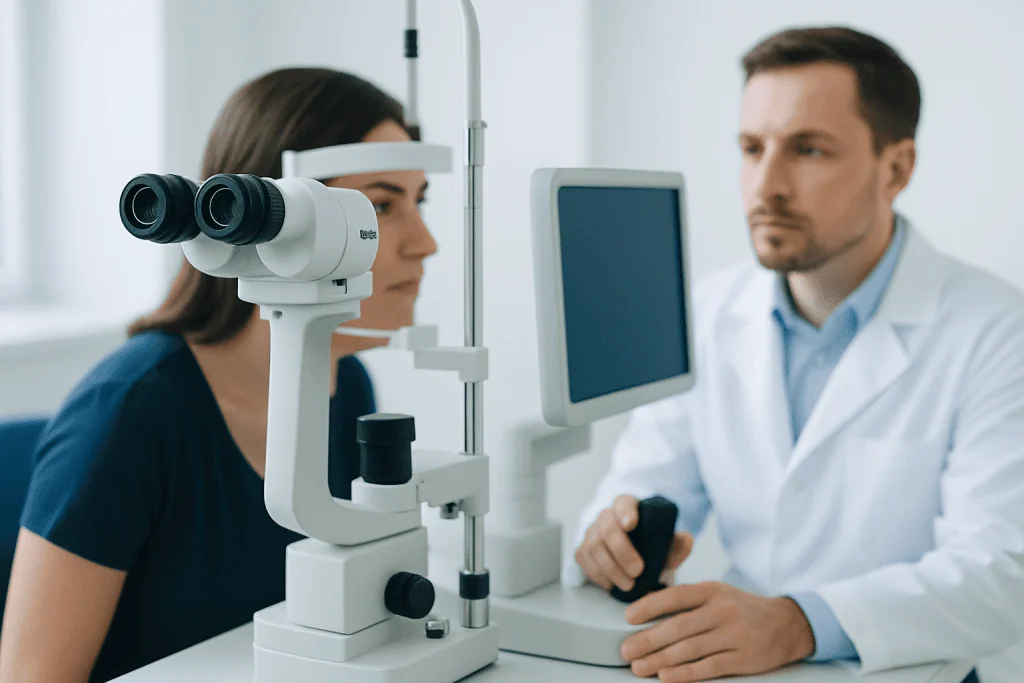
Neuro-ophthalmology assessment plays a central role in care. A neuro-ophthalmologist evaluates how the nervous system and the eyes are working together, checks for optic nerve swelling, measures visual function, and helps guide urgent treatment decisions. Because many conditions can mimic pseudotumor cerebri, careful evaluation is necessary before the diagnosis is confirmed.
Symptoms
The symptoms of pseudotumor cerebri can develop gradually or appear more suddenly. Headache is the most frequent complaint. It may be daily, often worse in the morning, and sometimes more noticeable when coughing, bending, or straining. Some people also feel nausea or neck and shoulder discomfort along with the headache.
Visual symptoms are particularly important. A person may notice brief episodes of blurred or dim vision, especially when standing up or changing position. Others describe double vision, reduced side vision, trouble focusing, or seeing flashing lights. In some cases, visual changes are subtle at first and are found mainly during a formal eye examination.
Another common symptom is pulsatile tinnitus, a rhythmic “whooshing” sound in the ears that seems to match the heartbeat. Some people also experience dizziness or pain behind the eyes. Because these symptoms can overlap with migraine and other neurological conditions, evaluation by a qualified doctor is important.
- Frequent or persistent headache
- Temporary visual dimming or blurring
- Double vision
- Pulsatile tinnitus
- Nausea or dizziness
- Episodes of reduced peripheral vision
Causes and Risk Factors

In many people, pseudotumor cerebri is called idiopathic, which means no single clear cause is identified. Doctors believe the condition relates to imbalance in the production, circulation, or absorption of cerebrospinal fluid, leading to increased pressure inside the skull. Researchers are still working to understand exactly why this happens.
Several risk factors are known. Obesity is one of the strongest associations, and recent weight gain may increase risk. The condition is also more common in females, especially during reproductive years. That said, it can occur in men, older adults, and children as well.
Some cases are linked to underlying medical problems or medicines. Doctors may look for hormonal disorders, sleep apnea, anemia, kidney disease, or blood clotting problems affecting the veins that drain the brain. Certain medications, including vitamin A derivatives and some antibiotics, may contribute in susceptible people. Because of this, the medical history is an important part of evaluation.
When a specific trigger or associated illness is found, treatment may include addressing that factor along with lowering intracranial pressure. If no cause is found after a thorough workup, the diagnosis is generally idiopathic intracranial hypertension.
How Neuro-Ophthalmology Assessment Helps Diagnose It
Diagnosis begins by ruling out other causes of raised intracranial pressure and symptoms such as headache or visual change. The doctor takes a detailed history and performs a neurological and eye examination. A dilated eye exam is especially important because it can reveal papilledema, the swelling of the optic disc caused by increased pressure around the optic nerve.
Neuro-ophthalmology testing helps measure whether vision is being affected. This may include visual acuity testing, color vision assessment, pupil examination, eye movement evaluation, and visual field testing to check for blind spots or side-vision loss. Optical coherence tomography may also be used to measure optic nerve swelling and follow changes over time.
Brain imaging is usually needed to exclude a mass, bleeding, hydrocephalus, or another structural problem. Magnetic resonance imaging of the brain, sometimes with venography, can help detect signs associated with high intracranial pressure and identify narrowing or clotting in the venous sinuses. After imaging has ruled out a dangerous mass effect, a lumbar puncture may be performed to measure the opening pressure and analyze the cerebrospinal fluid.
The diagnosis of pseudotumor cerebri is based on the overall picture: symptoms, papilledema or related eye findings, elevated cerebrospinal fluid pressure, normal fluid composition, and no other better explanation on imaging or examination. Since several conditions can look similar, accurate diagnosis often requires teamwork between neurology, eye specialists, and radiology.
Treatment Options
Treatment aims to protect vision, lower intracranial pressure, and relieve symptoms such as headache. The approach depends on how severe the condition is and whether visual function is stable. People with mild disease may be treated with careful monitoring, lifestyle changes, and medicine, while those with rapidly worsening vision may need urgent procedures.
Weight reduction is often an important part of treatment for people who are overweight, as even modest weight loss may improve pressure and symptoms. Doctors may also prescribe medication to reduce cerebrospinal fluid production. Headache management may involve a broader plan as well, since some patients have overlapping headache disorders such as migraine.
If vision is threatened despite medical treatment, procedures may be considered. These can include repeated pressure-lowering measures in selected situations, optic nerve sheath fenestration, or cerebrospinal fluid shunting. In some patients with venous sinus narrowing, specialists may evaluate whether venous sinus stenting is appropriate, though this is not suitable for everyone and requires careful selection.
Some people benefit from coordinated care involving neurology, neuro-ophthalmology, neurosurgery, and weight-management specialists. Where surgical intervention is needed, options may overlap with broader neurosurgical care depending on the cause, pressure pattern, and risk to vision. Treatment plans are individualized, and regular follow-up is essential to make sure vision remains safe.
Prevention and Self-Care
There is no guaranteed way to prevent pseudotumor cerebri, but some practical steps may lower risk or help reduce recurrence. Maintaining a healthy body weight is one of the most important. If weight loss is recommended, it is usually safest and most sustainable when done with guidance from a doctor or dietitian.
Patients should take medicines exactly as prescribed and should not stop or change treatment without medical advice. If a medication might be contributing to raised intracranial pressure, the treating doctor can review alternatives. Regular eye and follow-up appointments are important even when symptoms improve, because visual changes can sometimes progress quietly.
Keeping a symptom diary may help. Recording headaches, visual dimming, double vision, or pulsatile tinnitus can make it easier for the care team to track patterns and treatment response. Healthy sleep habits, management of sleep apnea when present, and attention to overall cardiovascular health may also support recovery and long-term control.
When to See a Doctor
Anyone with persistent new headaches, episodes of blurred or dim vision, double vision, or a pulse-like sound in the ears should arrange a medical assessment. These symptoms do not always mean pseudotumor cerebri, but they deserve attention because several neurological and eye conditions can cause them.
Urgent assessment is especially important if vision seems to be getting worse, side vision is narrowing, or there is severe headache with vomiting or major neurological symptoms. Rapid treatment can help protect eyesight when papilledema is present. It is also wise to seek review if symptoms continue despite treatment or if medication side effects become difficult to manage.
At centers with neuro-ophthalmology, neurology, imaging, and neurosurgical expertise, patients can often have diagnosis and follow-up coordinated more efficiently. Acibadem International’s multidisciplinary specialists and JCI-accredited hospitals diagnose and treat pseudotumor cerebri for international patients, with care tailored to the person’s symptoms, vision findings, and overall health.
Frequently asked questions
Is pseudotumor cerebri a real tumor?
No. Pseudotumor cerebri does not mean there is an actual brain tumor. The name refers to symptoms and pressure changes that can resemble those caused by a tumor, even though imaging does not show one.
Can pseudotumor cerebri cause permanent vision loss?
Yes, it can if the pressure damages the optic nerves over time. This is why prompt diagnosis, regular eye examinations, and follow-up visual field testing are so important.
What kind of doctor treats pseudotumor cerebri?
Care often involves more than one specialist. A neurologist and a neuro-ophthalmologist commonly lead assessment and monitoring, and neurosurgeons or other specialists may be involved when procedures are needed.
Does every person with pseudotumor cerebri need a lumbar puncture?
A lumbar puncture is commonly part of diagnosis because it helps measure cerebrospinal fluid pressure and checks that the fluid itself is normal. However, doctors first use imaging to make sure it is safe and to rule out other causes.
Can weight loss really help pseudotumor cerebri?
For many people who are overweight, weight loss can be an important part of treatment and may reduce pressure and symptoms. The amount needed varies, so it is best planned with medical guidance.
Do headaches go away once pressure is treated?
Not always immediately. Some patients improve significantly as pressure comes down, while others continue to have headaches that need separate management, especially if another headache disorder is also present.
References
- National Eye Institute
- National Institute of Neurological Disorders and Stroke
- American Academy of Ophthalmology
- European Headache Federation
- UpToDate
This article is for general information only and is not a substitute for professional medical advice. Please consult a qualified doctor about your individual situation.
Heart care in Turkey — expert evaluation and treatment
JCI-accredited · board-certified surgeons · reply within 24h
Add us as a Preferred Source to see more of our trusted health content across Google Search, AI Overviews and Discover.