Ptosis: Diagnosis, Outlook, and Modern Treatment Approaches

Ptosis means the upper eyelid sits lower than normal and may affect one or both eyes. It can be present from birth or develop later due to aging, nerve problems, muscle disorders, injury, or contact lens use.
Key Takeaways
- Ptosis means the upper eyelid sits lower than normal and may affect one or both eyes.
- It can be present from birth or develop later due to aging, nerve problems, muscle disorders, injury, or contact lens use.
- Careful eye examination helps determine whether ptosis is affecting vision and whether another medical condition is involved.
- Treatment depends on the cause and may include observation, managing the underlying condition, or eyelid surgery.
- Sudden ptosis, ptosis with double vision, or ptosis with weakness needs prompt medical assessment.
Ptosis is a drooping of the upper eyelid that may be mild, noticeable mainly for appearance, or significant enough to block vision. Diagnosis focuses on finding the cause and measuring how much the eyelid affects sight, with treatment ranging from monitoring to surgery.
Overview: What ptosis is and why it matters
Ptosis is the medical term for a drooping upper eyelid. The eyelid may hang only slightly lower than usual or may drop enough to partly cover the pupil and interfere with vision. Some people notice ptosis mainly because of facial asymmetry or a tired appearance, while others experience eye strain, brow lifting, or difficulty seeing clearly, especially when reading or looking straight ahead.
Ptosis can affect one eye or both eyes. It may be present at birth, called congenital ptosis, or appear later in life, called acquired ptosis. In many adults, the most common reason is weakening or stretching of the tendon that lifts the eyelid. However, ptosis can also be linked to nerve conditions, muscle disorders, trauma, eye surgery, or masses around the eyelid.
The outlook for ptosis depends largely on its cause. Some cases remain stable and mild, some improve when an underlying problem is treated, and some need a procedure to lift the eyelid to a more functional position. Because drooping can occasionally signal a neurological or muscular disorder, diagnosis is about more than appearance alone.
How ptosis can affect vision and daily life
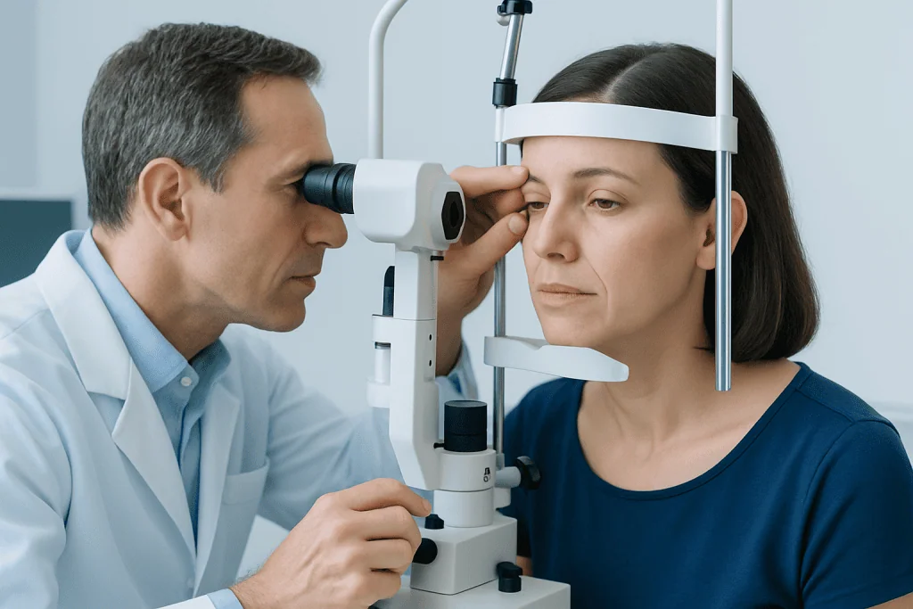
Even a small change in eyelid position can change how the eyes feel and function. When the eyelid covers part of the visual axis, a person may tilt the head backward or raise the eyebrows to see better. Over time, this can lead to forehead tension, headaches, and visual fatigue. Children with significant ptosis may be at risk of developing lazy eye if the eyelid blocks vision during visual development.
Ptosis can also make the eyes feel uneven. One eyelid may look heavier, and the face may appear less symmetrical in photographs or during conversation. In some people, the drooping is more noticeable at the end of the day or when they are tired, which can suggest a muscle-related cause. Others may also have a sensation of heaviness, reduced peripheral upper vision, or trouble keeping the eye open while driving or reading.
Because symptoms vary, clinicians usually consider both function and appearance. If ptosis is reducing vision, causing compensatory brow strain, or creating concern about an underlying disorder, a fuller ophthalmic assessment is appropriate. In some cases, ptosis appears alongside other eyelid or eye-surface concerns that need separate attention, such as glaucoma monitoring or dry-eye symptoms.
Symptoms and signs of ptosis

The main sign of ptosis is a visibly low upper eyelid. In mild cases, the lid sits lower than the other side but does not block the pupil. In more significant cases, the eyelid partly covers the eye, making the eye look smaller and reducing the upper field of vision. The drooping may be constant or may fluctuate during the day.
Common symptoms and associated signs include:
- One or both upper eyelids drooping
- Reduced upper vision or needing to lift the chin to see
- Frequent eyebrow raising to compensate
- Forehead tension or headaches from overusing the brow muscles
- Eye fatigue, especially with reading or screen use
- Facial asymmetry or a tired appearance
- In some neuromuscular conditions, worsening droop with fatigue
Some symptoms suggest a need for faster assessment. Sudden drooping, a new difference in pupil size, double vision, eye pain, severe headache, or weakness elsewhere in the body may point to a nerve or neurological problem rather than simple age-related ptosis. In children, a persistent droopy lid should always be evaluated because of its potential effect on vision development.
Causes and risk factors
Ptosis develops when the muscle that lifts the upper eyelid, or the structures that support it, do not work as they should. In adults, the most common form is aponeurotic ptosis, often related to age-related stretching of the levator tendon. It can also occur after long-term contact lens wear, previous eye surgery, or repeated eyelid rubbing. This type tends to develop gradually.
Congenital ptosis is usually due to incomplete development of the levator muscle. A child may have it in one eye or both eyes, and severity can vary. Acquired ptosis has a wider range of causes, including nerve disorders such as third cranial nerve palsy, sympathetic nerve disruption seen in Horner syndrome, and muscle diseases such as myasthenia gravis. Structural causes, including eyelid swelling, tumors, scarring, or trauma, may also weigh the lid down or change eyelid mechanics.
Important risk factors and associated conditions include aging, prior eyelid or cataract surgery, contact lens use, diabetes-related nerve problems, neurological disease, muscular disorders, and direct injury to the eyelid area. Sometimes ptosis appears with other eye conditions that also need care, such as a lens change requiring cataract surgery or a lid position problem related to previous operations. The key point is that ptosis is a sign with several possible causes, not a diagnosis by itself.
How ptosis is diagnosed
Diagnosis begins with a medical history and a detailed eye examination. The clinician asks when the drooping started, whether it has changed over time, and whether it varies with tiredness. Questions also focus on double vision, headache, trauma, contact lens use, prior eye surgery, and any generalized weakness. In children, the specialist assesses visual development and checks for amblyopia or compensatory head posture.
The examination measures eyelid position, eyelid crease, muscle function, brow compensation, and how much the drooping affects the pupil. Eye movement, pupil responses, and visual fields may be checked to look for nerve or brain-related causes. The eye surface is also examined because some people with ptosis have dryness or incomplete blinking that can influence treatment planning.
Additional tests depend on the suspected cause. If a neuromuscular disorder is considered, blood tests or specialist neurological evaluation may be needed. Imaging studies may be recommended if there is concern about a mass, trauma, or a neurological cause. When surgery is being considered, the evaluation helps determine which approach is most suitable, such as oculoplastic surgery for functional eyelid correction. The aim is to identify not only how much the eyelid droops, but why it is happening.
Modern treatment approaches
Treatment for ptosis is individualized. Mild cases that do not affect vision may simply be observed, especially if the eyelid position is stable and there are no warning signs of an underlying condition. If ptosis is caused by another medical problem, such as a nerve palsy or myasthenia gravis, treatment focuses first on the underlying disorder. In these situations, eyelid position may improve as the primary condition is managed.
When ptosis interferes with sight, causes significant brow strain, or is cosmetically important to the patient, surgery is often the most effective option. The technique depends on how well the eyelid-lifting muscle works and how severe the drooping is. Procedures may tighten or reattach the levator tendon, adjust muscles from the inside of the eyelid, or in more severe cases use a sling to help the forehead muscles lift the lid. Functional and reconstructive planning may overlap with blepharoplasty in selected patients, but the goals and techniques are not exactly the same.
Recovery and results depend on the cause, tissue quality, and preoperative eyelid function. Most people can expect improved eyelid height, better upper vision, and less need to raise the eyebrows. However, perfect symmetry is not always possible, and temporary swelling, dryness, or minor asymmetry can occur during healing. Children with congenital ptosis may need earlier intervention if the lid blocks vision or contributes to abnormal head posture.
At centers with multidisciplinary eye care, oculoplastic, ophthalmology, and neurology teams may work together when ptosis is linked to a broader condition. Acibadem International’s multidisciplinary specialists in JCI-accredited hospitals diagnose and treat ptosis for international patients when surgical or specialist evaluation is needed.
Prevention, self-care, and long-term outlook
Not all cases of ptosis can be prevented, especially congenital forms or those related to aging. Still, protecting eye health can lower the chance of avoidable eyelid problems. Gentle contact lens handling, avoiding excessive eye rubbing, using appropriate eye protection during sports or work, and following postoperative instructions carefully after eye surgery may reduce strain on eyelid tissues.
Self-care does not correct true ptosis, but it can help with comfort and symptom tracking. People may find it useful to note whether drooping changes through the day, whether it worsens with fatigue, and whether headaches, double vision, or brow strain occur. This information can help the specialist identify whether the problem is mechanical, age-related, or potentially neuromuscular.
The long-term outlook is generally good once the cause is identified. Age-related and structural ptosis often respond well to surgery, while prognosis in neurological or muscular cases depends on the underlying condition. Regular follow-up is important for children, for anyone whose vision is affected, and for patients with associated eye disease or fluctuating symptoms.
When to seek medical care
Medical evaluation is advisable if an upper eyelid has become noticeably droopy, especially if the change is new, getting worse, or affecting vision. Children with ptosis should be assessed promptly because early visual development matters. Adults should also arrange an eye examination if they are developing brow strain, head tilting, eye fatigue, or trouble seeing because of the lid position.
Urgent assessment is important if ptosis appears suddenly or comes with double vision, unequal pupils, severe headache, eye pain, recent trauma, or weakness in the face, arms, or legs. These features can point to a neurological or vascular problem that needs prompt attention. Seeking early care helps distinguish routine age-related ptosis from conditions that require more immediate treatment.
Frequently asked questions
Is ptosis the same as excess eyelid skin?
Not exactly. Ptosis means the upper eyelid margin itself sits too low, while excess eyelid skin involves loose skin above the eyelid. Some people have both conditions, which is why a careful eye examination is important before treatment.
Can ptosis go away on its own?
Sometimes, but it depends on the cause. Ptosis related to temporary swelling or certain short-term nerve problems may improve, while age-related or congenital ptosis usually does not correct itself. A doctor can help determine whether observation is reasonable or treatment is needed.
Is ptosis dangerous?
Ptosis itself is often not dangerous, but it should not be ignored. It can interfere with vision, and in some cases it may be a sign of a nerve, muscle, or brain-related condition. Sudden ptosis or ptosis with double vision or weakness needs prompt medical evaluation.
How is ptosis treated in children?
Treatment depends on whether the drooping blocks vision or affects visual development. Some children are monitored closely, while others need surgery earlier to reduce the risk of amblyopia or abnormal head posture. Pediatric eye specialists guide the timing based on the severity and the child’s vision.
What type of doctor treats ptosis?
Ptosis is often evaluated by an ophthalmologist, especially an oculoplastic specialist. Depending on the suspected cause, care may also involve pediatric ophthalmology, neurology, or neuro-ophthalmology. This team approach is useful when the drooping may be linked to a broader medical condition.
What is recovery like after ptosis surgery?
Recovery usually involves temporary swelling, bruising, and mild discomfort around the eyelid. The exact healing course depends on the procedure and the individual patient, but follow-up visits are important to check eyelid position, eye comfort, and symmetry. The surgeon will also watch for dryness or incomplete eyelid closure during early healing.
References
- American Academy of Ophthalmology
- National Eye Institute
- Merck Manual Professional Edition
- Mayo Clinic
- MedlinePlus
This article is for general information only and is not a substitute for professional medical advice. Please consult a qualified doctor about your individual situation.
Explore treatments in Turkey — costs, top hospitals & a free quote
JCI-accredited · board-certified surgeons · reply within 24h
Add us as a Preferred Source to see more of our trusted health content across Google Search, AI Overviews and Discover.

More from the Health Library


Related Specialists

Dr. Nurettin Ümit Özak
Orthopedic Surgery & Traumatology
Fzt. Sabit Kılıç
Physical Medicine & Rehabilitation
Dr. Murat Karakoç
Internal Medicine
Dr. Yeliz Kantürk
Otorhinolaryngology