Radiology: What Patients Need to Know
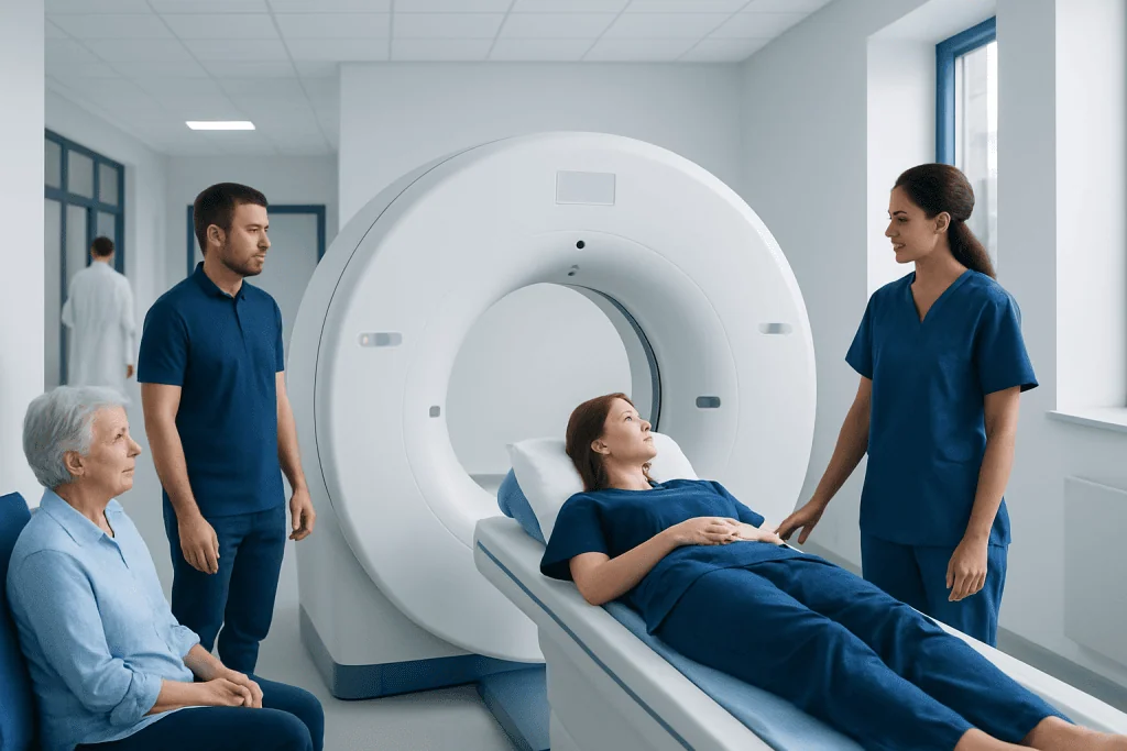
Radiology includes several imaging methods, and each test is selected for a different purpose. Some radiology exams use radiation, while others, such as ultrasound and MRI, do not.
Key Takeaways
- Radiology includes several imaging methods, and each test is selected for a different purpose.
- Some radiology exams use radiation, while others, such as ultrasound and MRI, do not.
- Preparation depends on the test and may include fasting, removing metal items, or using contrast material.
- Radiologists interpret images and share findings with the doctor who ordered the test.
- Many radiology exams are quick and noninvasive, though some may require injections or brief stillness.
- Patients should seek medical advice promptly for urgent symptoms, even if imaging has not yet been done.
Radiology is the medical specialty that uses imaging to look inside the body, helping doctors diagnose, monitor, and sometimes treat health conditions. For patients, radiology usually means a scan or image-based test such as an X-ray, ultrasound, CT, MRI, or mammogram, chosen to answer a specific medical question.
Overview: what radiology means for patients
Radiology is the branch of medicine that uses images to help doctors see structures and processes inside the body. In everyday care, this helps confirm a diagnosis, check the cause of symptoms, guide treatment decisions, and follow a condition over time. It is not one single test but a group of imaging methods, each with different strengths.
For patients, radiology often begins with a clear clinical question, such as whether a bone is broken, why abdominal pain is happening, or whether a lump needs closer assessment. The most suitable exam is then chosen based on the body area, the symptom, the patient’s age, pregnancy status, and how detailed the images need to be.
Radiology also includes image-guided procedures. In these cases, imaging is used not only to look at the body but to help doctors perform a targeted procedure more safely and accurately, such as a biopsy or drainage. In some settings, minimally invasive care may be planned through interventional radiology procedures when appropriate.
Types of radiology tests and how they differ
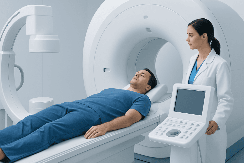
Common radiology tests include X-rays, ultrasound, computed tomography (CT), magnetic resonance imaging (MRI), mammography, and nuclear medicine scans. Each creates images in a different way. X-rays are often used for bones and chest problems, ultrasound is useful for soft tissues and pregnancy, CT gives fast detailed cross-sectional images, and MRI is especially helpful for the brain, spine, joints, and many soft tissues.
One important difference is whether the test uses ionizing radiation. X-rays, CT, mammography, and many nuclear medicine studies do use radiation. Ultrasound and MRI do not. This does not mean one type is always better than another. The right exam is the one most likely to answer the medical question accurately while keeping risks as low as reasonably possible.
Some tests may use a contrast material to make structures easier to see. Contrast can be swallowed, injected into a vein, or less commonly given in other ways, depending on the exam. Patients should tell their care team about kidney disease, allergies, asthma, pregnancy, breastfeeding, or prior reactions to contrast so the team can plan safely.
- X-ray: often used for fractures, chest conditions, and some abdominal concerns
- Ultrasound: commonly used for the abdomen, pelvis, blood flow, thyroid, and pregnancy
- CT scan: detailed images often used in emergency care, trauma, infection, and cancer evaluation
- MRI scan: detailed soft-tissue imaging, often used for the brain, nerves, spine, joints, and organs
- Mammography: breast imaging used for screening and diagnostic assessment
Why a doctor may recommend radiology

Radiology is usually recommended to answer a specific medical question rather than as a general check without a reason. A doctor may request imaging to investigate symptoms such as pain, swelling, shortness of breath, headaches, weakness, bleeding, or a suspected infection. Imaging can also help detect complications after surgery or injury, or monitor a known condition over time.
In some cases, radiology is used for screening before symptoms appear. Examples include mammography for breast cancer screening and certain imaging studies for people with higher risk based on age, family history, or other medical factors. Screening decisions are individualized and should be discussed with a qualified clinician.
Imaging may also be part of care planning for conditions that involve multiple specialists. For example, scans can support the assessment of brain tumors or help evaluate blood flow problems that may need vascular surgery. When cancer is suspected or confirmed, imaging often plays a central role in diagnosis, staging, treatment planning, and follow-up.
What to expect before, during, and after an imaging exam
Preparation depends on the type of exam. Many X-rays need little or no preparation. Ultrasound of the abdomen may require fasting, while pelvic ultrasound may require a full bladder. CT or MRI sometimes involves contrast, so patients may be asked about kidney function, medications, allergies, and implanted devices. Metal objects usually need to be removed before MRI, and some implanted devices may need special review for safety.
During the test, most patients lie still on a table while images are taken. Ultrasound involves a handheld probe and gel on the skin. CT is usually quick and may feel like passing through a short ring-shaped scanner. MRI takes longer and can be noisy, but ear protection is provided. Some people feel anxious in enclosed spaces, so it is helpful to tell the team in advance if claustrophobia is a concern.
After the exam, most patients can return to normal activities right away. If contrast was used, the care team may advise hydration or observation for a short time. Results are interpreted by a radiologist, a doctor trained to analyze medical images, and then shared with the referring physician. The timing of results varies depending on how urgent the exam is and what follow-up is needed.
Benefits, safety, and possible risks
The main benefit of radiology is that it gives doctors information that cannot be learned from symptoms or a physical exam alone. This can lead to earlier diagnosis, more accurate treatment planning, and better monitoring of recovery or progression. In some situations, imaging can help avoid unnecessary surgery by clarifying what is happening inside the body.
Radiology is generally safe, but every test has considerations. Imaging that uses radiation is performed only when the expected benefit outweighs the small potential risk. Radiology teams use the lowest appropriate dose and tailor exams to the patient’s size, age, and clinical need whenever possible. Patients should tell the team if they are pregnant or think they may be pregnant.
Contrast materials are also generally safe, but side effects and allergic-type reactions can occur. Kidney function may need checking before some contrast-enhanced CT or MRI studies. MRI is not harmful because it does not use radiation, but it may not be suitable for everyone with certain implants or metal fragments. Questions about safety are appropriate and should always be discussed before the test begins.
How radiology helps guide treatment
Radiology does more than detect disease. Imaging often helps determine how serious a condition is, whether treatment is working, and whether a different plan is needed. For example, repeated scans may show whether an infection is improving, whether a fracture is healing well, or whether inflammation has settled after treatment.
Imaging can also be used to guide procedures with precision. A radiologist may use ultrasound, CT, or fluoroscopy to place a needle for a biopsy, drain a collection of fluid, or support other targeted interventions. This image guidance can reduce damage to nearby tissues and help the doctor reach the correct area more accurately.
In complex conditions, radiology works together with many specialties. Patients with suspected vascular problems may need imaging before cardiovascular surgery, while people with neurological symptoms may undergo advanced MRI or CT to assess causes ranging from stroke to tumors. Near the end of a patient’s care pathway, multidisciplinary teams may review imaging together to decide on the most appropriate next steps. Acibadem International’s multidisciplinary specialists and JCI-accredited hospitals diagnose and treat a wide range of conditions for international patients using coordinated imaging and specialist care.
When to seek medical care
Radiology is an important tool, but urgent symptoms should not be delayed while waiting to arrange a scan. Immediate medical care is important for chest pain, sudden weakness, facial drooping, trouble speaking, severe shortness of breath, major injury, seizures, uncontrolled bleeding, or a sudden severe headache. These symptoms may need urgent assessment first, with imaging arranged quickly as part of emergency care.
Non-urgent medical review is appropriate for persistent pain, a new lump, ongoing cough, recurrent headaches, unexplained weight loss, abdominal swelling, blood in urine or stool, or symptoms that do not improve as expected. A doctor can decide whether imaging is necessary and which test is most useful. Self-requesting the wrong test may delay answers rather than speed them up.
Patients should also contact their doctor if they have questions after an imaging test, if symptoms worsen despite normal results, or if they develop a delayed reaction after contrast material. A normal scan can be reassuring, but it does not replace clinical follow-up when symptoms continue.
Frequently asked questions
What is radiology in simple terms?
Radiology is the medical specialty that uses imaging tests to look inside the body. It helps doctors diagnose problems, monitor conditions, and sometimes guide treatment procedures.
Does every radiology test use radiation?
No. X-rays, CT scans, mammograms, and many nuclear medicine tests use radiation, but ultrasound and MRI do not. The choice of test depends on what the doctor needs to assess.
Is radiology safe?
Radiology is generally safe when used appropriately. Imaging teams weigh the benefits and risks, use the lowest suitable radiation dose when needed, and check for factors such as pregnancy, kidney disease, implants, or prior contrast reactions.
Do patients need to prepare for a radiology exam?
Sometimes. Preparation depends on the exam and may include fasting, drinking water, avoiding metal items, or arriving early for a contrast-related safety check. The imaging center usually provides instructions ahead of time.
Who reads the images and explains the results?
A radiologist, who is a doctor specially trained in medical imaging, reviews the images and writes a report. The doctor who ordered the test then explains what the results mean in the context of the patient’s symptoms and overall care.
What happens if an imaging test is normal but symptoms continue?
A normal result can be reassuring, but it does not always explain every symptom. If symptoms persist, worsen, or change, follow-up with a doctor is important because another test, repeat imaging, or a different type of evaluation may be needed.
References
- Radiological Society of North America
- American College of Radiology
- National Institutes of Health
- World Health Organization
- U.S. Food and Drug Administration
This article is for general information only and is not a substitute for professional medical advice. Please consult a qualified doctor about your individual situation.
Explore treatments in Turkey — costs, top hospitals & a free quote
JCI-accredited · board-certified surgeons · reply within 24h
Add us as a Preferred Source to see more of our trusted health content across Google Search, AI Overviews and Discover.

More from the Health Library

Eyelid Exercises: What Patients Need to Know

Quervain’s: What Patients Need to Know

Testosterone Gel: An Evidence-Based Guide for Patients

Interferons: An Evidence-Based Guide for Patients

Erythematous: A Complete Medical Overview