

Radiomics Explained: How Advanced Scan Analysis May Support Cancer Diagnosis
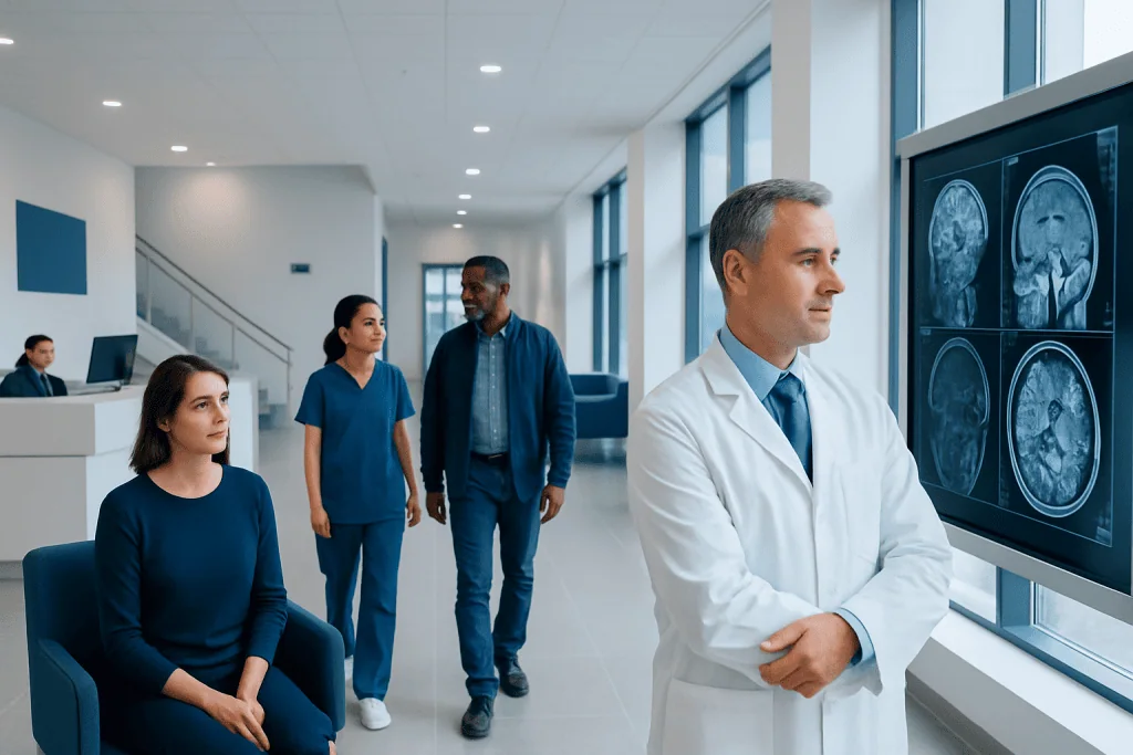
Radiomics converts CT, MRI, PET, and other scan images into measurable data features. It may help support cancer detection, tumor characterization, and treatment planning.
Key Takeaways
- Radiomics converts CT, MRI, PET, and other scan images into measurable data features.
- It may help support cancer detection, tumor characterization, and treatment planning.
- Radiomics is usually combined with radiology, pathology, and clinical information.
- Results can be affected by image quality, scanner differences, and data analysis methods.
- This technology is promising, but it is not a stand-alone diagnosis for most patients.
Radiomics is a method that extracts detailed data from routine medical scans to look for patterns the human eye may not easily detect. In cancer care, it may support diagnosis, risk assessment, and treatment planning, but it complements rather than replaces expert medical judgment.
Overview: What Radiomics Means
Radiomics is a form of advanced image analysis that turns medical scans into large amounts of measurable information. Instead of looking only at a scan as a picture, radiomics examines the image as data. It can measure features such as shape, texture, intensity, and the way these patterns vary across a tumor or another area of concern.
In cancer care, this approach may help doctors learn more from scans such as CT, MRI, or PET. Some image features may reflect how a tumor is behaving, how aggressive it may be, or how it might respond to treatment. These features are sometimes called imaging biomarkers because they may provide clues about disease characteristics.
Radiomics is different from routine image reading, but it does not replace it. A radiologist still interprets the scan, and the treating team still relies on symptoms, physical examination, laboratory findings, and often biopsy results. Radiomics is best understood as a supportive tool that may add another layer of information to the overall assessment.
How Advanced Scan Analysis Works
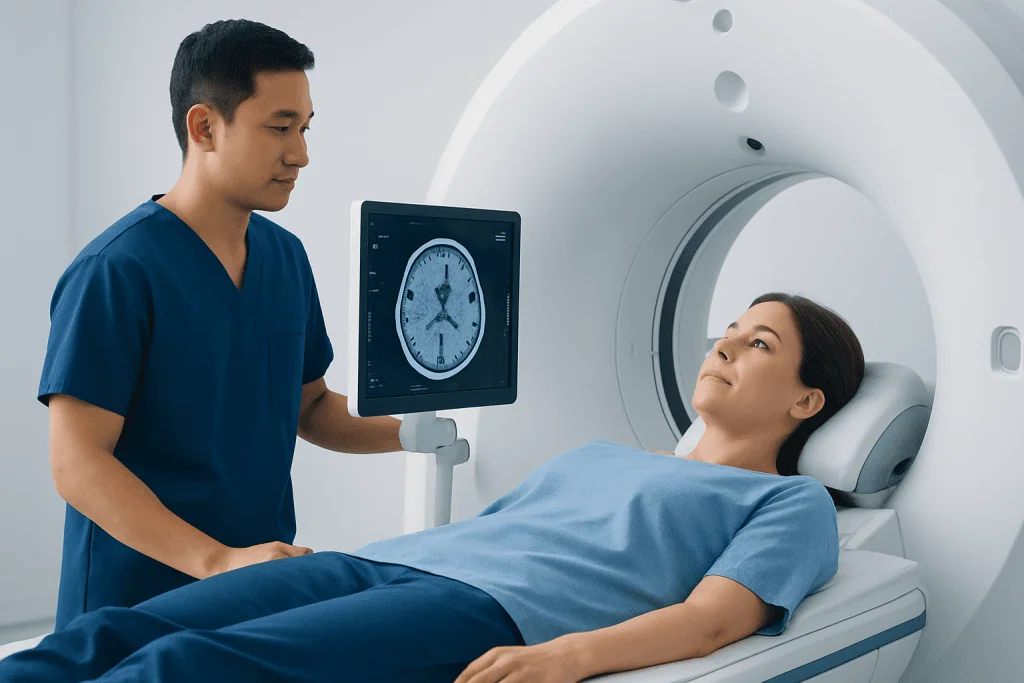
The process begins with a medical image, usually from a standard scan that a patient may already need as part of care. After the scan is performed, a specific region is selected for analysis. This might be the whole tumor, part of a tumor, lymph nodes, or nearby tissue. The selected area is then processed by specialized software.
The software extracts quantitative features from the image. These can include simple measurements such as size and shape, as well as more complex texture patterns that may suggest how uniform or irregular a lesion is. In some systems, these data are then combined with machine learning methods to look for patterns linked to diagnosis, prognosis, or treatment response.
Several steps are important for reliable results, including consistent scanning technique, careful selection of the area being measured, and appropriate data analysis. Small differences in how an image is taken or processed can affect the numbers produced. For that reason, radiomics works best in settings that use standardized protocols and expert review.
Radiomics is often discussed alongside artificial intelligence, but they are not exactly the same. Radiomics focuses on extracting interpretable image features, while AI may use those features, or the image itself, to build predictive models. In practice, the two approaches often overlap in modern imaging research and clinical innovation.
How Radiomics May Support Cancer Diagnosis
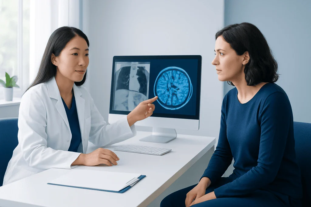
Radiomics may help support cancer diagnosis by revealing image patterns that are too subtle to identify visually. For example, it may help distinguish between tissue that appears more likely benign and tissue that appears more suspicious, although it does not confirm cancer on its own. In many cases, a tissue diagnosis from pathology remains the standard way to confirm malignancy.
Another possible role is tumor characterization. Two tumors can look similar on a routine scan yet behave differently. Radiomic analysis may help estimate whether a tumor is more likely to be slow growing or more aggressive, and whether there may be areas of biological variation within the same tumor. This can be useful because cancers are often not uniform throughout.
Radiomics is also being studied for its ability to support treatment planning and follow-up. It may help identify patients who could benefit from closer monitoring, additional imaging, or discussion in a multidisciplinary tumor board. In some settings, it may complement PET-CT imaging or MRI evaluation when doctors need a more detailed picture of disease behavior.
Importantly, radiomics is usually one part of a larger decision process. Doctors compare scan findings with pathology, molecular testing, and the patient’s overall condition. This integrated approach is especially important in complex diseases such as lung cancer or breast cancer, where treatment decisions depend on many factors.
Potential Benefits and Current Limitations
One of the main potential benefits of radiomics is that it uses information already present in routine imaging. This means it may provide additional insight without always requiring a new invasive procedure. Because scans can be repeated over time, radiomics may also help track changes during treatment or recovery in a noninvasive way.
Another advantage is that radiomics may capture tumor heterogeneity, meaning differences within a tumor that may matter clinically. A biopsy samples only a small area, while imaging can assess a larger region. Although a biopsy is still essential in many cases, radiomics may contribute a broader view of the tumor environment.
At the same time, there are important limitations. Results can vary depending on the scanner type, image resolution, contrast use, and software methods. Different hospitals may produce somewhat different radiomic features from similar scans if protocols are not standardized. This is one reason radiomics is still being actively validated.
There is also the risk of overinterpreting data. A mathematical pattern is not automatically clinically meaningful. To be useful, radiomic models must be tested carefully across different patient groups and compared with established standards of care. Patients should know that while radiomics is promising, it is not yet a replacement for expert imaging review, pathology, or evidence-based cancer assessment.
Which Scans and Cancers Are Most Commonly Studied
Radiomics can be applied to several types of medical imaging. CT is widely studied because it is common in cancer diagnosis and staging. MRI is particularly useful for soft tissues and organs such as the brain, liver, prostate, and breast. PET, often combined with CT, can provide metabolic information that may be especially valuable when paired with radiomic analysis.
Researchers have explored radiomics in many cancers, including lung, breast, brain, colorectal, prostate, and liver cancers. It is also being studied in lymphomas and in some noncancer conditions. The goal is not only to detect disease, but also to estimate likely behavior, treatment sensitivity, and the chance of recurrence.
In real-world care, radiomics may be most helpful when used in diseases where imaging already plays a central role. For instance, detailed analysis may be added to CT scan assessment or to serial imaging follow-up over time. In selected cases, it may also be discussed alongside procedures such as biopsy when the care team is trying to match imaging findings with tissue results.
Even when a cancer type has been widely studied, radiomics does not work in isolation. Every patient is different, and imaging features must be interpreted in context. The most reliable use comes from a multidisciplinary approach that includes radiologists, oncologists, pathologists, surgeons, and other specialists as needed.
What Patients Can Expect in Practice
For most patients, radiomics does not change the basic experience of having a scan. The imaging test itself is usually performed in the usual way. If radiomic analysis is used, it typically happens afterward, using specialized software to evaluate the images that have already been collected.
Patients may not always see the word radiomics in a report, because some centers use these methods mainly in research programs or as part of advanced multidisciplinary review. When it is used clinically, the result may appear as an additional assessment that helps estimate risk, characterize a lesion, or support treatment planning. It is usually one factor among many rather than the only basis for a decision.
It is reasonable for patients to ask how the scan results will be interpreted and whether advanced image analysis is part of the process. Helpful questions include whether the findings are certain or uncertain, whether more imaging or a biopsy is needed, and how the scan information will affect the treatment plan. Clear discussion can help patients understand both the value and the limits of this technology.
At centers with advanced imaging and cancer programs, radiomics may be reviewed by teams experienced in complex image interpretation. Near the end of the care pathway, some international patients may seek evaluation at institutions such as Acibadem International, where multidisciplinary specialists and JCI-accredited hospitals diagnose and treat cancer-related conditions using modern imaging and treatment approaches.
Safety, Privacy, and Questions to Discuss with a Doctor
Radiomics itself does not add radiation or discomfort beyond the scan that is already being performed. Safety considerations are therefore mainly related to the imaging test involved. For example, CT uses ionizing radiation, MRI may involve contrast agents or special screening for metal implants, and PET requires a radiotracer. The medical team weighs these factors before recommending a test.
Because radiomics depends on digital data, privacy and data handling are also important. Hospitals and research centers should follow appropriate standards for storing and analyzing imaging information. When AI or machine learning tools are involved, patients may wish to ask whether the tool has been clinically validated and how results are reviewed by specialists.
Useful questions for a doctor include:
- What does my scan show, and how certain is the interpretation?
- Would advanced image analysis add meaningful information in my case?
- Do I still need biopsy or other tests to confirm the diagnosis?
- How will these imaging findings affect my treatment options or follow-up plan?
Patients should seek timely medical advice if they have persistent symptoms, a new mass, unexpected weight loss, unexplained bleeding, or scan findings that require follow-up. Early evaluation does not always mean cancer is present, but it helps ensure that the right tests and next steps are considered without delay.
Frequently asked questions
Is radiomics the same as artificial intelligence?
Not exactly. Radiomics focuses on extracting measurable features from medical images, while artificial intelligence may use those features or the raw images to build prediction models. In many modern systems, the two methods are used together.
Can radiomics diagnose cancer by itself?
Usually no. Radiomics may support cancer diagnosis by adding information from imaging, but it does not replace a doctor's assessment or, when needed, a biopsy. Pathology remains the main way to confirm many cancers.
Which scans can be used for radiomics?
Radiomics can be applied to CT, MRI, and PET scans, among others. The choice depends on the organ being studied and the clinical question. These scans are often already part of standard cancer evaluation.
Does radiomics change the scan experience for patients?
Usually it does not. The analysis often takes place after the scan is completed, using software to study the images in more detail. Most patients undergo the same imaging test they would have had anyway.
Why is radiomics not used everywhere yet?
One reason is that results can vary if scan protocols and software methods are not standardized. Another is that predictive models need careful validation in different hospitals and patient groups. As evidence grows, clinical use may become more consistent.
Can radiomics help choose cancer treatment?
It may help support treatment planning by suggesting how a tumor is behaving or how it changes over time. However, treatment decisions are still based on the full clinical picture, including pathology, staging, general health, and patient preferences.
References
- World Health Organization
- National Cancer Institute
- Radiological Society of North America
- European Society for Medical Oncology
- American College of Radiology
This article is for general information only and is not a substitute for professional medical advice. Please consult a qualified doctor about your individual situation.
Oncology care in Turkey — second opinion and treatment plan
JCI-accredited · board-certified surgeons · reply within 24h
Add us as a Preferred Source to see more of our trusted health content across Google Search, AI Overviews and Discover.

More from the Health Library



Related Specialists

Dr. Halil Algan
Treatment of Pain Algology
Dr. Sevim Buzkan Ertuğrul
Acibadem Life Clinical Service
Dr. Hamza Furkan Şen
Gynecology & Obstetrics
Assoc. Prof. Dr. Nefise Nazlı Yenigül
Gynecology & Obstetrics