Retinoschisis: An Evidence-Based Guide for Patients

Retinoschisis means splitting within the retinal tissue; it is not the same as retinal detachment, although the conditions can be related. The two main forms are inherited X-linked juvenile retinoschisis and age-related degenerative retinoschisis.
Key Takeaways
- Retinoschisis means splitting within the retinal tissue; it is not the same as retinal detachment, although the conditions can be related.
- The two main forms are inherited X-linked juvenile retinoschisis and age-related degenerative retinoschisis.
- Symptoms may be absent, especially in peripheral disease, but central retinal involvement can reduce detailed or side vision.
- Dilated eye examination and retinal imaging help an ophthalmologist distinguish retinoschisis from retinal detachment and other retinal disorders.
- New flashes, a sudden increase in floaters, a shadow or curtain in vision, or sudden vision loss require urgent eye assessment.
Retinoschisis is an eye condition in which layers of the retina, the light-sensitive tissue at the back of the eye, separate from one another. Many cases remain stable and require regular monitoring, while others need treatment if complications such as retinal detachment develop.
What Is Retinoschisis?
Retinoschisis is a condition in which the retina separates into layers. The retina is the thin, light-sensitive tissue lining the back of the eye. It converts light into signals that travel through the optic nerve to the brain, allowing a person to see. When retinal layers split, small fluid-filled spaces can form within the retina.
Retinoschisis can affect the central retina, called the macula, which supports sharp reading and facial recognition vision. It can also affect the outer, or peripheral, retina, which contributes to side vision. The impact varies widely: some people have no noticeable symptoms, while others experience reduced clarity, blind spots, or problems with side vision.
Importantly, retinoschisis is different from a retinal detachment. In retinal detachment, the retina lifts away from the supporting tissue beneath it. In retinoschisis, the split occurs within the retina itself. However, in a minority of cases, breaks in retinal layers can allow a retinal detachment to develop, which is why continued ophthalmic follow-up is important.
The Main Types of Retinoschisis
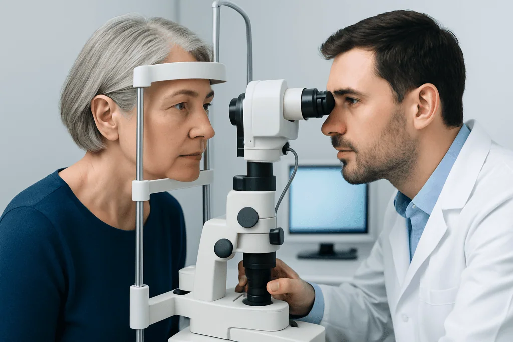
The two most recognized forms have different causes, typical ages of onset, and patterns of retinal involvement. An ophthalmologist can identify the likely type by considering a person’s age, family history, symptoms, and detailed retinal examination.
X-linked juvenile retinoschisis is an inherited condition that mainly affects boys and men. It commonly becomes apparent in childhood, although severity differs substantially between individuals. It is usually related to changes in the RS1 gene, which is important for normal retinal structure. The central retina often has a spoke-like pattern of small cystic spaces, and vision may be reduced from an early age.
Degenerative retinoschisis, also called acquired or senile retinoschisis, usually occurs later in adulthood. It tends to involve the far peripheral retina and may affect both eyes. It is often found during a routine dilated eye examination in someone without symptoms. Most people with degenerative retinoschisis retain stable central vision and do not require a procedure.
Symptoms and How Vision May Be Affected
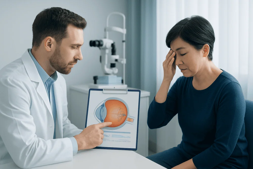
Retinoschisis does not always cause symptoms. This is particularly common in degenerative retinoschisis because peripheral retinal changes may not affect the macula or central visual field. A person may only learn about the condition after an optometrist or ophthalmologist notices it during a dilated retinal examination.
When symptoms occur, they depend on the area of retina involved. Juvenile retinoschisis may cause reduced sharpness of vision, difficulty reading small print, reduced contrast sensitivity, or problems with schoolwork that relies on fine visual detail. Some people notice a patch of missing or distorted vision. Strabismus, also known as an eye turn, or involuntary eye movements can occur in more severe childhood cases.
Peripheral retinoschisis can occasionally cause a gradual area of reduced side vision. Symptoms such as flashes of light, a sudden shower of new floaters, a dark curtain or shadow moving across vision, or sudden loss of vision are not typical signs to ignore. They can indicate a retinal tear or detachment and should be assessed urgently.
- Blurring or reduced sharp vision, particularly in childhood-onset disease
- Difficulty with reading, recognizing faces, or seeing contrast
- Areas of missing or reduced side vision
- Flashes, new floaters, or a curtain-like shadow when a retinal complication occurs
Causes and Risk Factors
X-linked juvenile retinoschisis is caused by an inherited genetic change, most often involving the RS1 gene on the X chromosome. Because males have one X chromosome, they are more likely to develop the condition when they inherit an altered RS1 gene. Females may carry the genetic change and can pass it to their children, but they are usually unaffected or have minimal findings. Genetic counseling can help families understand inheritance patterns and discuss testing when appropriate.
Degenerative retinoschisis is associated with aging changes in the peripheral retina. Its precise cause is not fully understood. It is not considered contagious and is not caused by reading, screen use, or ordinary physical activity. It may be identified more often in people with farsightedness, but it can occur in others as well.
Having retinoschisis does not automatically mean a person will develop retinal detachment. Risk becomes more relevant if there are retinal holes in both layers of the schisis area, progressive fluid beneath the retina, or symptoms suggesting a tear or detachment. A specialist evaluates these features individually rather than predicting risk from the diagnosis name alone.
How Retinoschisis Is Diagnosed and Monitored
Diagnosis begins with a review of vision changes, family history, previous eye conditions, and general health. The ophthalmologist checks visual acuity and examines the retina after dilating the pupils with eye drops. Dilation gives a clearer view of the central and peripheral retina and helps distinguish retinoschisis from retinal detachment, retinal tears, cysts, and other causes of retinal changes.
Optical coherence tomography, often called OCT, is especially useful when the macula is involved. This noninvasive scan produces cross-sectional images of retinal layers and can show the location and extent of splitting. Wide-field retinal photography, visual field testing, and ultrasound may also be used in selected situations, particularly when the far peripheral retina needs evaluation or the view inside the eye is limited.
For suspected juvenile retinoschisis, genetic testing may confirm a change in the RS1 gene and support family counseling. Eye specialists may also recommend baseline vision testing and periodic assessments to monitor changes over time. Follow-up intervals vary according to the type of retinoschisis, symptoms, retinal findings, age, and whether complications are present.
Treatment Options and Everyday Eye Care
There is no single treatment needed for every person with retinoschisis. Many cases of degenerative retinoschisis are stable and are managed with observation, scheduled dilated examinations, and clear advice about warning symptoms. Preventive laser treatment is not routinely used for uncomplicated retinoschisis because most cases do not progress to retinal detachment.
When retinal detachment develops or is judged likely to progress, a retinal specialist may recommend treatment. Depending on the findings, this can include laser therapy, cryotherapy, scleral buckling, vitrectomy, or a combination of approaches. The purpose is to repair or support the retina and preserve as much vision as possible. The most suitable approach depends on the exact location and extent of retinal breaks or detachment.
Management of X-linked juvenile retinoschisis focuses on monitoring vision, treating complications when they occur, and supporting daily function. In selected patients with cystic changes in the macula, a specialist may consider medicines that can reduce retinal fluid; response and appropriateness vary, and ongoing monitoring is required. Low-vision support, updated glasses, educational accommodations, and visual aids can be valuable when central vision is affected.
People with retinoschisis should attend recommended eye appointments and tell their eye specialist about any significant visual change. Ordinary daily activity is often possible, but an individual with a recent retinal complication or surgery should follow their specialist’s guidance about work, sports, travel, and recovery. Acibadem International’s multidisciplinary eye specialists at JCI-accredited hospitals assess and treat retinal conditions for international patients when specialist care is needed.
When to Seek Medical Care
Anyone with diagnosed retinoschisis should continue routine follow-up with an ophthalmologist, even when vision feels unchanged. Regular examinations allow the care team to compare retinal findings over time and identify uncommon complications early. Children with suspected inherited retinal disease should be assessed by an ophthalmologist experienced in pediatric or inherited retinal conditions.
Urgent same-day eye care is recommended for a sudden onset or marked increase in floaters, repeated flashes of light, a dark shadow, a curtain-like area in the visual field, or sudden worsening of vision. These symptoms may be caused by a retinal tear or detachment and need prompt assessment. A person should not wait for a routine appointment if these warning signs appear.
A gradual change in reading vision, increasing distortion, new difficulty seeing in dim light, or a new patch of missing vision should also be discussed with an eye care professional soon. These symptoms can have several causes, and only a full examination can determine whether retinoschisis or another retinal condition is responsible.
Frequently asked questions
Is retinoschisis the same as retinal detachment?
No. Retinoschisis is a split between layers within the retina, while retinal detachment occurs when the retina separates from the tissue beneath it. Retinoschisis can occasionally be associated with retinal detachment, so ongoing eye monitoring is important.
Can retinoschisis cause blindness?
Many people with retinoschisis, especially the degenerative peripheral form, maintain useful vision and have stable disease. Significant vision loss is more likely when the macula is affected or when complications such as retinal detachment occur. Regular specialist care helps identify changes that may need treatment.
Is juvenile retinoschisis inherited?
Yes. X-linked juvenile retinoschisis is generally caused by changes in the RS1 gene and is most often inherited through families. It mainly affects boys and men, while women may carry the genetic change. Genetic counseling may help explain risks for relatives and future children.
Does degenerative retinoschisis require surgery?
Usually not. Most cases are observed with regular dilated eye examinations because they remain stable and do not threaten central vision. Surgery or other procedures may be considered only if a retinal detachment or another significant complication develops.
Can glasses correct vision loss from retinoschisis?
Glasses can correct refractive errors such as short-sightedness, farsightedness, or astigmatism, which may improve overall vision. However, glasses cannot repair the retinal splitting itself. If retinoschisis affects the macula, low-vision aids and practical visual support may also be helpful.
What symptoms should prompt urgent evaluation?
A sudden increase in floaters, flashes of light, a curtain or shadow over vision, or sudden vision loss should be assessed urgently by an eye professional. These symptoms may signal a retinal tear or detachment, which requires timely evaluation. Even if symptoms improve, medical advice should still be sought.
References
- American Academy of Ophthalmology
- National Eye Institute
- National Organization for Rare Disorders
- Orphanet
- Royal College of Ophthalmologists
This article is for general information only and is not a substitute for professional medical advice. Please consult a qualified doctor about your individual situation.
Explore treatments in Turkey — costs, top hospitals & a free quote
JCI-accredited · board-certified surgeons · reply within 24h
Add us as a Preferred Source to see more of our trusted health content across Google Search, AI Overviews and Discover.