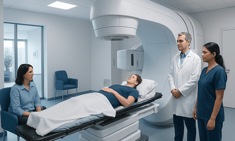
Retrograde Urogram Procedure: An Evidence-Based Patient Guide

A retrograde urogram usually refers to a retrograde pyelogram, which images the ureters and kidney drainage system through a cystoscope. The procedure is often performed when CT, ultrasound, or other imaging does not fully explain a urinary tract problem.
Key Takeaways
- A retrograde urogram usually refers to a retrograde pyelogram, which images the ureters and kidney drainage system through a cystoscope.
- The procedure is often performed when CT, ultrasound, or other imaging does not fully explain a urinary tract problem.
- Most people go home the same day and have mild burning with urination or blood-tinged urine briefly afterward.
- Possible risks include urinary infection, bleeding, contrast reaction, and rarely injury to the urinary tract.
- A retrograde urethrogram is a different test that examines the urethra, usually with X-ray contrast introduced at the urinary opening.
A retrograde urogram procedure is an imaging examination in which a urologist places contrast dye into the urinary tract through the bladder and takes X-ray images. It can help identify a blockage, stone, narrowing, injury, or other concern affecting urine flow from the kidneys to the bladder.
Overview: what the retrograde urogram procedure shows
A retrograde urogram procedure is a specialized X-ray examination of the urinary tract. In everyday clinical use, the term often describes a retrograde pyelogram: contrast dye is introduced through a thin tube passed into the bladder and then into one or both ureters, the tubes that carry urine from the kidneys to the bladder. X-ray images show the inside outline of the ureter and the kidney’s urine-collecting system.
The test is generally performed by a urologist during cystoscopy, often under sedation or anesthesia. It may be used to clarify an abnormality found on ultrasound, CT, or other imaging, or when a clinician needs detailed information before treating a stone, placing a ureteral stent, or investigating a possible obstruction.
Although the names sound similar, a retrograde urogram or retrograde pyelogram is not the same as a retrograde urethrogram. A retrograde urethrogram evaluates the urethra, the channel through which urine leaves the body. The appropriate test depends on where the suspected problem is located.
How the procedure works and who may need it
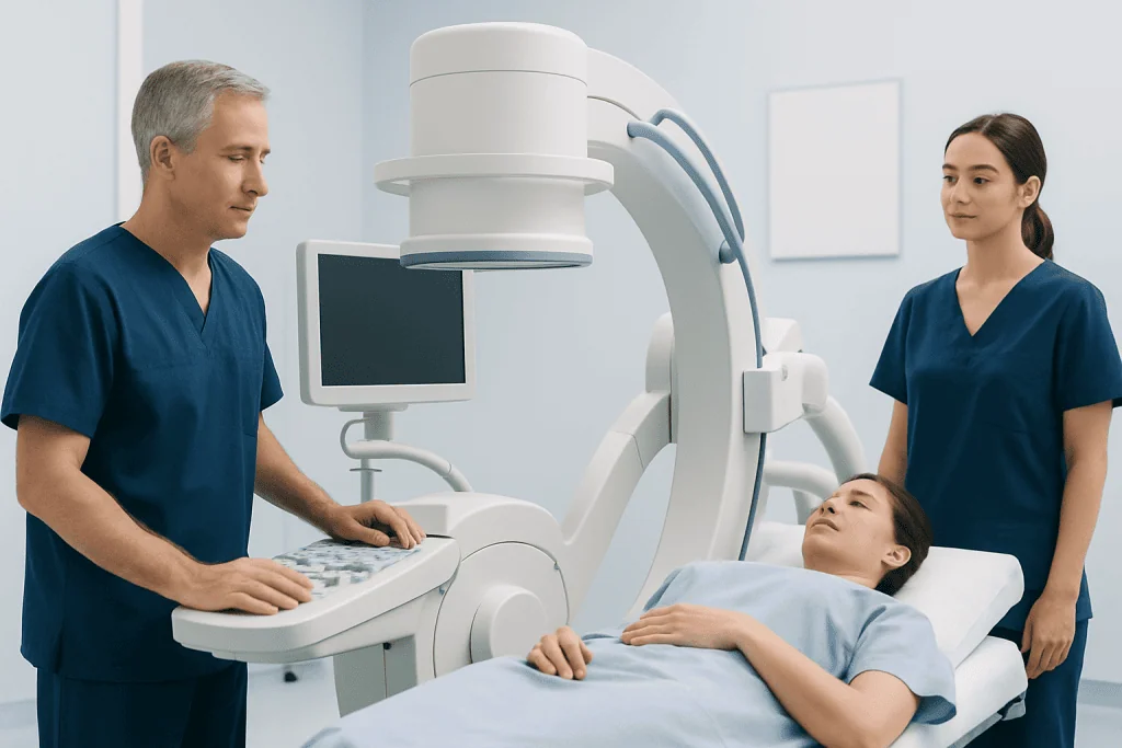
During a retrograde pyelogram, contrast travels “retrograde,” meaning in the opposite direction of normal urine flow. Rather than dye reaching the kidneys through a vein, it is placed directly into the ureter through a small catheter. This approach can provide clear, targeted images while avoiding intravenous contrast in selected situations.
A clinician may recommend the examination when there is concern for a ureteral stone, narrowing (stricture), blockage, leakage, injury, unexplained dilation of the kidney drainage system, or an abnormal finding that needs further assessment. It may also be part of planning or carrying out a urologic procedure, such as stone treatment or ureteral stent placement.
Before the test, the care team reviews medical history, current medicines, pregnancy possibility, prior reactions to contrast material, bleeding conditions, kidney problems, and signs of urinary infection. Active infection may need treatment before an elective procedure. People taking blood-thinning medicine should receive individualized instructions and should not stop prescribed medicine without guidance from the clinician managing it.
- It can be useful when detail about the ureter or renal pelvis is needed.
- It may help guide treatment during the same anesthetic session.
- It is not always necessary when less invasive imaging has already answered the clinical question.
Step by step: what happens during a retrograde urogram procedure

Preparation varies by setting and anesthesia plan. The patient may be asked not to eat or drink for a period before sedation or general anesthesia. On arrival, the team confirms identity, reviews allergies and medications, checks vital signs, and explains the planned procedure. A urine test may be obtained if infection is suspected.
The patient lies on an X-ray table, usually on their back. After anesthesia or sedation is given, the urologist inserts a cystoscope through the urethra into the bladder. This slim instrument contains a camera that allows the clinician to inspect the bladder and identify the openings of the ureters.
A very small catheter is guided through the cystoscope into the ureter opening. Contrast dye is introduced slowly while X-ray images, often fluoroscopic images, are taken. The clinician may repeat this on the other side if needed. If the findings require action, a stent may be placed or another planned treatment may proceed. The instruments are removed once the examination is complete.
The imaging portion itself is commonly brief, but the total time in the procedure area depends on anesthesia, whether one or both sides are examined, and whether treatment is performed at the same time. The care team can provide the best estimate for an individual situation.
Benefits, limitations and possible risks
The main benefit of a retrograde pyelogram is direct, detailed visualization of the ureters and kidney drainage system. It can help distinguish between a true blockage and other causes of urinary tract dilation, locate a narrowing or filling defect, and support safe planning for a procedure. Because the contrast is administered directly into the urinary tract, it may be considered when intravenous contrast is not suitable for a particular patient.
Like any invasive examination, it has limitations and risks. It does not assess every cause of urinary symptoms, and it may not replace CT, ultrasound, urine testing, or other evaluation. The result needs to be interpreted alongside symptoms, examination findings, laboratory results, and previous imaging.
Temporary burning during urination, frequent urination, mild lower abdominal discomfort, and a small amount of blood in the urine can occur after cystoscopy. Less common risks include urinary tract infection, more significant bleeding, injury or perforation of the urethra, bladder, or ureter, and a reaction to contrast material. Radiation exposure from fluoroscopy is kept as low as reasonably achievable, but it is still considered when deciding whether the test is appropriate.
Patients should contact their clinical team promptly if they develop fever, chills, worsening pain, inability to pass urine, heavy bleeding or blood clots in the urine, persistent vomiting, rash, breathing difficulty, or symptoms that do not improve as expected.
Recovery timeline and self-care after cystoscopy with retrograde pyelogram
Most people recover at home after a same-day retrograde pyelogram. If sedation or general anesthesia was used, they will need a responsible adult to take them home and should avoid driving, alcohol, important decisions, and operating machinery until the effects have fully worn off, according to the discharge instructions.
Mild stinging when passing urine, urinary urgency, and light pink urine may last for a day or two after cystoscopy. Drinking fluids, if the clinician has not advised fluid restriction, can support normal urine flow and may reduce irritation. Rest is sensible on the day of the procedure, and many people return to usual light activity within 24 to 48 hours.
If a ureteral stent was inserted, recovery can feel different. Stents may cause urinary frequency, urgency, flank or bladder discomfort, or intermittent blood in the urine until removal. The urology team will explain stent care, planned removal, activity advice, and medicines that may be appropriate for symptom relief.
For people seeking coordinated assessment of urinary obstruction, stones, strictures, or related concerns, Acibadem International’s multidisciplinary specialists and JCI-accredited hospitals provide diagnostic and treatment services for international patients.
What is a retrograde urogram?
A retrograde urogram is a contrast X-ray study in which dye is placed into the urinary tract through the bladder rather than injected into a vein. In many clinical settings, it refers specifically to a retrograde pyelogram, which outlines the ureters and the renal pelvis and calyces, the urine-collecting spaces within the kidneys.
The procedure is commonly combined with cystoscopy. It may reveal a ureteral blockage, narrowing, stone, leak, or other structural change that affects urine drainage. A urologist may also use it during an intervention, for example to guide placement of a ureteral stent or to plan treatment for a urinary stone.
The phrase “urogram” can be used differently across countries and institutions. Patients can ask the ordering clinician exactly which structures will be examined, whether cystoscopy is included, what anesthesia is planned, and whether treatment might occur during the same visit.
Is a retrograde urethrogram painful?
A retrograde urethrogram may cause temporary pressure, discomfort, or a burning sensation, but it is usually designed to be tolerable. For this test, contrast is gently introduced through the opening of the urethra while X-ray images are taken. Local anesthetic gel may be used, and the care team will explain what sensations to expect.
Discomfort can vary depending on urethral irritation, injury, narrowing, anxiety, and the technique needed for the examination. The test is typically shorter than a procedure involving cystoscopy and ureter catheterization, although the individual preparation and imaging experience will vary.
Patients should tell the radiology or urology team if they have severe pain, a history of urethral surgery or trauma, active urinary symptoms, latex allergy, or a previous contrast reaction. Afterward, mild burning with urination can occur briefly, but severe pain, fever, significant bleeding, or inability to urinate should be medically assessed.
How much does a retrograde urethrogram cost?
The cost of a retrograde urethrogram varies substantially by country, hospital, insurance coverage, imaging setting, whether a specialist consultation is included, and whether additional tests or treatment are needed. It is therefore not possible to give one reliable price that applies to every patient.
Before scheduling, patients can ask the hospital or insurer for an individualized estimate. Helpful questions include whether the estimate includes the clinician’s fee, imaging and contrast materials, facility charges, anesthesia if applicable, laboratory testing, follow-up visits, and treatment of any finding discovered during the evaluation.
A retrograde urethrogram and a retrograde pyelogram are different tests and may have different resource requirements. The ordering clinician can explain which examination is medically indicated; choosing a test should be based on the clinical question rather than cost alone.
When to seek medical care
Urgent medical assessment is important for severe flank or abdominal pain, fever or chills with urinary symptoms, inability to urinate, vomiting that prevents fluid intake, visible blood clots in the urine, or symptoms of a serious allergic reaction such as swelling of the face or throat, wheezing, or difficulty breathing. These symptoms can have several causes and should not be managed only with home care.
After a retrograde urogram procedure, the treating team should be contacted if burning, light bleeding, or discomfort becomes worse rather than gradually improving; if fever develops; or if there is persistent heavy bleeding, increasing abdominal or back pain, or trouble passing urine. Clear discharge instructions should take priority because they are tailored to the procedure performed and the patient’s health needs.
For non-urgent questions about results, recovery, or persistent urinary symptoms, follow-up with a urologist is appropriate. The next steps may include observation, urine tests, further imaging, medication, stone management, stenting, or another treatment depending on the cause identified.
Frequently asked questions
How long does it take to recover from a cystoscopy with retrograde pyelogram?
Most people return home the same day and feel well enough for light activities within 24 to 48 hours. Mild burning with urination, urgency, or light blood-tinged urine may occur briefly. Recovery may take longer if a stent is placed or another treatment is performed during the procedure.
What is a retrograde urogram?
A retrograde urogram is an X-ray contrast examination of the urinary tract performed through the bladder. It commonly refers to a retrograde pyelogram, which shows the ureters and the kidney drainage system. It is often done during cystoscopy by a urologist.
Is a retrograde urethrogram painful?
A retrograde urethrogram can cause pressure, temporary burning, or discomfort as contrast is introduced into the urethra. Local anesthetic gel may be used to improve comfort. Severe pain is not expected and should be reported to the care team.
How much does a retrograde urethrogram cost?
Costs differ by location, hospital, insurance coverage, specialist fees, and whether additional testing or care is required. A hospital billing team or insurer can provide an individualized estimate before the examination. Patients should ask what is included in the quoted amount.
Will a retrograde pyelogram show kidney stones?
A retrograde pyelogram can show a filling defect or blockage pattern that suggests a stone in the ureter or kidney drainage system. However, CT is often more effective for identifying and characterizing urinary stones. The best imaging test depends on symptoms, medical history, and the clinical question.
Do I need anesthesia for a retrograde urogram procedure?
Many retrograde pyelograms are done with sedation or anesthesia because cystoscopy and ureter catheterization can be uncomfortable. The exact plan depends on the setting, the patient’s health, and whether treatment is planned at the same time. The anesthesia team and urologist will discuss preparation and safety considerations beforehand.
References
- American Urological Association
- Radiological Society of North America
- National Institute of Diabetes and Digestive and Kidney Diseases
- National Health Service
This article is for general information only and is not a substitute for professional medical advice. Please consult a qualified doctor about your individual situation.
Explore treatments in Turkey — costs, top hospitals & a free quote
JCI-accredited · board-certified surgeons · reply within 24h
Add us as a Preferred Source to see more of our trusted health content across Google Search, AI Overviews and Discover.