Skeletal System Labeled: A Complete Medical Overview
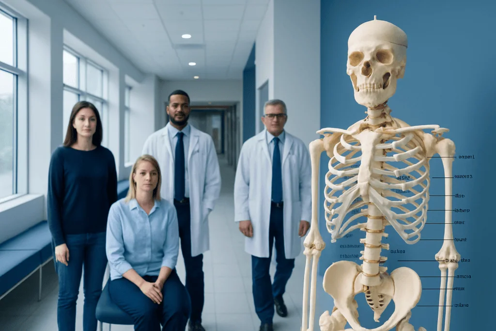
The adult human skeleton has 206 bones organized into axial and appendicular parts. A labeled skeleton is usually studied by body region, bone type, and function.
Key Takeaways
- The adult human skeleton has 206 bones organized into axial and appendicular parts.
- A labeled skeleton is usually studied by body region, bone type, and function.
- Bones do more than provide shape; they protect organs, store minerals, and support blood cell production.
- Joints, cartilage, ligaments, and bone marrow are essential parts of the skeletal system.
- Persistent bone pain, deformity, swelling, or limited movement should be assessed by a doctor.
A skeletal system labeled overview identifies the major bones, joints, and supporting tissues of the human body by region and function. Understanding how the skeleton is organized can make anatomy clearer and also help people recognize when bone or joint symptoms may need medical attention.
Overview: What a skeletal system labeled diagram shows
A skeletal system labeled diagram shows the major bones and supporting structures of the body, usually grouped into the skull, spine, chest, upper limbs, pelvis, and lower limbs. In simple terms, it is a map of the human skeleton that names each bone and helps explain how the body is supported, protected, and able to move.
For medical learning, the skeleton is not only a list of bones. A complete overview also includes joints, cartilage, ligaments, and bone marrow. These structures work together so the body can stand upright, protect vital organs, and perform daily movements such as walking, lifting, turning, and chewing.
Most anatomy charts divide the skeleton into two major parts: the axial skeleton and the appendicular skeleton. This regional approach is often the clearest way to understand a labeled skeleton because it connects each bone to its function in posture, balance, and motion.
How the skeletal system is organized
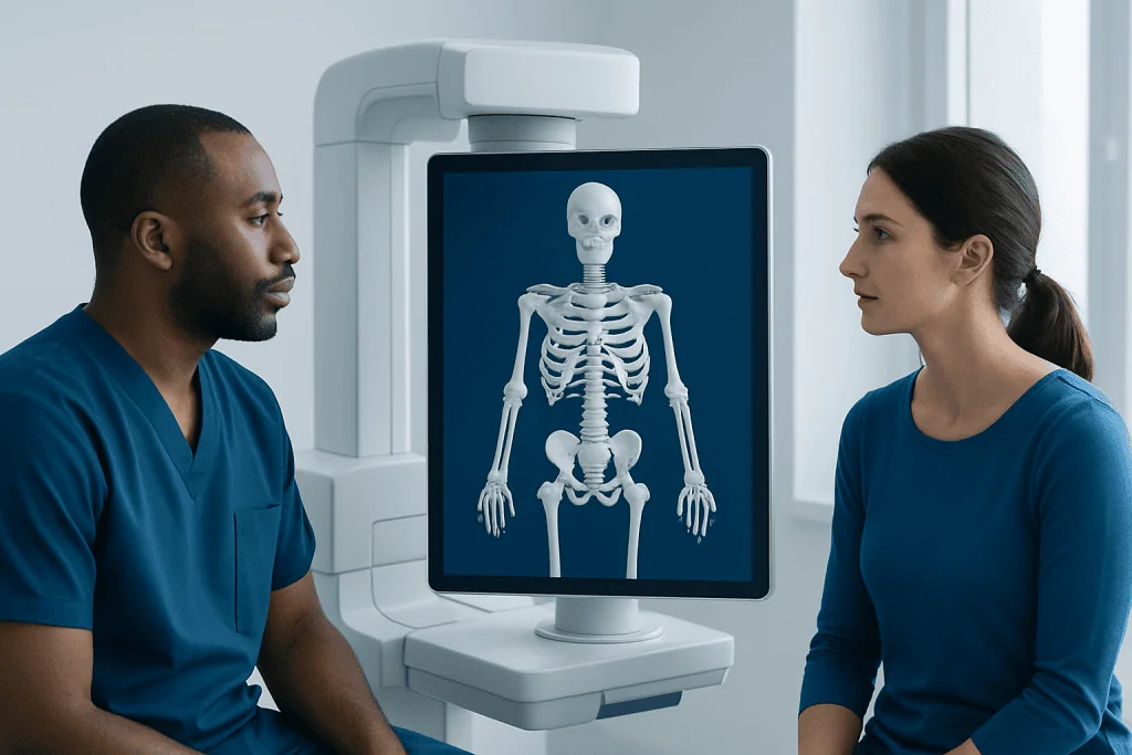
The adult skeletal system contains 206 bones, although this number is higher in infants because some bones fuse during growth. Bones vary in shape and size, but they are generally classified as long bones, short bones, flat bones, irregular bones, and sesamoid bones. This classification helps explain why different bones have different strengths and functions.
The axial skeleton forms the body’s central framework. It includes the skull, the vertebral column, and the rib cage. These bones mainly protect the brain, spinal cord, heart, and lungs while also helping maintain posture.
The appendicular skeleton includes the shoulder girdles, arms, hands, pelvis, legs, and feet. These bones are more directly involved in movement and weight-bearing. When clinicians evaluate fractures, joint problems, or spinal disorders, understanding whether the issue involves the axial or appendicular skeleton helps guide diagnosis and treatment.
- Axial skeleton: skull, hyoid bone, spine, ribs, sternum
- Appendicular skeleton: clavicle, scapula, arms, hands, pelvis, legs, feet
- Support tissues: cartilage, ligaments, joints, bone marrow
Major bones in a labeled human skeleton
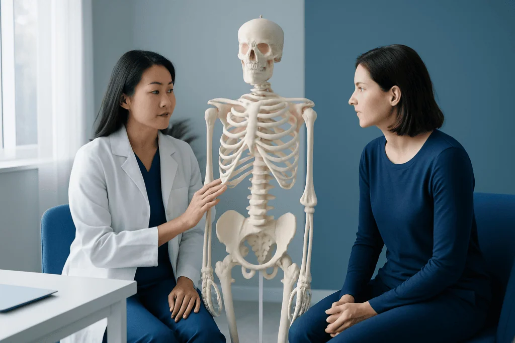
In the head, a labeled skeleton usually identifies the cranium, which protects the brain, and the facial bones, which shape the face and support the eyes, nose, and mouth. The mandible, or lower jaw, is the main movable bone of the skull. The hyoid bone in the neck is also important because it supports the tongue and muscles used in swallowing.
In the trunk, the vertebral column is divided into the cervical, thoracic, lumbar, sacral, and coccygeal regions. The rib cage includes the ribs and sternum and protects the heart and lungs. The pelvis, though often discussed with the appendicular skeleton, is a key weight-bearing structure linking the spine to the lower limbs.
In the upper limbs, the clavicle and scapula form the shoulder girdle. The arm includes the humerus, the forearm contains the radius and ulna, and the hand is made up of carpals, metacarpals, and phalanges. In the lower limbs, the femur is the thigh bone, the patella protects the knee, the tibia and fibula support the lower leg, and the foot includes tarsals, metatarsals, and phalanges.
These labels are not only useful for study. They also help patients understand imaging reports, fracture locations, and treatment plans, especially when discussing problems such as spinal disease, pelvic injury, or joint damage.
Functions of the skeletal system beyond body support
The most familiar function of the skeleton is support. Bones give the body structure, maintain height and posture, and provide attachment points for muscles. Without this framework, coordinated movement would not be possible.
The skeleton also protects vital organs. The skull protects the brain, the vertebrae protect the spinal cord, and the ribs shield the heart and lungs. The pelvis helps protect parts of the lower abdominal and reproductive organs. This protective role is one reason injuries to the skeleton can affect more than bones alone.
Bones are also living tissue with important metabolic functions. They store minerals such as calcium and phosphorus and release them as the body needs. Bone marrow, found inside many bones, produces blood cells. Red marrow contributes to red blood cells, white blood cells, and platelets, while yellow marrow is richer in fat storage.
The skeleton also plays a role in movement through joints and muscle attachment. Bones act as levers, while muscles generate force. If a bone is weak, a joint is damaged, or a ligament is torn, everyday movement may become painful or limited.
Joints, cartilage, and ligaments: the structures often missed on simple labels
A complete skeletal system labeled overview should include more than bone names. Joints are the places where bones meet, and they allow different types of motion. Some joints are fixed or nearly fixed, while others are highly mobile, such as the shoulder, hip, knee, and fingers.
Cartilage is smooth connective tissue that covers the ends of many bones within joints. It reduces friction and helps absorb impact. Ligaments are strong bands of connective tissue that connect bone to bone and help stabilize joints. Tendons, while part of the musculoskeletal system more broadly, connect muscle to bone and support movement.
Clinically, these structures matter because pain is not always caused by a broken bone. Joint inflammation, cartilage wear, ligament injury, and degenerative conditions may all cause symptoms. For example, osteoarthritis can affect cartilage and joint function, while spinal conditions may involve discs, vertebrae, and surrounding tissues together.
Understanding these details makes a labeled skeleton more practical. It helps explain why two people with pain in the same area may have very different causes, ranging from a stress fracture to a soft tissue injury or age-related joint disease.
How doctors assess skeletal system problems
Doctors assess skeletal complaints by combining a medical history, physical examination, and imaging or laboratory tests when needed. Symptoms such as pain, swelling, deformity, stiffness, limited motion, numbness, or difficulty bearing weight can help point to the affected area and likely cause. The evaluation also considers trauma, sports activity, posture, age, and medical conditions that affect bone strength.
Imaging is often central to diagnosis. Standard X-rays are commonly used to identify fractures, alignment problems, and many joint changes. In more complex cases, advanced studies such as MRI or CT scanning may help show soft tissues, spinal structures, subtle fractures, or detailed bone anatomy.
Bone density testing may be recommended when bone loss is suspected, especially in older adults or people with fracture risk factors. Blood tests can also be useful when a doctor is considering infection, inflammatory disease, vitamin deficiency, or metabolic bone disorders. If symptoms suggest the spine is involved, evaluation may focus on posture, reflexes, strength, and gait, especially in disorders such as scoliosis.
The aim of diagnosis is not only to name the affected bone or joint, but also to understand why the problem developed and whether it affects function, safety, or long-term bone health.
Common skeletal system conditions and treatment approaches
Skeletal system problems include fractures, sprains, dislocations, degenerative joint disease, spinal curvature disorders, bone infections, and low bone density. Some conditions happen suddenly after injury, while others develop gradually over time. Symptoms can range from mild stiffness to significant pain and difficulty moving.
Treatment depends on the cause, severity, age, general health, and whether the problem affects a bone, joint, or surrounding tissues. Many conditions improve with rest, activity modification, pain management, bracing, or physical therapy and rehabilitation. Structured rehabilitation can help restore strength, flexibility, balance, and function after injury or surgery.
Some skeletal conditions need more specialized care. Fractures may require immobilization or surgery. Advanced joint damage can sometimes be treated with procedures such as knee replacement when conservative care is no longer effective. Low bone density and osteoporosis often require long-term management to reduce fracture risk and support bone strength.
Near the end of the care pathway, some patients also need coordinated support from orthopedics, radiology, rehabilitation, and internal medicine specialists. Acibadem International’s multidisciplinary specialists in JCI-accredited hospitals diagnose and treat skeletal system disorders for international patients when advanced evaluation or treatment is needed.
Bone health, self-care, and when to seek medical care
Everyday bone health starts with balanced nutrition, regular physical activity, and fall prevention. Adequate calcium, vitamin D, and protein support bone maintenance, while weight-bearing and resistance exercises help preserve strength and balance. Avoiding smoking and limiting excess alcohol can also support healthier bones over time.
Good posture, safe lifting habits, protective sports equipment, and prompt attention to overuse symptoms may reduce strain on bones and joints. People at higher risk of bone loss, including older adults and those with certain medical conditions or long-term medication use, may benefit from individualized medical advice and screening.
Medical care should be sought promptly for severe pain after an injury, inability to bear weight, visible deformity, sudden swelling, suspected fracture, numbness, weakness, or loss of bladder or bowel control with back symptoms. A doctor should also assess persistent bone pain, repeated fractures, progressive curvature, or joint stiffness that interferes with daily life.
Although a skeletal system labeled chart is a useful learning tool, it cannot replace a professional evaluation. If symptoms do not improve, recur frequently, or raise concern, consulting a qualified doctor is the safest next step.
Frequently asked questions
What does skeletal system labeled mean?
It refers to a diagram or explanation that names the bones and related structures of the body. A good labeled overview usually includes major bones, joints, cartilage, and the division between the axial and appendicular skeleton.
How many bones are in the adult human skeleton?
Most adults have 206 bones. Babies have more because some bones are separate at birth and gradually fuse as the body grows.
What is the difference between the axial and appendicular skeleton?
The axial skeleton forms the body's central axis and includes the skull, spine, and rib cage. The appendicular skeleton includes the shoulders, arms, pelvis, legs, and feet, which are more involved in movement.
Why are joints and cartilage important in the skeletal system?
Joints allow bones to move in relation to one another, while cartilage helps reduce friction and absorb force. Problems in these structures can cause pain and stiffness even when the bones themselves are intact.
Can bone pain happen without a fracture?
Yes. Bone or joint pain can result from overuse, inflammation, arthritis, infection, low bone density, or soft tissue injury around the area. Persistent or unexplained pain should be evaluated by a doctor.
How can a person keep the skeletal system healthy?
Bone health is supported by regular exercise, adequate calcium and vitamin D, good nutrition, and avoiding tobacco. Fall prevention, proper posture, and timely medical advice for persistent symptoms are also important.
References
- World Health Organization
- National Institute of Arthritis and Musculoskeletal and Skin Diseases
- MedlinePlus
- American Academy of Orthopaedic Surgeons
- Mayo Clinic
This article is for general information only and is not a substitute for professional medical advice. Please consult a qualified doctor about your individual situation.
Joint and spine care in Turkey — expert assessment & treatment
JCI-accredited · board-certified surgeons · reply within 24h
Add us as a Preferred Source to see more of our trusted health content across Google Search, AI Overviews and Discover.

More from the Health Library

Mindbodygreen Podcast: An Evidence-Based Guide for Patients
Thoracic Duct: What Patients Need to Know

Cat Nap: A Complete Medical Overview

Clonazepam: An Evidence-Based Guide for Patients

White Spots on Lips: An Evidence-Based Guide for Patients

Herxheimer Reaction: An Evidence-Based Guide for Patients
Related Specialists

Assoc. Prof. Dr. Serap Aktaş Yıldırım
Anesthesiology
Prof. Dr. Cahide Soydaş Çınar
Cardiology
Dr. Murat Sarıkaş
Orthopedic Surgery & Traumatology
Prof. Dr. Hülya Dede
Gynecology & Obstetrics