Skin Cancer Screening What to Expect: How It Works, Results and What to Expect

A skin cancer screening usually involves a head-to-toe visual skin examination, including areas that are not easily self-checked. Patients may be asked to undress to underwear or wear a gown; clinicians protect privacy and explain the examination as they go.
Key Takeaways
- A skin cancer screening usually involves a head-to-toe visual skin examination, including areas that are not easily self-checked.
- Patients may be asked to undress to underwear or wear a gown; clinicians protect privacy and explain the examination as they go.
- A dermatologist can often identify lesions that need further assessment, but a biopsy is needed to confirm or rule out skin cancer.
- People with changing spots, a personal or family history of skin cancer, many moles, or substantial sun exposure may benefit from regular professional checks.
- Early assessment of a suspicious skin change can support timely diagnosis and treatment when needed.
Skin cancer screening is a clinical examination in which a dermatologist or qualified healthcare professional checks the skin for suspicious moles, spots, sores or changing lesions. Most appointments are quick and painless, and a biopsy is only needed if an area requires a closer laboratory diagnosis.
Skin Cancer Screening: What to Expect
Skin cancer screening is a focused skin examination performed by a dermatologist or another trained clinician to look for signs of skin cancer or precancerous changes. The clinician examines visible skin, asks about any spots that are new, changing, itchy, painful, bleeding or slow to heal, and may use a handheld magnifying device called a dermatoscope.
For most people, the appointment is straightforward and does not involve needles, scans or special preparation. The clinician may identify normal moles, explain which areas should be watched, or recommend monitoring, photography, removal or biopsy of a particular lesion. A screening examination does not itself diagnose every lesion; laboratory examination of a tissue sample is the usual way to confirm whether cancer is present.
Screening is especially useful because skin changes may develop in places a person cannot easily see, such as the scalp, back, behind the ears, soles, nails and genital region. It also provides an opportunity to discuss sun protection and individual risk factors.
Who May Benefit From a Professional Skin Check
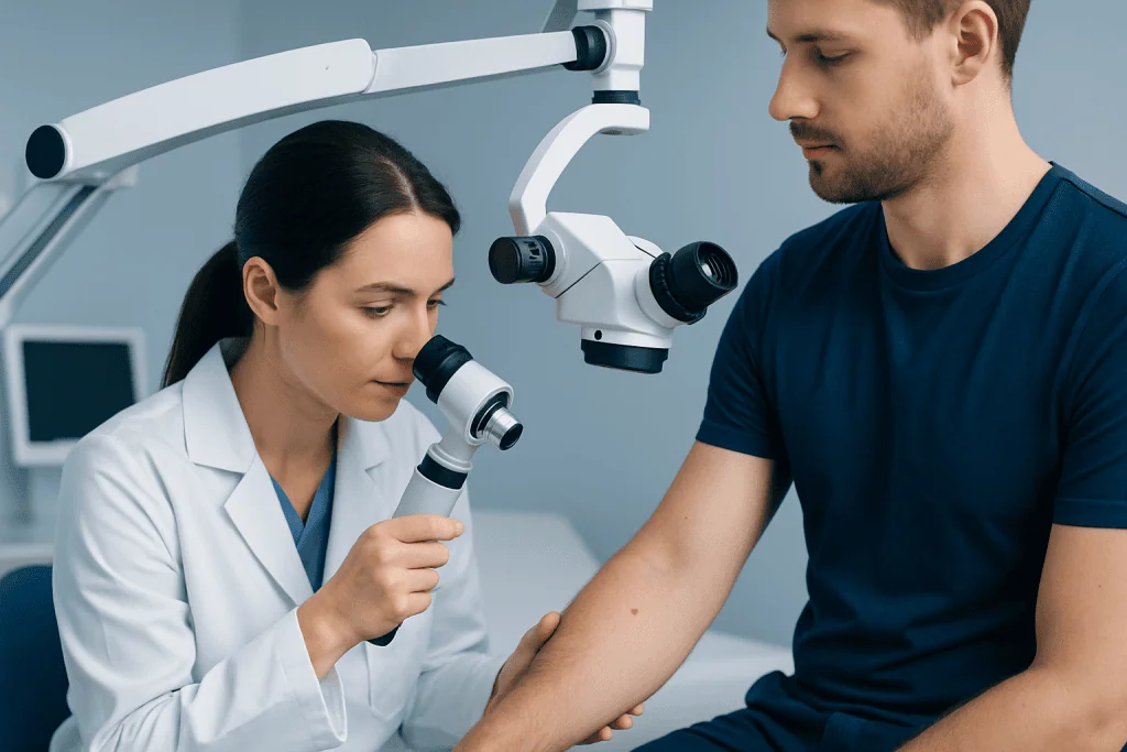
Anyone can develop skin cancer, and a person does not need to have very fair skin or a history of sunburn to seek an assessment. A professional skin check can be appropriate for someone who has noticed a concerning spot, has difficulty monitoring their own skin, or simply wants an expert baseline examination.
Regular checks may be particularly helpful for people with a previous skin cancer, a close family history of melanoma, numerous or unusual-looking moles, a weakened immune system, or a history of substantial ultraviolet exposure from sunlight or tanning devices. People who have received radiation treatment, immunosuppressive medicines or organ transplantation may also need individualized advice about surveillance.
The right screening interval varies. Some people may only need an examination when they notice a change, while others may be advised to return periodically because of their risk profile. A dermatologist can recommend a follow-up plan based on the skin findings, medical history and ability to perform self-checks.
How a Skin Cancer Screening Works: Step by Step
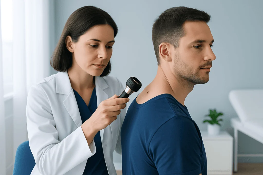
Before the examination, the clinician generally asks about personal and family history of skin cancer, severe sunburns, tanning bed use, medicines, immune conditions and previous biopsies. Patients should mention any specific lesion of concern, including how long it has been present and whether it has changed. Removing nail polish and heavy makeup beforehand can help the clinician examine nails and facial skin more completely.
During a full-body check, the patient may be asked to change into a gown or undress to underwear. The clinician typically examines the face, ears, scalp, neck, trunk, arms, hands, legs, feet and spaces between toes. Depending on symptoms, risk and consent, the examination may also include the buttocks, breasts and groin. A chaperone can usually be requested, and patients may ask the clinician to explain each part of the examination before it happens.
A dermatoscope shines light through a magnifying lens, allowing the clinician to see patterns of pigment and blood vessels below the surface of a lesion. The examination is painless. The clinician may measure a mole, document it in the medical record, or use clinical photographs for comparison at a later visit when appropriate.
- Most screening visits last only as long as needed to perform a careful examination and discuss findings.
- There is usually no recovery time after a visual skin check.
- If a lesion needs sampling, the clinician will explain the recommended biopsy technique, local anesthetic and wound care.
Results, Biopsy and the Recovery Timeline
Results of the visual examination are often discussed at the same visit. Many spots are benign, such as common moles, seborrheic keratoses or cherry angiomas. If a lesion looks concerning, the clinician may recommend observation over time, specialized imaging, removal, or a skin biopsy. A biopsy is a minor procedure in which a small sample or the entire lesion is removed and examined by a pathologist.
Local anesthetic is commonly used for a biopsy, so patients may feel a brief sting from the numbing injection but should not feel pain during tissue removal. Depending on the method, the area may be covered with a dressing, closed with stitches or left to heal naturally. Mild tenderness, redness or bruising can occur for a short time. The care team provides instructions about keeping the site clean, changing dressings and when stitches may need removal.
Pathology results can take several days or longer depending on the laboratory process and whether additional review is needed. If cancer is identified, treatment depends on the type, size, depth and location of the lesion. Options may include surgical removal, topical therapies, cryotherapy, radiation treatment or systemic treatment in selected cases. The benefit of screening is that it can help identify lesions early, when management may be simpler; the main limitations are that not every suspicious-looking lesion is cancer and a biopsy can leave a small scar.
Do You Wear a Bra for a Skin Check?
Patients may wear a bra to the appointment, but a complete skin check may require the clinician to examine skin that the bra covers. The goal is to inspect all skin surfaces, because suspicious lesions can occur on the chest, beneath the breasts, along the bra line and on the back.
Privacy should be respected throughout the visit. A patient can usually change into a gown, keep covered areas covered until they are being examined, and ask for a chaperone. It is reasonable to tell the clinician about preferences or concerns before the examination begins so the visit can be conducted respectfully and comfortably.
If a person prefers not to have a particular area examined, they can say so. However, it is important to mention any new, changing or symptomatic spot in that area, as the clinician can explain why an examination may be medically useful and discuss options.
Do Female Dermatologists Examine the Groin Area?
Yes. Female dermatologists, like all dermatologists, may examine the groin area when it is clinically relevant or as part of a full-body skin examination. Skin cancer and other skin conditions can occur in the genital area, inner thighs, buttocks and nearby skin, so excluding these areas may leave lesions unnoticed.
The examination should be professional, brief and based on informed consent. The clinician should explain what will be examined, preserve draping and privacy as much as possible, and respond to questions. Patients may request a chaperone regardless of the clinician’s gender, and they may ask to pause or stop the examination at any time.
A person does not need to feel embarrassed about reporting a mole, sore, dark patch, bleeding spot or persistent irritation in the groin. These concerns are part of routine dermatology care, and early assessment helps clarify the cause.
Can a Dermatologist Tell If You Have Skin Cancer Just by Looking?
A dermatologist can often recognize features that make a lesion more or less suspicious based on its appearance, location, history and dermatoscopic pattern. For example, asymmetry, irregular borders, varied color, enlargement, an evolving mole, or a sore that does not heal may warrant closer assessment. Clinical experience and dermatoscopy improve the accuracy of an examination.
However, looking alone cannot confirm every diagnosis. Some harmless lesions can resemble cancer, and some skin cancers can appear subtle. When there is uncertainty or concern, a biopsy and pathology review provide the definitive diagnosis. Patients should not assume a spot is harmless simply because it does not hurt or because it has been present for a long time.
Keeping track of changes between appointments is valuable. A person can check their skin regularly in good light, use mirrors or ask a trusted person to help inspect difficult-to-see areas, and report any lesion that changes in size, shape, color, sensation or behavior.
What Is the 2 Week Rule for Skin Cancer?
The “2 week rule” is often used as a practical prompt to seek medical advice for a skin sore, scab, ulcer, rash-like patch or other change that does not heal or improve within about two weeks. It is not a diagnostic test and does not mean every lesion present for two weeks is skin cancer. Many common skin problems take longer to resolve for non-cancerous reasons.
Still, a persistent lesion deserves evaluation, especially if it repeatedly bleeds, crusts, grows, becomes painful, changes color, or returns after appearing to heal. A new or changing pigmented mole should also be assessed rather than watched for a fixed two-week period.
Prompt assessment is particularly important for people with a history of skin cancer or immune suppression. A clinician can examine the area and determine whether reassurance, treatment for a benign condition, follow-up monitoring or biopsy is appropriate.
When to Seek Medical Care
Medical care should be sought for a new or changing mole, a spot with irregular shape or multiple colors, a lesion that enlarges, or a sore that repeatedly crusts, bleeds or does not heal. It is also sensible to arrange an appointment for persistent itching, tenderness or pain in a particular skin lesion, especially when there is no clear explanation.
People should not wait for a routine screening date if they notice a concerning change. A dermatologist can decide whether the lesion needs monitoring or testing. If a suspicious lesion is confirmed, care may involve dermatology, pathology, surgical specialists and oncology teams according to the diagnosis.
Acibadem International’s multidisciplinary specialists and JCI-accredited hospitals diagnose and treat skin conditions and skin cancers for international patients. A qualified clinician can provide individualized recommendations based on the appearance of the lesion and the person’s health history.
Frequently asked questions
How should someone prepare for a skin cancer screening?
It can help to arrive with clean skin and remove nail polish so the nails can be examined. Patients should note any spots that have changed and be ready to discuss their medical history, sun exposure and family history. They should avoid relying on makeup or self-tanner to cover a lesion of concern.
Is a skin cancer screening painful?
A routine visual skin examination is painless. If the clinician recommends a biopsy, local anesthetic is usually used to numb the area first. Mild soreness or tenderness may occur afterward, depending on the biopsy method.
How often should skin cancer screening be done?
There is no single schedule that suits everyone. The timing depends on personal risk factors, previous skin findings and whether a person has a history of skin cancer. A dermatologist can recommend an appropriate interval after an individual assessment.
What happens if a dermatologist finds a suspicious mole?
The dermatologist may examine it with a dermatoscope, photograph or measure it for follow-up, or recommend a biopsy. A biopsy allows a pathologist to examine cells under a microscope and provide a definitive diagnosis. The next steps depend on the pathology result.
Can skin cancer occur in areas not exposed to the sun?
Yes. Although ultraviolet exposure is an important risk factor for many skin cancers, suspicious lesions can occur on the soles of the feet, under nails, on the scalp, genitals and other less sun-exposed areas. This is one reason a full skin examination can be useful.
Should a person be worried about every new mole?
Not every new mole or spot is dangerous, and many skin changes are benign. However, a new lesion in adulthood or any spot that changes in appearance, bleeds, itches persistently or does not heal should be assessed by a healthcare professional. Early evaluation provides clarity and appropriate guidance.
References
- American Academy of Dermatology
- American Cancer Society
- National Cancer Institute
- World Health Organization
- NHS
This article is for general information only and is not a substitute for professional medical advice. Please consult a qualified doctor about your individual situation.
Skin Cancer in Turkey — costs, top hospitals & a free quote
JCI-accredited · board-certified surgeons · reply within 24h
Add us as a Preferred Source to see more of our trusted health content across Google Search, AI Overviews and Discover.