

Squamosal Suture: What Patients Need to Know

The squamosal suture is a normal skull seam between the temporal and parietal bones. In adults, it usually causes no symptoms and is often mentioned only on scans or after injury.
Key Takeaways
- The squamosal suture is a normal skull seam between the temporal and parietal bones.
- In adults, it usually causes no symptoms and is often mentioned only on scans or after injury.
- In infants and children, doctors may assess skull sutures if head shape seems unusual or growth patterns raise concern.
- Pain near the temple is more often related to muscles, the jaw, nerves, or injury than to the suture itself.
- Urgent medical care is important after significant head trauma or when neurological symptoms develop.
The squamosal suture is a normal fibrous joint on the side of the skull, where the temporal bone meets the parietal bone. Most people never notice it, but it can become relevant in head injury, imaging reports, or rare cases of abnormal skull suture fusion in infants and children.
What is the squamosal suture?
The squamosal suture is a natural joint made of tough fibrous tissue on each side of the skull. It connects the squamous part of the temporal bone, near the temple and above the ear, to the parietal bone. Like other skull sutures, it helps the skull form properly in infancy and childhood and then remains as a stable seam in later life.
For most patients, the squamosal suture is simply a normal anatomical feature rather than a medical problem. It may appear on an X-ray, CT scan, or MRI report, especially after a head injury or when doctors are evaluating headaches, skull shape, or facial pain. Seeing the term in a report does not automatically mean anything is wrong.
In babies and young children, skull sutures are especially important because they allow the skull to expand as the brain grows. If a suture closes too early, this can affect head shape and sometimes brain development. That broader condition is called craniosynostosis, and it may involve one or more skull sutures.
Where it is located and why it matters
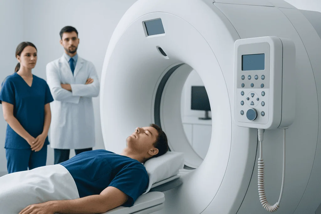
The squamosal suture runs along the side of the head in the temple region. Because of this location, patients may wonder whether pain above the ear or at the side of the head comes from the suture itself. In practice, the answer is usually no. The structures in this area include scalp tissues, jaw muscles, the temporomandibular joint, nerves, and blood vessels, all of which can also cause symptoms.
The suture matters most in three situations: normal skull growth in childhood, interpretation of imaging, and evaluation after trauma. On scans, doctors distinguish a normal suture from a skull fracture by looking at its shape, edges, and expected location. This is one reason specialist review of head imaging can be important after injury.
In children, the skull sutures work together rather than in isolation. A concern involving the squamosal suture often leads doctors to examine the entire skull, facial bones, and head growth pattern. If needed, further evaluation may involve craniosynostosis surgery planning or other pediatric craniofacial care, depending on the cause.
Can the squamosal suture cause symptoms?

By itself, a normal squamosal suture usually does not cause symptoms. Many people are unaware of it for their entire lives. When discomfort is felt in the temple area, doctors often first consider more common explanations such as tension headaches, migraine, jaw clenching, temporomandibular joint strain, scalp tenderness, sinus-related pressure, or a recent blow to the head.
Symptoms become more medically relevant when there has been trauma, visible swelling, a change in head shape, or neurological complaints. After injury, tenderness over the side of the skull may reflect a bruise, soft tissue injury, or in some cases a fracture near a normal suture line. In infants, a ridge along a skull suture or an unusual head shape may prompt assessment, although many head-shape changes have harmless causes.
Possible warning features that deserve attention include:
- Persistent or worsening pain after a head injury
- Vomiting, confusion, fainting, or unusual sleepiness
- A soft spot that seems tense or a head shape that is changing unexpectedly in a baby
- Seizures, weakness, vision changes, or difficulty speaking
- Swelling, bruising, or fluid leaking from the ear or nose after trauma
Conditions linked to the squamosal suture
The most important condition patients may hear about is premature fusion of skull sutures, known as craniosynostosis. Although the more commonly discussed sutures are coronal, sagittal, metopic, and lambdoid, the squamosal suture can also be involved in rare or complex cases. When this happens, specialists assess whether the finding is isolated, part of a broader skull growth pattern, or related to a syndrome.
Another reason the squamosal suture appears in medical care is trauma. A fracture in the temporal or parietal region can lie close to a normal suture, and this may be difficult for non-specialists to interpret. CT imaging is often used to clarify whether a line on a scan is a normal suture or an injury. In selected cases, patients may need neurosurgical care if there is bleeding, pressure on the brain, or a complex skull injury.
Less commonly, findings near the suture are part of a wider skull, ear, or craniofacial evaluation. In children with developmental concerns, specialists may combine clinical examination with imaging and growth measurements. If symptoms such as recurrent headaches or neurological signs suggest a brain-related cause rather than a skull-seam issue, doctors may also evaluate for disorders that affect the brain or surrounding structures, though these are not typical causes of isolated temple pain.
How doctors evaluate it
Evaluation starts with a careful history and physical examination. Doctors ask about age, symptoms, recent trauma, headaches, vision changes, developmental milestones, and whether there has been any change in head shape. In infants and children, they also measure head circumference and compare growth over time.
Imaging is chosen based on the reason for concern. CT scans are especially useful for viewing skull bones and sutures, while MRI is better for soft tissues and the brain itself. In young children, clinicians balance the need for accurate diagnosis with the goal of limiting unnecessary radiation exposure, so not every child with an unusual head shape needs immediate CT imaging.
When the main question is whether there is an intracranial problem, a doctor may recommend MRI to examine the brain and nearby structures in more detail. Radiologists and specialists look at the pattern of the suture, bone edges, surrounding tissues, and any associated signs of pressure or injury. This helps separate normal anatomical variation from true disease.
Treatment and follow-up
Treatment depends entirely on the underlying cause. A normal squamosal suture seen on imaging does not need treatment. If the issue is muscle tension, a jaw problem, or a mild soft tissue injury, care may focus on rest, posture, hydration, avoiding further trauma, and managing the contributing condition under medical guidance.
If there is a skull fracture, management ranges from observation to hospital care, depending on the type of fracture and whether the brain or nearby blood vessels are affected. Children with suspected premature suture fusion are usually assessed by a multidisciplinary team that may include pediatrics, neurosurgery, plastic or craniofacial surgery, and radiology. Treatment is not based on the suture name alone, but on head growth, symptoms, imaging, and overall development.
When surgery is needed for craniosynostosis or a related craniofacial problem, the goal is to support healthy skull shape and brain growth while reducing complications. Near the end of a patient’s care journey, some families may seek evaluation at centers experienced in complex pediatric skull conditions. Acibadem International’s multidisciplinary specialists and JCI-accredited hospitals diagnose and treat such conditions for international patients.
Self-care and when to seek medical care
For mild temple-area discomfort without red-flag symptoms, general self-care may include rest, staying hydrated, limiting jaw clenching, and avoiding another head impact. Patients should not assume pain is coming from the squamosal suture itself, especially if the pain is recurrent. A clinician can help distinguish headache, jaw, nerve, sinus, and injury-related causes.
Medical advice should be sought promptly if a person has persistent pain after a head injury, increasing swelling, vomiting, severe headache, confusion, fainting, weakness, speech changes, or seizures. In infants and children, parents should arrange medical review if they notice a new or unusual head shape, a firm ridge along the skull, developmental concerns, or a head circumference that seems to be changing unusually fast or slowly.
Urgent or emergency care is needed after significant trauma, especially when there is loss of consciousness, drowsiness, worsening headache, bleeding behind the ear, fluid from the nose or ear, or any neurological change. Early assessment helps doctors identify whether the concern is a simple soft tissue injury, a fracture, or a deeper problem that needs specialist treatment.
Frequently asked questions
Is the squamosal suture a normal part of the skull?
Yes. The squamosal suture is a normal fibrous joint between the temporal and parietal bones on the side of the skull. In most people, it does not cause symptoms or require treatment.
Can adults have problems with the squamosal suture?
Adults usually do not have problems from the suture itself. It is more often mentioned after head trauma or as an incidental finding on imaging. Pain in the temple area is commonly caused by other nearby structures rather than the suture.
Is squamosal suture the same as a skull fracture?
No. A squamosal suture is a normal anatomical seam, while a skull fracture is a break in the bone. On imaging, doctors use the line's location, appearance, and surrounding findings to tell the difference.
What does it mean if a baby's skull suture closes too early?
Early closure of a skull suture may suggest craniosynostosis. This can affect head shape and, in some cases, how the skull grows with the brain. A pediatric specialist can decide whether observation, imaging, or treatment is needed.
Should a patient worry if a scan mentions the squamosal suture?
Usually not. Reports often describe normal anatomy in detail, and the squamosal suture may simply be named because it was seen clearly. The meaning depends on the full report and the reason the scan was done.
When should someone seek urgent care for pain near the squamosal suture?
Urgent care is important after a head injury if there is severe headache, vomiting, confusion, loss of consciousness, seizures, weakness, or fluid from the ear or nose. These symptoms may point to a more serious injury and should be assessed promptly.
References
- American Academy of Pediatrics
- National Institute of Neurological Disorders and Stroke
- Radiological Society of North America
- Johns Hopkins Medicine
- MedlinePlus
This article is for general information only and is not a substitute for professional medical advice. Please consult a qualified doctor about your individual situation.
Joint and spine care in Turkey — expert assessment & treatment
JCI-accredited · board-certified surgeons · reply within 24h
Add us as a Preferred Source to see more of our trusted health content across Google Search, AI Overviews and Discover.

More from the Health Library



Related Specialists

Dr. Zeynep Göksu Saydam
Emergency Service
Prof. Dr. Tevfik Rifki Evrenkaya
Internal Medicine
Dr. Fatih Kaya
General Surgery
Assoc. Prof. Dr. Nefise Nazlı Yenigül
Gynecology & Obstetrics