Stereotactic Breast Biopsy Explained: Causes, Management, and When to See a Doctor
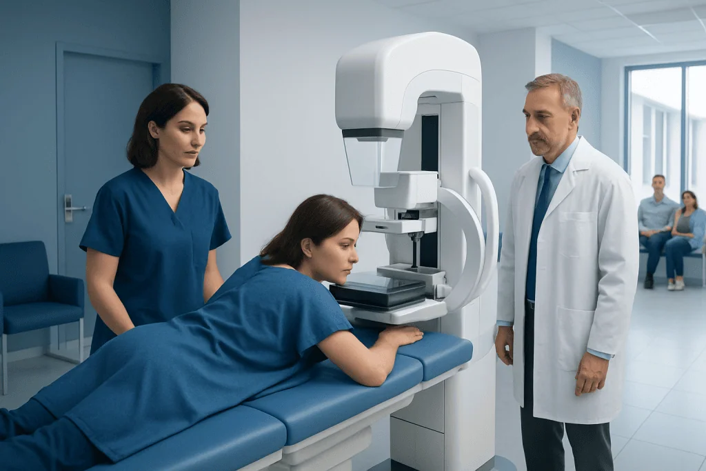
Stereotactic breast biopsy uses mammogram images from different angles to guide a needle to a precise area in the breast. It is commonly recommended for suspicious calcifications or other abnormalities seen on a mammogram but not clearly felt on examination.
Key Takeaways
- Stereotactic breast biopsy uses mammogram images from different angles to guide a needle to a precise area in the breast.
- It is commonly recommended for suspicious calcifications or other abnormalities seen on a mammogram but not clearly felt on examination.
- The procedure is usually done with local anesthetic, takes a short time, and most people return to normal activities within a day.
- Results may show benign changes, high-risk lesions, or breast cancer, which helps guide the next step in care.
- Medical advice should be sought promptly for a new breast lump, bloody nipple discharge, skin changes, or concerning biopsy-site symptoms.
Stereotactic breast biopsy is a minimally invasive procedure that uses mammogram-guided imaging to remove small tissue samples from an area seen on breast imaging, often tiny calcifications or a subtle abnormality that cannot be felt. It helps doctors find out whether a breast change is benign or needs further treatment, while usually avoiding surgery for diagnosis.
Overview: what a stereotactic breast biopsy is
A stereotactic breast biopsy is a needle biopsy that uses mammography images taken from different angles to locate a breast abnormality with high precision. It is most often used when a mammogram shows an area that needs closer evaluation, especially tiny calcium deposits called microcalcifications or a small area of architectural distortion that may be difficult to see on ultrasound.
In practical terms, the procedure allows a radiologist to take small tissue samples without open surgery. The breast is compressed, imaging is used to calculate the exact position of the target, and a biopsy needle is guided to that spot. The tissue is then sent to a pathology laboratory, where it is examined under a microscope.
This type of biopsy is considered minimally invasive and is often preferred when the abnormality is visible on mammography but not easy to feel. It can provide a diagnosis with less scarring, less recovery time, and lower procedural burden than a surgical biopsy.
Why it may be recommended
Doctors usually recommend a stereotactic breast biopsy after an imaging test shows a change that cannot be confidently labeled benign. The goal is not to assume cancer, but to identify the cause of the imaging finding as accurately as possible. Many biopsies ultimately show noncancerous conditions, but tissue sampling is sometimes the only reliable way to know.
Common reasons include suspicious clusters of calcifications, a small density seen on a screening mammogram, or a subtle distortion in the breast tissue pattern. These findings may be too small or too deep to guide by touch alone. When an abnormality is better seen on mammography than on ultrasound, stereotactic guidance can be especially useful.
Sometimes the biopsy is part of a broader evaluation of breast cancer risk or diagnosis after an abnormal screening result. It may also be used to clarify findings that remain uncertain after additional mammogram views. The recommendation depends on the radiologist’s assessment, breast density, the appearance of the lesion, and whether other imaging methods can target the area.
What happens before, during, and after the procedure

Before the biopsy, the medical team reviews breast imaging, current medications, allergies, and any bleeding risks. Patients are often advised to mention blood thinners, aspirin use, pregnancy, or a history of fainting during medical procedures. The team may suggest avoiding certain products such as deodorant or powders on the day of breast imaging if additional scans are needed.
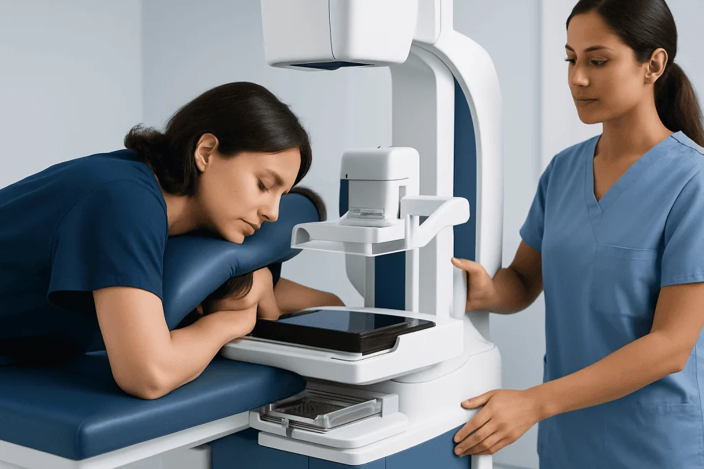
During the procedure, the patient may lie face down on a specialized table with the breast positioned through an opening, or sit upright depending on the equipment used. The breast is compressed similarly to a mammogram, and local anesthetic is used to numb the skin and deeper tissue. The radiologist then uses computer-guided coordinates from mammographic images to place the needle accurately. Several tissue samples are usually taken, and a tiny marker clip may be left at the site so the area can be identified on future imaging.
Afterward, pressure is applied to reduce bleeding, and the site is covered with a small dressing. Mild bruising, soreness, or swelling is common for a few days. Most people can go home soon after the procedure and resume light daily activities the same day, though heavy exercise and strenuous lifting are often postponed briefly. If a suspicious area later requires surgery, the biopsy clip helps the surgeon identify the exact location and can support further planning such as breast cancer surgery.
What the results can show
Biopsy results usually fall into several broad categories. A benign result means no cancer is found and the tissue change may be explained by noncancerous conditions such as fibrocystic changes, fibroadenoma, fat necrosis, or benign calcifications. In these cases, the doctor compares the pathology with the imaging to make sure the result fully explains what was seen on the mammogram.
Some biopsies identify high-risk or borderline changes, such as atypical ductal hyperplasia, lobular neoplasia, radial scar, or papilloma. These findings are not always cancer, but they can increase future risk or suggest that a nearby area may need further evaluation. A surgeon or breast specialist may recommend additional imaging, closer surveillance, or surgery depending on the pathology and imaging findings.
If the biopsy shows cancer, the result helps determine the next steps in staging and treatment. Additional tests may assess the type of cancer and whether it is invasive or noninvasive. Further care may involve a multidisciplinary breast team, and treatment can include surgery, medical oncology, and radiation planning such as radiation oncology when appropriate.
Benefits, limits, and possible risks
The main benefit of stereotactic breast biopsy is precision. It can target small abnormalities that are visible on mammography but not easily found with a physical exam. Compared with open surgical biopsy, it usually causes less discomfort, requires only local anesthetic, leaves a smaller scar, and shortens recovery time.
Like all procedures, it also has limits. Not every breast abnormality can be sampled this way. If the lesion is better seen on ultrasound or MRI, a different biopsy method may be more suitable. In some cases, if the target is very close to the chest wall or difficult to access safely, the doctor may recommend another approach. Rarely, the sample may not fully explain the imaging finding, and additional biopsy or surgery is needed.
Risks are generally low but can include bruising, bleeding, pain, swelling, or infection. A small hematoma, or collection of blood under the skin, may develop and usually resolves with time. Very rarely, the biopsy may miss part of the abnormal area, which is why radiology-pathology correlation and follow-up imaging are important. This procedure is one of several methods used within modern breast health services to reach an accurate diagnosis while minimizing unnecessary surgery.
Recovery, self-care, and follow-up
Most people recover quickly after a stereotactic breast biopsy. Mild tenderness, a pulling sensation, and skin discoloration can occur for a short period. Applying a cold pack intermittently during the first day and wearing a supportive bra may help improve comfort. The care team usually gives clear instructions about bathing, dressing changes, and when regular exercise can be restarted.
Follow-up matters because the biopsy result must match the imaging appearance. If the pathology is benign and clearly explains the mammogram finding, the doctor may recommend routine screening or a short-term follow-up mammogram. If the pathology is high-risk, discordant, or malignant, additional consultations are arranged to discuss the next step.
Patients should keep copies of pathology reports and imaging recommendations when possible. This can be especially helpful if care continues across different clinics. Near the end of the diagnostic process, some people benefit from evaluation in a specialized center; Acibadem International’s multidisciplinary specialists and JCI-accredited hospitals diagnose and treat breast conditions for international patients.
When to seek medical care
Medical advice should be sought if a mammogram report recommends tissue sampling or if there is uncertainty about the next step after an abnormal breast imaging result. A new breast lump, persistent focal pain, bloody or spontaneous nipple discharge, skin dimpling, redness that does not improve, or nipple inversion should also be assessed by a qualified doctor.
After a stereotactic breast biopsy, prompt medical review is important if there is increasing redness, warmth, pus, fever, severe swelling, ongoing bleeding, or pain that is worsening rather than improving. These symptoms do not always mean a serious problem, but they can suggest infection or a larger hematoma and should not be ignored.
It is also reasonable to ask for clarification if the pathology report is difficult to understand, if the biopsy result and imaging findings seem not to match, or if a second opinion is desired. Clear communication with a breast radiologist, surgeon, or breast specialist can help patients understand the diagnosis and the plan ahead.
Frequently asked questions
Is a stereotactic breast biopsy painful?
Most people feel pressure rather than sharp pain because local anesthetic is used to numb the area. Some soreness or bruising afterward is common, but it is usually mild and temporary.
How long does a stereotactic breast biopsy take?
The procedure itself is often completed within about an hour, though timing can vary by imaging setup and how easily the area is reached. Patients should allow extra time for preparation, positioning, and post-procedure observation.
Why would a doctor choose stereotactic biopsy instead of ultrasound-guided biopsy?
Stereotactic guidance is often chosen when the abnormality is best seen on mammography, especially microcalcifications or subtle tissue distortion. If a lesion is clearly visible on ultrasound, ultrasound-guided biopsy may be preferred because it is often simpler and does not require breast compression.
Can a benign result still need more testing?
Yes. If the pathology result does not adequately explain what was seen on imaging, additional biopsy, closer follow-up, or surgery may be recommended. This is called imaging-pathology correlation and helps reduce the chance of a missed diagnosis.
Will a marker clip be left in the breast?
In many cases, a tiny marker clip is placed at the biopsy site. It is usually safe, does not set off security systems, and helps doctors identify the exact location on future mammograms or if surgery is later needed.
How soon are results available?
Results are often available within several business days, depending on the pathology laboratory and whether special testing is required. The care team explains the findings and whether routine follow-up, additional imaging, or treatment is recommended.
References
- American Cancer Society
- National Cancer Institute
- Radiological Society of North America
- American College of Radiology
- Breastcancer.org
This article is for general information only and is not a substitute for professional medical advice. Please consult a qualified doctor about your individual situation.
Oncology care in Turkey — second opinion and treatment plan
JCI-accredited · board-certified surgeons · reply within 24h
Add us as a Preferred Source to see more of our trusted health content across Google Search, AI Overviews and Discover.