

Thoracic Cavity — Explained by Medical Evidence, Not Myths
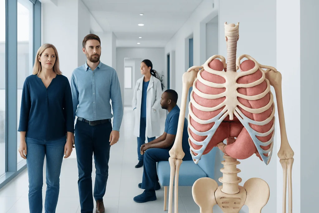
The thoracic cavity is the chest compartment between the neck and abdomen, enclosed by the ribs, sternum, thoracic spine, and diaphragm. It houses the lungs, heart, pleura, mediastinum, major blood vessels, esophagus, thymus, nerves, and lymphatic structures.
Key Takeaways
- The thoracic cavity is the chest compartment between the neck and abdomen, enclosed by the ribs, sternum, thoracic spine, and diaphragm.
- It houses the lungs, heart, pleura, mediastinum, major blood vessels, esophagus, thymus, nerves, and lymphatic structures.
- Its main roles are protecting vital organs, supporting breathing, and allowing the heart and lungs to move and function normally.
- Symptoms related to thoracic cavity problems can include chest pain, breathlessness, cough, fever, palpitations, or trouble swallowing.
- Diagnosis may involve a physical exam, chest X-ray, CT, MRI, ultrasound, blood tests, lung function testing, or endoscopy depending on the cause.
- Urgent care is important for sudden severe chest pain, major breathing difficulty, bluish lips, fainting, or symptoms after chest injury.
The thoracic cavity is the space inside the chest that contains and protects the heart, lungs, major blood vessels, esophagus, and other vital structures. Understanding its anatomy and function helps explain common symptoms such as chest pain, shortness of breath, and cough, and why some conditions need prompt medical evaluation.
Overview: what the thoracic cavity is
The thoracic cavity is the space inside the chest. It sits between the neck and the abdomen and is surrounded by the rib cage, breastbone, thoracic spine, and diaphragm. This enclosed space protects several organs that are essential for breathing, circulation, and swallowing.
In simple terms, the thoracic cavity is much more than a hollow chest area. It is a carefully organized compartment that allows the lungs to expand, the heart to beat, and major blood vessels and nerves to pass safely between different parts of the body. Because so many vital structures are located here, symptoms that seem similar at first—such as chest discomfort or shortness of breath—can come from different organs within the same region.
The thoracic cavity is usually divided into the right and left pleural cavities, which contain the lungs, and the mediastinum, the central space between them. The mediastinum contains the heart, great vessels, trachea, esophagus, thymus, lymph nodes, and important nerves. This structure-based view is useful because it helps explain how doctors think about chest symptoms and imaging findings.
Anatomy and organs inside the thoracic cavity
The bony and muscular walls of the thoracic cavity provide both protection and movement. The ribs and sternum form the front and sides, the thoracic vertebrae form the back, and the diaphragm forms the floor. Between the ribs are intercostal muscles, which help expand and contract the chest during breathing.
Inside this space are several major structures. The lungs sit in the right and left pleural cavities, each surrounded by a thin lining called the pleura. The heart lies centrally within the mediastinum and is enclosed by the pericardium. Nearby are the aorta, vena cava, pulmonary arteries and veins, trachea, bronchi, esophagus, lymphatic channels, and nerves.
The main contents of the thoracic cavity include:
- Lungs and pleura
- Heart and pericardium
- Trachea and major bronchi
- Aorta and other major blood vessels
- Esophagus
- Thymus, especially more prominent in childhood
- Lymph nodes and lymphatic vessels
- Important nerves, including the vagus and phrenic nerves
Although the thoracic cavity is often associated mainly with the lungs, the central chest structures are just as important. Conditions affecting the mediastinum, pleura, or chest wall can also cause symptoms and may require imaging or specialist evaluation.
How the thoracic cavity works
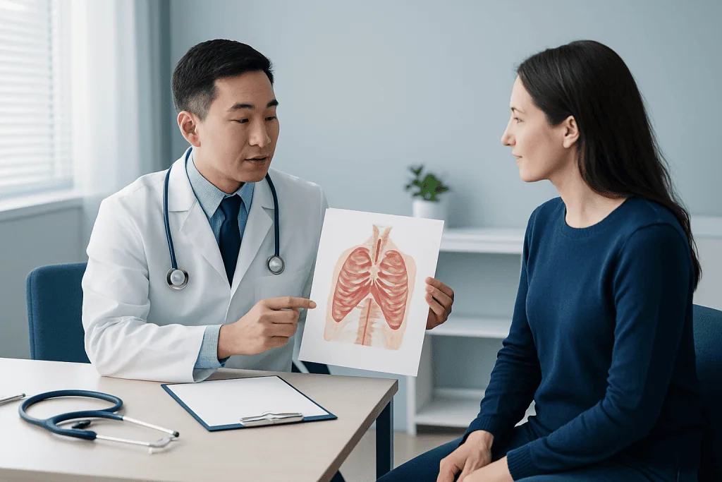
The thoracic cavity supports breathing through pressure changes. When the diaphragm contracts and moves downward, and the intercostal muscles lift and expand the chest wall, the volume inside the thoracic cavity increases. This lowers pressure and allows air to flow into the lungs. When these muscles relax, the chest recoils and air moves out.
The cavity also protects the heart and major vessels while allowing them enough room to function. The lungs need a smooth, lubricated pleural surface to glide against the chest wall, and the heart needs space to expand and pump. The diaphragm plays a dual role by separating the chest from the abdomen and helping drive normal breathing.
Another important function is as a passageway. The thoracic cavity contains structures that connect the mouth and neck to the lungs, stomach, and abdominal organs, including the trachea and esophagus. Because of this, symptoms such as cough, wheezing, reflux-related discomfort, or trouble swallowing can all be linked to the same general region even when the causes differ.
Common symptoms and conditions involving the thoracic cavity
Problems in the thoracic cavity may affect the lungs, heart, pleura, chest wall, airways, esophagus, or mediastinum. Symptoms vary depending on which structure is involved, but common complaints include chest pain, pressure, cough, shortness of breath, wheezing, fever, palpitations, or difficulty swallowing. Some symptoms are mild and temporary, while others need prompt medical care.
Examples of conditions that may involve the thoracic cavity include infections such as pneumonia, inflammation of the pleura or pericardium, fluid around the lungs, collapsed lung, trauma, rib fractures, tumors, blood clots in the lungs, and disorders of the esophagus. Certain symptoms may overlap with heart or lung disease, which is why self-diagnosis can be unreliable. Related conditions may include lung cancer and pneumonia when symptoms or imaging suggest a disease affecting lung tissue within the chest.
Some thoracic cavity conditions develop suddenly, such as a pneumothorax after injury or severe coughing, while others progress gradually, such as chronic inflammatory or growth-related conditions. Pain can also come from the chest wall muscles or joints rather than internal organs, but chest symptoms should still be assessed in context, especially in older adults or people with known heart or lung disease.
Doctors often look for symptom patterns, including whether pain changes with breathing, movement, eating, exertion, or body position. That information helps narrow the possible source within the thoracic cavity and guides further testing.
Causes and risk factors
Thoracic cavity problems do not have a single cause because this part of the body contains many organ systems. Infections may be caused by bacteria, viruses, or fungi. Breathing-related conditions can result from smoking, environmental exposures, asthma, chronic lung disease, or blood clots. Cardiac causes may relate to coronary artery disease, inflammation, rhythm problems, or structural heart disease.
Injuries are another important cause. Falls, road traffic accidents, sports trauma, or blunt impacts can affect the ribs, lungs, pleura, heart, or large vessels. Less obvious factors such as severe acid reflux, repeated vomiting, or swallowing disorders can also cause chest symptoms by affecting the esophagus within the thoracic cavity.
Risk factors depend on the underlying condition but may include:
- Smoking or secondhand smoke exposure
- Air pollution or occupational dust and chemical exposure
- Recent infection or weakened immunity
- Older age
- High blood pressure, diabetes, or high cholesterol
- Recent surgery, immobility, or a history of blood clots
- Chest trauma
- A personal or family history of heart or lung disease
Knowing these risk factors can help patients and doctors decide how urgently symptoms should be evaluated. Even when symptoms seem mild, the combination of chest discomfort with significant risk factors often justifies medical review.
How doctors diagnose thoracic cavity problems
Diagnosis starts with a medical history and physical examination. A doctor may ask where the symptom is felt, when it began, whether it is linked to breathing or activity, and whether there are associated symptoms such as fever, cough, fainting, or leg swelling. On examination, they may listen to the lungs and heart, check oxygen levels, and look for tenderness or signs of injury.
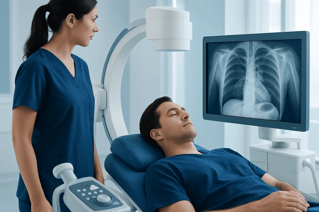
Imaging is often central to evaluating the thoracic cavity. A chest X-ray can show pneumonia, fluid around the lungs, an enlarged heart, or some forms of lung collapse. CT scans provide more detailed images of the lungs, pleura, vessels, mediastinum, and chest wall. In selected cases, doctors may also use MRI, ultrasound, echocardiography, or nuclear medicine tests.
Other tests depend on the likely cause. Blood tests may check for infection, inflammation, anemia, or cardiac injury markers. Lung function tests can help assess airway or chronic lung conditions. Endoscopy may be useful for swallowing symptoms or suspected esophageal disease. If a procedure is needed for diagnosis or treatment, doctors may consider bronchoscopy to view the airways or sample tissue.
Because chest symptoms can overlap across different diseases, diagnosis often involves ruling out urgent causes first. The goal is not only to identify a condition but also to determine how serious it is and whether treatment is needed quickly.
Treatment options and supportive care
Treatment for a thoracic cavity problem depends entirely on the underlying cause. Infections may be managed with appropriate antimicrobial therapy and supportive care. Inflammatory conditions may respond to rest, monitoring, and medications recommended by a clinician. Breathing problems can require inhaled medicines, oxygen, or hospital-based care if symptoms are more severe.
Some conditions need procedures rather than medication alone. Fluid around the lungs may require drainage, and a collapsed lung may need observation or intervention. Suspected growths or complex abnormalities may need tissue sampling, surgery, or specialist procedures. In carefully selected cases, doctors may use thoracic surgery for chest conditions affecting the lungs, pleura, mediastinum, or chest wall, or lung transplant evaluation for advanced end-stage lung disease.
Self-care has a role, but it should not replace medical assessment for significant chest symptoms. Rest, hydration, avoiding smoking, and following a clinician’s advice can support recovery. Patients should avoid taking unprescribed antibiotics or ignoring persistent symptoms, since delayed diagnosis can make treatment more difficult.
Near the end of the care pathway, some people may benefit from a multidisciplinary approach involving pulmonology, cardiology, thoracic surgery, radiology, and rehabilitation. Acibadem International’s multidisciplinary specialists in JCI-accredited hospitals diagnose and treat thoracic conditions for international patients when specialist evaluation is needed.
Prevention, self-care, and when to seek medical care
Not every thoracic cavity condition can be prevented, but healthy habits can lower risk. Avoiding tobacco, staying physically active, keeping vaccinations up to date, using protective equipment in hazardous workplaces, and managing chronic conditions such as high blood pressure or asthma all support chest health. Good posture, core strength, and safe lifting can also help reduce some chest wall strain and discomfort.
People with known heart or lung disease should follow their treatment plan and report any change from their usual symptoms. Ongoing cough, recurrent chest infections, exercise intolerance, or new swallowing problems should not be ignored. Early evaluation can help identify treatable causes before complications develop.
Medical care should be sought promptly for chest pain that is severe, sudden, or associated with shortness of breath, sweating, fainting, bluish lips, confusion, or pain spreading to the arm, back, jaw, or upper abdomen. Urgent assessment is also important after chest injury, if breathing becomes difficult, or if there is coughing up blood. If symptoms are mild but persistent, arranging a non-emergency medical visit is still the safest approach.
Patients who need a specialist workup may be referred for advanced imaging, esophageal manometry for selected swallowing-related symptoms, or organ-specific care depending on the suspected source of the problem within the chest.
Frequently asked questions
What is the thoracic cavity in simple terms?
The thoracic cavity is the space inside the chest. It protects the lungs, heart, and other important structures and helps the body breathe and circulate blood normally.
Which organs are in the thoracic cavity?
The thoracic cavity contains the lungs, heart, pleura, trachea, major bronchi, esophagus, thymus, major blood vessels, lymphatic structures, and important nerves. These structures are arranged within the pleural cavities and the mediastinum.
Is the thoracic cavity the same as the lungs?
No. The lungs are two organs located within the thoracic cavity, but the cavity also includes the heart, blood vessels, airway passages, esophagus, and supporting tissues. This is why chest symptoms can have many different causes.
What causes pain in the thoracic cavity?
Pain in the thoracic cavity can come from the lungs, pleura, heart, esophagus, ribs, muscles, or nerves. Causes range from muscle strain and infection to heart disease, blood clots, or trauma, so the full symptom pattern matters.
How do doctors check the thoracic cavity?
Doctors usually begin with a history, physical exam, and basic tests such as oxygen measurement and chest X-ray. Depending on the symptoms, they may also use CT, MRI, ultrasound, heart tests, blood tests, lung function tests, or endoscopy.
When is a thoracic cavity problem an emergency?
Emergency care is needed for sudden severe chest pain, serious trouble breathing, fainting, bluish lips, coughing up blood, or symptoms after significant chest injury. These symptoms can point to conditions that require urgent assessment.
References
- National Heart, Lung, and Blood Institute
- American Lung Association
- Merck Manual Consumer Version
- Mayo Clinic
- MedlinePlus
This article is for general information only and is not a substitute for professional medical advice. Please consult a qualified doctor about your individual situation.
Joint and spine care in Turkey — expert assessment & treatment
JCI-accredited · board-certified surgeons · reply within 24h
Add us as a Preferred Source to see more of our trusted health content across Google Search, AI Overviews and Discover.

More from the Health Library

Bronchoalveolar Lavage: An Evidence-Based Guide for Patients

Clobazam: An Evidence-Based Guide for Patients

Carotene: A Complete Medical Overview

Fitness for Hips: An Evidence-Based Guide for Patients

Ashwagandha Benefits — Explained by Medical Evidence, Not Myths

Carbidopa Levodopa — Explained by Medical Evidence, Not Myths
Oral & Dental Health Specialists at Acibadem

Dt. Akif Aydın Şen
Oral & Dental Health
Dt. Alara Güler
Oral & Dental Health
Dt. Alara Naz Kenir Çelik
Oral & Dental Health
Dr. Alen Palancıoğlu
Oral Dental & Maxillofacial Surgery